Abstract
Introduction:
In this study, the cleaning ability of a stainless-steel rotary instrument called Gentlefile, was compared with three nickel-titanium rotary instruments.
Materials and Methods:
In this in vitro study, forty mandibular single-rooted premolars were randomly assorted into four groups: Gentlefile, ProTper Universal, RaCe files, and XP-Endo Finisher/ProTaper Universal system (n=10). Final instrumentation was done using the aforementioned files with 5.25% sodium hypochlorite and normal saline for root canal irrigation. Debris and smear layers were observed by the scanning electron microscope on the canal walls in the coronal, middle, and apical third of the root level, through a 4-point scoring system. The chi-square test and Kruskal-Wallis were used for data analysis.
Results:
The Gentlefile demonstrated a promising outcome in smear layer clearance and debris removal compared with the other three rotary systems (P<0.05), specifically at the apical third of the root canal. Based on chi-square test results, there was a significant relationship between root canal cleaning (three levels of cleanliness) in ProTaper Universal (P=0.004) and Gentlefile (P=0.04) groups. Neither of the investigated systems achieved complete cleanliness.
Conclusion:
The Gentlefile rotary system can be capable of cleaning the apical third of root canals more than the other three groups including Protaper Universal, RaCe, and XP-Endo Finisher.
Key Words: Cleanliness, Instrumentation, Root Canal Preparation, Smear Layer
Introduction
One of the crucial principles of endodontic treatment is properly preparing the root canal by removing pulp tissue, all microorganisms, and shaping the canal system while preserving its original structure and creating an apical seal [1, 2]. This process increases the chances of successful endodontic treatment [3, 4]. Initially, manual files were used for removing organic matter and shaping the canal during instrumentation [2, 5]. However, studies have shown that manual files are not very effective in comprehensive debridement and may result in debris accumulation on the canal walls [6], making the procedure lengthy, especially for complex canals [7]. Furthermore, stainless steel (SS) hand files are not very efficient in removing dentin from the apical section of the canal; although they are effective in removing dentin form the coronal section of the canal [5, 8]. The possible benefit of rotary instrumentation over other instrumentation techniques such as manual preparation with SS hand files regarding cleaning and disinfecting effects would be irrigant warming and/or turbulence caused by the mechanical rotation of instruments [9]. Recently, Neelkantan et al. [3] showed that supplementary irrigation agitation with the Finisher GF Brush during the final phase of root canal treatment significantly enhanced the efficacy of canal debridement achieved through the use of Gentlefile and a 25-gauge, 0.04 taper rotary nickel-titanium instrument.
As a result, achieving a successful endodontic treatment can be a difficult process [10]. However, the introduction of Nickel-Titanium (NiTi) files has led to significant advancements in these tools over the past 20 years. NiTi files possess a unique super-elasticity property due to the use of memory shape alloys. Nonetheless, it has been observed that these files can cause microcracks in dentin [11-13]. During preparation, these instruments may fracture due to torsion and cyclic fatigue. In teeth with complex anatomy where the cutting tip does not contact the canal wall, the use of rotary files can be challenging. In such situations, irrigation is preferred over mechanical removal by filing [14-16].
A major challenge in root canal therapy is removing the organic particle debris, which forms during the cleaning and shaping of the canal using different endodontic file systems [17, 18]. It is a superficial layer that consists of organic materials, dentin debris, pulp residues, microorganisms, and their products [19, 20]. Researchers have found that smear layer removal helps to improve the root canal irrigation [6, 21]. Furthermore, it prevents coronal and apical leaks while ensuring a better bond between dentin and sealer [21, 22]. However, a review study by Torabinejad et al. showed conflicting results obtained from numerous in vitro studies regarding the significance of the presence or the removal of the smear layer [23].
Increased canal curvature results in heightened stress on the rotary file system employed during root canal therapy and, subsequently, on the root canal itself [24]. The concentration of stress in the root canal can lead to canal transportation, straightening, and deviation, which in turn result in thinner areas of dentin. The weakening of the root structure due to thinner dentin heightens the risk for apical cracking, which is a type of vertical root fracture (VRF) [11]. Due to NiTi's limitations in rotary systems, SS files have drawn researchers' attention. Among SS rotary system, Gentle file (GF, Tornado, Houston, TX, USA), which offers more flexibility, is a special SS rotary system. Compared to NiTi instruments, it is more flexible [25], abrades the dental canal walls, and prevents cutting through the dentin relatively to NiTi instruments [3].
GF system includes six files: one coronal file (18 mm, gray) and five 25 mm final files (yellow, red, blue, green, and black). Each file is made of three SS wires, which separates the file into a bilayer apical section and a three-layer upper section [26]. These files have an inactive non-working tip with a 4% taper in the apical section. A large amount of debris is removed by pulling the file gently along the canal walls. Using this file requires a speed of 6500 rpm handpiece. Its speed changes automatically without the operator's control. A unique system, not every handpiece with that speed, can be used [4]. ProTaper Universal files (Dentsply Maillefer, Ballaigues, Switzerland) have been designed with a progressive taper which improves both flexibility and cutting efficiency. Furthermore, reduced torsional loading, canal transportation, and cyclic fatigue have been reported by using this system in root canal treatments [27, 28]. The RaCe (reamer with alternating cutting edges) system (FGK Dentaire, La-Chaux-de-Fonds, Switzerland) is easy to apply with the special design of the files which prevents the screw-in effects and provides a better control of the instrument advancement for the endodontist, with proper flexibility and improvement in curved root canals [29]. The XP-Endo Finisher (FGK Dentaire, La Chaux-de-Fonds, Switzerland) was introduced to be used after any root canal instrumentation, as a final step to improve root canal cleaning while conserving dentin [30]. It is a # 25 tip non-tapered rotary NiTi instrument made of a special alloy (MaxWire; Martensite-Austenite Electropolish Flex, FKG Dentaire, Switzerland) [31].
Only a few studies have been conducted to evaluate the effectiveness GF files compared to other rotary systems. Regarding debris formation, Nouroloyouni et al. [32] demonstrated that the Gentlefile system did not demonstrate reduced apical debris extrusion or faster instrumentation time compared to ProTaper Universal. In addition, Gentlefile yielded no significant difference compared with two single-file nickel-titanium instruments of different kinematics when evaluating accumulated hard tissue debris [33]. A recent study by Godiny et al. [34] concluded that Gentlefile demonstrated better bacterial reduction percentage from root canals than ProTaper Universal. Also, Htun et al. [4] showed that instrumentation of canals with Gentlefile resulted in less transportation at the mid-root level and better cleanliness than those instrumented with HyFlex EDM (Coltene/Whaledent, Altstätten, Switzerland) and ProTaper Next (PTN; Dentsply Sirona, Ballaigues, Switzerland). Therefore, this study compared four types (Gentlefile, ProTaper, RaCe, and XP-Endo Finisher) based on their cleaning efficacy, specifically their performance in three parts of the root canal without considering any other factors. The null hypothesis of this study was that the Gentlefile system removes more impurities and residue than other rotary systems. Ultimately, this study aims to establish that the Gentlefile group is more effective in cleaning the apical third of canals compared to those treated with ProTaper, RaCe, and XP-Endo Finisher using other instrumentation methods.
Methods and Materials
Sampling of teeth and endodontic preparation
Specimen group contains 40 extracted single-rooted human mandibular premolars. This research was conducted at the Department of Endodontics within the Faculty of Dentistry at Urmia University of Medical Sciences in Iran. The study protocol was reviewed by the Urmia University of Medical Sciences ethical committee and got approved with this code: IR.UMSU.REC.1397.402. As all of the used samples of this study were removed as a result of periodontal problems, and the samples do not belong to any individual anymore, there was no need for patient informed consent. However, the utilized samples' consent was obtained from the Department of Oral and Maxillofacial Surgery at the Dentistry Faculty of Urmia University of Medical Sciences, Urmia, Iran. The teeth with straight non- calcified canals were confirmed; however, fractured, cracked, root resorbed, and open apex teeth were excluded. Other exclusion criteria were teeth with more than one root canal or one apical foramen and those with immature apex, root curvature>10° and previous endodontic treatment. Extracted teeth were stored in 3% chloramine-T solution as a storage media at 4°C for disinfection. The crowns were cut from the cemento-enamel junction (CEJ) using diamond discs. After that, access cavities were prepared with a tapered carbide bur. The working length was examined with the #15 K-Flexofile (Maillefer, Dentsply, Ballaigues, Switzerland), 1 mm distant from the apical foramen.
Following initial filing with K-files, rotary files were used to prepare the root canals with the crown down technique, according to the manufacturers’ instructions. Samples were randomly assorted into 4 groups (n=10). In group 1, Gentle file were used in the preparation up to number 30, with the MedicNRG handpiece (Gentlefile Drive, MedicNRG). In group 2, ProTaper Universal files (Dentsply, Maillefer, Switzerland) were used to instrumentation. A Silver Reciproc endodontic motor (VDW, Gmbh-Munich, Germany) was used at 350 rpm and 2.5 N.cm torque. The canals were prepared in a crown-down manner. For this, first the coronal thirds of canals were prepared with Sx files and then S1, S2, F1, F2, and F3 were used. The third group was prepared by RaCe files (FKG Dentaire, La Chaux-de-Fonds, Switzerland). The filing was performed at a speed of 500 rpm and torques of 1.5 N.cm. Canals was prepared using crown-down technique. According to manufacturers’ instructions the RaCe files used were as follows: #40, 10%; #35, 8%; #30, 6%; #25, 4%; and #25, 2%. Protaper Universal (Dentsply Maillefer, Ballaigues, Switzerland) and XP-Endo Finisher files (FKG Dentaire, La Chaux-de-Fonds, Switzerland) were used to prepare the fourth group. ProTaper Universal files were used with the same protocol as the second group and XP-Endo Finisher files at 900 rpm speed and 1 N.cm torque. In all groups, irrigation between filing steps were performed using 10 mL 2.5% sodium hypochlorite (Paksan, Tehran, Iran) with a 27-gauge needle (Taj, Tehran, Iran), which was inserted 1 mm short of the working length. Final rinsing was accomplished by 2 mL of normal saline. Apical patency was checked using #10 K-Flexofile. All samples were kept in distilled water after preparation.
Scanning electron microscopic (SEM) investigation
Two longitudinal grooves were created with a depth of 0.5 to 1 mm by a diamond disc on the buccal and lingual surfaces of the root [35]. If the root canal was perforated with a disk during the creation of the groove, the target sample was replaced with a new one. After that, the roots were divided to two halves (mesial and distal) by using a chisel and hammer. The intact half of the samples was taken to the laboratory for microscopic examination. Specimens were gold-coated [36] and tested with scanning electron microscope (Phenom ProX, Eindhoven, Netherlands). To be specific, in order to make it easier to split the roots into two halves, grooves were made longitudinally using a diamond disc without entering the root canals. Then, a large gutta-percha cone was inserted into the root canal to prevent small root fragments from covering the walls of the canal. The half of the root with the most visible part of the endodontic wall was kept and labeled. These labeled specimens were attached to metal stubs, dried, and examined using the mentioned scanning electron microscope. The cleanliness of the canals was blindly assessed by two calibrated investigators, utilizing a four-point scoring system, in each three regions of the root (coronal, middle, apical).
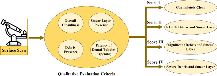
In order to define the scoring classification, at first, the entire surface of the specimen was thoroughly scanned. The derived images were evaluated based on the following criteria:
Level of cleanliness overall
Presence or absence of a smear layer and debris
Patency of the opening of dentinal tubules.
The images were then rated on a scoring scale of I to IV as follows (Figure 1):
Figure 1.
Basis of Defining the Scoring System
Grade I : Completely clean with no debris or smear layer present, and patent dentinal tubules.
Grade II : Mild debris and smear layer, with many patent dentinal tubules.
Grade III : Moderate debris and smear layer, with few patent dentinal tubules.
Grade IV : Severe debris and smear layer, with no patent dentinal tubules.
The scores were recorded directly onto a coding sheet, and the goal was to determine if there was any statistically significant difference in cleaning efficacy between the four examined experimental groups. In this paper, the remaining debris amount and smear layer was specifically evaluated and analyzed by the following scoring method [37]:
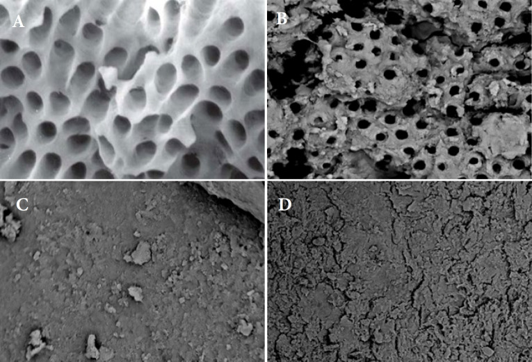
Score I: Open Dentin tubules. There is almost no debris or smear layer (Figure 2A).
Score II: A little debris and smear layer presence (Figure 2B).
Score III: A significant debris and smear layer presence (covers less than 50% of the surface) (Figure 2C).
Score IV: A lot of debris and smears are present (it covers more than 50% of the surface) (Figure 2D).
Figure 2.
A) Clean wall and open dentine tubules (2000× magnification); B) Thin layer of debris and smears (2000× magnification); C) The wall is covered with less than 50% debris (1000× magnification); D) A lot of debris left behind (1000× magnification)
The magnification of score's SEM images are (2000×) for Figures 2A and 2B, and (1000×) for Figures 2C and 2D. However, to better show the cleanliness, Figures 2A and 2B portray an almost clean wall and open dentine tubules with larger magnification, whereas Figures 2C and 2D display a significant amount of debris and smears comparing to the previous scoring SEM images.
Statistical analysis
Statistical analysis was carried out by the use of Statistical Package for Social Science (SPSS, version 20.0, Chicago, IL., USA). Two methods were conducted to determine the relationship between canal cleaning degrees in four types of files (XP; Protaper; Race; and Gentlefile) within three parts of canals (Coronal; Middle; and Apical). First, the amount of cleaning was classified as a qualitative item (I to IV), and then quantified as a mean±standard deviation. The chi-square test was implemented for comparing the degree of cleanliness among four groups in the first case, while Kruskal-Wallis was implemented to compare the amount of cleanliness in the second case. Due to non-normality of data distribution, it was not possible to do Two-way ANOVA analysis. The level of significance was set at 0.05.
Results
Table 1 summarizes the percentages of cleanliness in the experimental groups. There was no complete cleanliness (Score I) achieved by any of the systems examined. Based on chi-square test results, there was a significant relationship between root canal cleaning (three levels of cleanliness) in ProTaper (P=0.004) and Gentlelfile (P=0.04) groups.
Table 1.
The frequency of debris and Smear layer presence scores across four instruments at three root levels
| Group | Score | Coronal | Middle | Apical | P-value |
|---|---|---|---|---|---|
| XP-Endo Finisher | ii | 4(40%) | 6(60%) | 4(40%) | 0.585 |
| iii | 6(60%) | 4(40%) | 6(60%) | ||
| iv | 0 | 0 | 0 | ||
| ProTaper | ii | 4(40%) | 8(80%) | 0 | 0.004 |
| iii | 6(60%) | 2(20%) | 8(80%) | ||
| iv | 0 | 0 | 2(20%) | ||
| RaCe | ii | 4(40%) | 7(70%) | 5(50%) | 0.413 |
| iii | 6(60%) | 3(30%) | 4(40%) | ||
| iv | 0 | 0 | 1(10%) | ||
| Gentlefile | ii | 2(20%) | 3(30%) | 6(60%) | 0.040 |
| iii | 8(80%) | 7(70%) | 2(20%) | ||
| iv | 0 | 0 | 2(20%) |
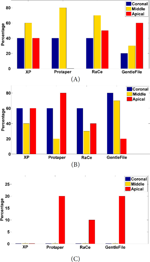
Based on Figure 3, the Gentlefile experimental group shows a better cleanliness results as a whole. Specifically, in the second score (Figure 3A), the cleanliness of GF is higher than other experimental groups in the apical third. Furthermore, in the third scoring classification (III) (Figure 3B), the GF group shows a greater percentage of cleanliness in the coronal and middle third. Finally, in the fourth score (IV), based on Figure 3C, GF group has a better cleaning capability in comparison with the three other experimental groups (XP, ProTaper, and RaCe).
Figure 3.
Cleanliness bar graph of four experimental groups in the coronal, middle, and apical third of II, III, and IV scoring method: A) Score II, B) Score III, C) Score IV
According to Table 1, the GF group shows greater cleanliness in the apical third as compared to the other three groups, meanwhile among the other 3 groups, the middle part of the canal had the highest cleaning rate. In general, the Gentlefile group achieved the most effective cleaning rate of 60% in the apical third.
Normality test
The results of Kolmogorov-Smirnov test, used for assessment of normal distribution of the data, are mentioned in Table 2. Considering the meaningfulness of all variables, which are lower than 0.05, and proving the zero-assumption by underscoring that all data are not normalized, it can be concluded that all variables have non-normalized distribution. Consequently, non-parametric tests can be implemented for their assessment.
Table 2.
The Kolmogorov-Smirnov test results
| Statistic | Degree of freedom | Meaningfulness (P-value) |
|
|---|---|---|---|
| Cleanliness rate | 0.329 | 120 | 0.001 |
Comparison of the root cleanliness by four experimental groups
Through all parts of the teeth and various experimental groups in this research, the 1st grade cleaning was not observed, and the achieved cleanliness were all between II and IV scoring grades. In order to investigate the effect of the position of the teeth root, and the utilized groups in the cleaning extent of the teeth, the Kruskal Wallis non-parametric test was utilized, and the obtained results are indicated in Table 3.
Table 3.
The Kruskal Wallis non-parametric test results
| Root position | Cleanliness of the files | |
|---|---|---|
| K2 statistic | 0.218 | 7.24 |
| Degrees of freedom | 3 | 3 |
| Meaningfulness | 0.169 | 0.045 |
The K2 (chi-square distribution) for 0.218 root position and 3 degrees of freedom has a meaningful level 0.169. Considering that this value is higher than 0.05, the root position has no significant effect in the cleanliness efficacy. As a result, regardless of the root position, in order to grade the various files, the mean grade ratings are calculated and mentioned in Table 4. Therefore, the results showed that the Gentlefile group has the highest cleaning grade rating with the value of 72.5, and RaCe group has the lowest cleaning mean grade among the evaluated groups.
Table 4.
The mean cleaning grade ratings of four experimental groups
| Group | Mean cleaning grade rating |
|---|---|
| XP-Endo Finisher | 61.1 |
| ProTaper | 54.9 |
| RaCe | 53.5 |
| Gentlefile | 72.5 |
Discussion
This study found that the Gentlefile group had better smear layer removal and less debris at the apical-root level compared to other groups. This partially rejected the assumption that all types of instruments should have no differences in cleanliness variables. The GF system's properties, such as flexibility, high rotation speeds, and abrading, contributed to improved cleanliness, according to a comparison of our findings with NiTi instruments. The scanning electron microscope is suitable technology for analyzing debris and smear layers [38-40]. The study showed that after instrumentation with the GF system, there was a considerable amount of debris and smear layer in the coronal and middle thirds, but over 80% of the smear layer was removed from the apical third, similar to previous studies [4, 41]. The high degree of flexibility in the GF system allowed it to cover all canal walls, especially at the apical third. Therefore, it is recommended to use the GF system for optimal canal shaping and cleaning, particularly at the apical portion of the canal. After instrumentation with the RaCe system, there was a significant amount of debris in the coronal third but little debris in the middle and apical thirds. In contrast, Bidar et al. [5] showed that use of Race files resulted in significantly more residual debris compared with other rotary files. This can be attributed to different types of assessed root canals and sample sizes.
Additionally, our study revealed that the ProTaper Universal system resulted in a notably lower organization of debris and smear layer in the middle third compared to other studies such as Htun et al. [4] and Sabet et al. [42]. This outcome is consistent
with the finding that the ProTaper system is more compatible with the anatomy of the middle part of the root canal, as stated Nishad et al. [43] . In contrast, some previous studies, including Sharma et al. [44]and Jayakumaar et al. [45], found that there was less smear layer remaining in the coronal third than in the middle third, and less in the middle third than in the apical third. The discrepancy between these findings and ours may be attributed to differences in sample size and irrigation solutions. Interestingly, after using the XP Endo Finisher system, we found that both the coronal and apical areas had significant amounts of smear layer remaining, but not in the middle third, which is similar to the results of Elnaghi et al.’s [46] study, which was conducted on extracted molar teeth with curved root canals. This difference can be attributed to the different root canal morphology of premolar and molar teeth, particularly in the coronal third, and to the different files used with the XP-Endo Finisher. According to Espinoza et al. [47], this system removes considerably more smear layers from the middle third compared to the apical third, which is consistent with our findings.
Azimian et al. [48] assessed the effect of XP-Endo finisher on the amount of residual debris and smear layer on the root canal walls, and concluded that debris removal was equal in all three coronal, middle, and apical parts, which is in contrast with our results. It is confirmed by the manufacturer that the XP-Endo Finisher File has shape memory capabilities. Since the file is formed during the cooling of the metal, it has no curvature when it is used for the first time. When the file comes in contact with canal walls, it becomes elastic and adapts to its shape. Similar reasons explain the high clearance of this system in the middle third compared to the coronal third. However, in the root canal, there is no required temperature to change the shape. For this reason, smear layer removal in the apical third is less than the middle third. One reason for this significant difference is the smaller root canal space in the apical third, and another is the unfeasibility of using an irrigating agent within the canal [48, 49]. It is necessary to keep in mind that samples used for this study have a moderate curvature, which would be a limitation. Consequently, to evaluate GF's performance, further studies will need to be conducted on severely-curved canals. Other limitation of this study is that no smear layer removal irrigation protocol has been used in this study which could be considered for the future studies for better similarity of the clinical studies.
Conclusion
Regarding the results of this study, it can be deduced that Gentlefile system eliminates more smears and debris than the other rotary systems. A lower cleaning rate was also seen with the Race and ProTaper systems as compared to the other rotary systems. To conclude, the Gentlefile group is more capable of cleaning the apical third of the canals than others instrumented by Protaper Universal, RaCe, and XP-Endo Finisher.
Acknowledgement
The authors thank the Research Vice Chancellor of Urmia University of Medical Sciences.
Conflict of Interest
None.
Funding Support
None.
Contributions
AAA: Concept and designing of the study, Writing, review and editing, FGR: Supervision, review and editing, RA: Data analysis, Writing, review and editing, MS: Investigation, Data collection, Writing and editing. All authors contributed to the study and approved the final manuscript
References
- 1.Tomson PL, Simon SR. Contemporary cleaning and shaping of the root canal system. Primary dental journal. 2016;5(2):46–53. doi: 10.1308/205016816819304196. [DOI] [PubMed] [Google Scholar]
- 2.Krokidis A, Nicola B, Antonio C, Panopoulos P. Comparison of Two Reciprocating and Anatomical Single File Techniques in Cleaning Oval Anatomies. Iran Endod J. 2023;18(1):41–6. doi: 10.22037/iej.v18i1.33623. [DOI] [PMC free article] [PubMed] [Google Scholar]
- 3.Neelakantan P, Khan K, Li KY, Shetty H, Xi W. Effectiveness of supplementary irrigant agitation with the Finisher GF Brush on the debridement of oval root canals instrumented with the Gentlefile or nickel titanium rotary instruments. Int Endod J. 2018;51(7):800–7. doi: 10.1111/iej.12892. [DOI] [PubMed] [Google Scholar]
- 4.Htun PH, Ebihara A, Maki K, Kimura S, Nishijo M, Okiji T. Cleaning and Shaping Ability of Gentlefile, HyFlex EDM, and ProTaper Next Instruments: A Combined Micro-computed Tomographic and Scanning Electron Microscopic Study. J Endod. 2020;46(7):973–9. doi: 10.1016/j.joen.2020.03.027. [DOI] [PubMed] [Google Scholar]
- 5.Bidar M, Moradi S, Forghani M, Bidad S, Azghadi M, Rezvani S, Khoynezhad S. Microscopic evaluation of cleaning efficiency of three different nickel-titanium rotary instruments. Iran Endod J. 2010;5(4):174–8. [PMC free article] [PubMed] [Google Scholar]
- 6.Shabahang S, Pouresmail M, Torabinejad M. In vitro antimicrobial efficacy of MTAD and sodium hypochlorite. J Endod. 2003;29(7):450–2. doi: 10.1097/00004770-200307000-00006. [DOI] [PubMed] [Google Scholar]
- 7.De-Deus G, Garcia-Filho P. Influence of the NiTi rotary system on the debridement quality of the root canal space. Oral Surg Oral Med Oral Pathol Oral Radiol Endod. 2009;108(4):e71–6. doi: 10.1016/j.tripleo.2009.05.012. [DOI] [PubMed] [Google Scholar]
- 8.Bergmans L, Van Cleynenbreugel J, Wevers M, Lambrechts P. Mechanical root canal preparation with NiTi rotary instruments: rationale, performance and safety Status report for the American Journal of Dentistry. Am J Dent. 2001;14(5):324–33. [PubMed] [Google Scholar]
- 9.Siddique R, Nivedhitha MS. Effectiveness of rotary and reciprocating systems on microbial reduction: A systematic review. J Conserv Dent. 2019;22(2):114–22. doi: 10.4103/JCD.JCD_523_18. [DOI] [PMC free article] [PubMed] [Google Scholar]
- 10.Zanza A, Reda R, Pagnoni F, Patil S. Future Trends in Endodontics: How Could Materials Increase the Long-Term Outcome of Root Canal Therapies? Materials (Basel) 2022;15:10. doi: 10.3390/ma15103473. [DOI] [PMC free article] [PubMed] [Google Scholar]
- 11.Li SH, Lu Y, Song D, Zhou X, Zheng QH, Gao Y, Huang DM. Occurrence of Dentinal Microcracks in Severely Curved Root Canals with ProTaper Universal, WaveOne, and ProTaper Next File Systems. J Endod. 2015;41(11):1875–9. doi: 10.1016/j.joen.2015.08.005. [DOI] [PubMed] [Google Scholar]
- 12.Nagendrababu V, Ahmed HMA. Shaping properties and outcomes of nickel-titanium rotary and reciprocation systems using micro-computed tomography: a systematic review. Quintessence Int. 2019;50(3):186–95. doi: 10.3290/j.qi.a41977. [DOI] [PubMed] [Google Scholar]
- 13.Khoshbin E, Donyavi Z, Abbasi Atibeh E, Roshanaei G, Amani F. The Effect of Canal Preparation with Four Different Rotary Systems on Formation of Dentinal Cracks: An In Vitro Evaluation. Iran Endod J. 2018;13(2):163–8. doi: 10.22037/iej.v13i2.16416. [DOI] [PMC free article] [PubMed] [Google Scholar]
- 14.Caviedes-Bucheli J, Rios-Osorio N, Usme D, Jimenez C, Pinzon A, Rincon J, Azuero-Holguin MM, Zubizarreta-Macho A, Gomez-Sosa JF, Munoz HR. Three-dimensional analysis of the root canal preparation with Reciproc Blue(R), WaveOne Gold(R) and XP EndoShaper(R): a new method in vivo. BMC Oral Health. 2021;21(1):88. doi: 10.1186/s12903-021-01450-1. [DOI] [PMC free article] [PubMed] [Google Scholar]
- 15.Samiei M, Shahi S, Abdollahi AA, Eskandarinezhad M, Negahdari R, Pakseresht Z. The Antibacterial Efficacy of Photo-Activated Disinfection, Chlorhexidine and Sodium Hypochlorite in Infected Root Canals: An in Vitro Study. Iran Endod J. 2016;11(3):179–83. doi: 10.7508/iej.2016.03.006. [DOI] [PMC free article] [PubMed] [Google Scholar]
- 16.Eshagh Saberi A, Ebrahimipour S, Saberi M. Apical Debris Extrusion with Conventional Rotary and Reciprocating Instruments. Iran Endod J. 2020;15(1):38–43. doi: 10.22037/iej.v15i1.23823. [DOI] [PMC free article] [PubMed] [Google Scholar]
- 17.Kum KY, Kazemi RB, Cha BY, Zhu Q. Smear layer production of K3 and ProFile Ni-Ti rotary instruments in curved root canals: a comparative SEM study. Oral Surg Oral Med Oral Pathol Oral Radiol Endod. 2006;101(4):536–41. doi: 10.1016/j.tripleo.2005.02.079. [DOI] [PubMed] [Google Scholar]
- 18.Ashraf H, Zargar N, Zandi B, Azizi A, Amiri M. The Evaluation of Debris and Smear Layer Generated by Three Rotary Instruments Neo NiTi, 2Shape and Revo_S: An Ex-vivo Scanning Electron Microscopic Study. Iran Endod J. 2023;18(2):96–103. doi: 10.22037/iej.v18i2.39966. [DOI] [PMC free article] [PubMed] [Google Scholar]
- 19.Sen BH, Wesselink PR, Turkun M. The smear layer: a phenomenon in root canal therapy. Int Endod J. 1995;28(3):141–8. doi: 10.1111/j.1365-2591.1995.tb00289.x. [DOI] [PubMed] [Google Scholar]
- 20.Cheung GS, Liu CS. A retrospective study of endodontic treatment outcome between nickel-titanium rotary and stainless steel hand filing techniques. J Endod. 2009;35(7):938–43. doi: 10.1016/j.joen.2009.04.016. [DOI] [PubMed] [Google Scholar]
- 21.Violich DR, Chandler NP. The smear layer in endodontics - a review. Int Endod J. 2010;43(1):2–15. doi: 10.1111/j.1365-2591.2009.01627.x. [DOI] [PubMed] [Google Scholar]
- 22.Kennedy WA, Walker WA, Gough RW. Smear layer removal effects on apical leakage. J Endod. 1986;12(1):21–7. doi: 10.1016/S0099-2399(86)80277-1. [DOI] [PubMed] [Google Scholar]
- 23.Torabinejad M, Handysides R, Khademi AA, Bakland LK. Clinical implications of the smear layer in endodontics: a review. Oral Surg Oral Med Oral Pathol Oral Radiol Endod. 2002;94(6):658–66. doi: 10.1067/moe.2002.128962. [DOI] [PubMed] [Google Scholar]
- 24.Delvarani A, Mohammadzadeh Akhlaghi N, Aminirad R, Tour Savadkouhi S, Vahdati SA. In vitro Comparison of Apical Debris Extrusion Using Rotary and Reciprocating Systems in Severely Curved Root Canals. Iran Endod J. 2017;12(1):34–7. doi: 10.22037/iej.2017.07. [DOI] [PMC free article] [PubMed] [Google Scholar]
- 25.Moreinos D, Dakar A, Stone NJ, Moshonov J. Evaluation of Time to Fracture and Vertical Forces Applied by a Novel Gentlefile System for Root Canal Preparation in Simulated Root Canals. J Endod. 2016;42(3):505–8. doi: 10.1016/j.joen.2015.12.023. [DOI] [PubMed] [Google Scholar]
- 26.Chen X, Ge J. Gentlefile Versus Protaper in the Shaping Ability in Simulated J-shaped Root Canals. J Res Square. 2020;12(3):1–12. [Google Scholar]
- 27.Wu J, Lei G, Yan M, Yu Y, Yu J, Zhang G. Instrument separation analysis of multi-used ProTaper Universal rotary system during root canal therapy. J Endod. 2011;37(6):758–63. doi: 10.1016/j.joen.2011.02.021. [DOI] [PubMed] [Google Scholar]
- 28.Labbaf H, Nazari Moghadam K, Shahab S, Mohammadi Bassir M, Fahimi MA. An In vitro Comparison of Apically Extruded Debris Using Reciproc, ProTaper Universal, Neolix and Hyflex in Curved Canals. Iran Endod J. 2017;12(3):307–11. doi: 10.22037/iej.v12i3.13540. [DOI] [PMC free article] [PubMed] [Google Scholar]
- 29.Tofangchiha M, Ebrahimi A, Adel M, Kermani F, Mohammadi N, Reda R, Testarelli L. In vitro evaluation of Kedo-S and RaCe rotary files compared to hand files in preparing the root canals of primary molar teeth. Front Biosci (Elite Ed) 2022;14(2) doi: 10.31083/j.fbe1402014. [DOI] [PubMed] [Google Scholar]
- 30.Vaz-Garcia ES, Vieira VTL, Petitet N, Moreira EJL, Lopes HP, Elias CN, Silva E, Antunes HDS. Mechanical Properties of Anatomic Finishing Files: XP-Endo Finisher and XP-Clean. Braz Dent J. 2018;29(2):208–13. doi: 10.1590/0103-6440201801903. [DOI] [PubMed] [Google Scholar]
- 31.Trope M, Debelian G. XP-3D Finisher™ file the next step in restorative endodontics. Endod Pract US. 2015;8(6):22–4. [Google Scholar]
- 32.Nouroloyouni A, Safavi Hir F, Farhang R, Noorolouny S, Salem Milani A, Alyali R. Evaluating In Vitro Performance of a Novel Stainless Steel Rotary System (Gentlefile) Based on Debris Extrusion and Instrumentation Time. Biomed Res Int. 2023;2023:9945236. doi: 10.1155/2023/9945236. [DOI] [PMC free article] [PubMed] [Google Scholar]
- 33.Chan CW, Romeo VR, Lee A, Zhang C, Neelakantan P, Pedulla E. Accumulated Hard Tissue Debris and Root Canal Shaping Profiles Following Instrumentation with Gentlefile, One Curve, and Reciproc Blue. J Endod. 2023;49(10):1344–51. doi: 10.1016/j.joen.2023.07.019. [DOI] [PubMed] [Google Scholar]
- 34.Godiny M, Mohammadi B, Norooznezhad M, Chalabi M. Endodontic Rotary Systems: Comparison between Gentlefile and Pro Taper Universal for removal of Enterococcus faecalis. J Clin Exp Dent. 2023;15(4):e277–e80. doi: 10.4317/jced.60147. [DOI] [PMC free article] [PubMed] [Google Scholar]
- 35.Wu MK, Wesselink PR. Efficacy of three techniques in cleaning the apical portion of curved root canals. Oral Surg Oral Med Oral Pathol Oral Radiol Endod. 1995;79(4):492–6. doi: 10.1016/s1079-2104(05)80134-9. [DOI] [PubMed] [Google Scholar]
- 36.Asgary S, Parirokh M, Eghbal MJ, Ghoddusi J, Eskandarizadeh A. SEM evaluation of neodentinal bridging after direct pulp protection with mineral trioxide aggregate. Aust Endod J. 2006;32(1):26–30. doi: 10.1111/j.1747-4477.2006.00004.x. [DOI] [PubMed] [Google Scholar]
- 37.Raisingani D, Meshram GK. Cleanliness in the Root Canal System: An Scanning Electron Microscopic Evaluation of Manual and Automated Instrumentation using 4% Sodium Hypochlorite and EDTA (Glyde File Prep)-An in vitro Study. Int J Clin Pediatr Dent. 2010;3(3):173–82. doi: 10.5005/jp-journals-10005-1073i. [DOI] [PMC free article] [PubMed] [Google Scholar]
- 38.Zargar N, Naseri M, Gholizadeh Z, Mehrabinia P. Evaluation of Residual Debris and Smear layer After Root Canal Preparation by Three Different Methods: A Scanning Electron Microscopy Study. Iran Endod J. 2022;17(3):138–45. doi: 10.22037/iej.v17i3.36525. [DOI] [PMC free article] [PubMed] [Google Scholar]
- 39.Mirseifinejad R, Tabrizizade M, Davari A, Mehravar F. Efficacy of Different Root Canal Irrigants on Smear Layer Removal after Post Space Preparation: A Scanning Electron Microscopy Evaluation. Iran Endod J. 2017;12(2):185–90. doi: 10.22037/iej.2017.36. [DOI] [PMC free article] [PubMed] [Google Scholar]
- 40.Khademi A, Saatchi M, Shokouhi MM, Baghaei B. Scanning Electron Microscopic Evaluation of Residual Smear Layer Following Preparation of Curved Root Canals Using Hand Instrumentation or Two Engine-Driven Systems. Iran Endod J. 2015;10(4):236–9. doi: 10.7508/iej.2015.04.005. [DOI] [PMC free article] [PubMed] [Google Scholar]
- 41.Nguyen TA, Kim Y, Kim E, Shin SJ, Kim S. Comparison of the Efficacy of Different Techniques for the Removal of Root Canal Filling Material in Artificial Teeth: A Micro-Computed Tomography Study. J Clin Med. 2019;8:7. doi: 10.3390/jcm8070984. [DOI] [PMC free article] [PubMed] [Google Scholar]
- 42.Sabet NE, Lutfy RA. Ultrastructural morphologic evaluation of root canal walls prepared by two rotary nickel-titanium systems: a comparative study. Oral Surg Oral Med Oral Pathol Oral Radiol Endod. 2008;106(3):e59–66. doi: 10.1016/j.tripleo.2008.05.005. [DOI] [PubMed] [Google Scholar]
- 43.Nishad SV, Shivamurthy GB. Comparative Analysis of Apical Root Crack Propagation after Root Canal Preparation at Different Instrumentation Lengths Using ProTaper Universal, ProTaper Next and ProTaper Gold Rotary Files: An In vitro Study. Contemp Clin Dent. 2018;9(Suppl 1):S34–S8. doi: 10.4103/ccd.ccd_830_17. [DOI] [PMC free article] [PubMed] [Google Scholar]
- 44.Sharma G, Kakkar P, Vats A. A Comparative SEM Investigation of Smear Layer Remaining on Dentinal Walls by Three Rotary NiTi Files with Different Cross Sectional Designs in Moderately Curved Canals. J Clin Diagn Res. 2015;9(3):ZC43–7. doi: 10.7860/JCDR/2015/11569.5710. [DOI] [PMC free article] [PubMed] [Google Scholar]
- 45.Jayakumaar A, Ganesh A, Kalaiselvam R, Rajan M, Deivanayagam K. Evaluation of debris and smear layer removal with XP-endo finisher: A scanning electron microscopic study. Indian J Dent Res. 2019;30(3):420–3. doi: 10.4103/ijdr.IJDR_655_17. [DOI] [PubMed] [Google Scholar]
- 46.Elnaghy AM, Mandorah A, Elsaka SE. Effectiveness of XP-endo Finisher, EndoActivator, and File agitation on debris and smear layer removal in curved root canals: a comparative study. Odontology. 2017;105(2):178–83. doi: 10.1007/s10266-016-0251-8. [DOI] [PubMed] [Google Scholar]
- 47.Espinoza I, Conde Villar AJ, Lorono G, Estevez R, Plotino G, Cisneros R. Effectiveness of XP-Endo Finisher and Passive Ultrasonic Irrigation in the Removal of the Smear Layer Using two Different Chelating Agents. J Dent (Shiraz) 2021;22(4):243–51. doi: 10.30476/DENTJODS.2021.86680.1204. [DOI] [PMC free article] [PubMed] [Google Scholar]
- 48.Azimian S, Bakhtiar H, Azimi S, Esnaashari E. In vitro effect of XP-Endo finisher on the amount of residual debris and smear layer on the root canal walls. Dent Res J (Isfahan) 2019;16(3):179–84. [PMC free article] [PubMed] [Google Scholar]
- 49.Karamifar K, Mehrasa N, Pardis P, Saghiri MA. Cleanliness of Canal Walls following Gutta-Percha Removal with Hand Files, RaCe and RaCe plus XP-Endo Finisher Instruments: A Photographic in Vitro Analysis. Iran Endod J. 2017;12(2):242–7. doi: 10.22037/iej.2017.47. [DOI] [PMC free article] [PubMed] [Google Scholar]


