



Abstract
Per- and polyfluoroalkyl substances (PFAS) are synthetic chemicals known for their environmental persistence and resistance to biodegradation. This study investigated the impact of adolescent exposure to a PFAS mixture on adult ovarian function. Female CD-1 mice were orally exposed to vehicle control or a PFAS mixture (comprised of perfluorooctanoic acid, perfluorooctanesulfonic acid, undecafluoro-2-methyl-3-oxahexanoic acid, and perfluorobutanesulfonic acid) for 15 d. After a 42-d recovery period, reproductive hormones, ovarian fibrosis, and ovarian gene and protein expression were analyzed using ELISA, Picrosirius red staining, qPCR, and immunoblotting, respectively. Results revealed that PFAS exposure did not affect adult body or organ weight, although ovarian weight slightly decreased. PFAS-exposed mice exhibited a disturbed estrous cycle, with less time spent in proestrus than control mice. Follicle counting indicated a reduction in primordial and primary follicles. Serum analysis revealed no changes in steroid hormones, follicle-stimulating hormone, or anti-Müllerian hormone, but a significant increase in luteinizing hormone was observed in PFAS-treated mice. Ovaries collected from PFAS-treated mice had increased mRNA transcripts for steroidogenic enzymes and fatty acid synthesis-related genes. PFAS exposure also increased collagen content in the ovary. Additionally, serum tumor necrosis factor-α levels were higher in PFAS-treated mice. Finally, transcripts and protein abundance for Hippo pathway components were upregulated in the ovaries of the PFAS-treated mice. Overall, these findings suggest that adolescent exposure to PFAS can disrupt ovarian function in adulthood.
Keywords: PFAS, mixtures, ovary, steroidogenesis, fibrosis
Per- and polyfluoroalkyl substances (PFAS) are a large group of synthetic chemicals with widespread use in industrial, commercial, and consumer products. Due to their lipo- and hydrophobic characteristics and distinctive fluorocarbon bonds, PFAS exhibit strong resistance against hydrolysis, photolysis, and degradation through thermal, chemical, or microbial processes (Glüge et al. 2020). Thus, PFAS are persistent in the environment. Humans are exposed to PFAS through contaminated food and water, as well as inhalation or dermal exposure from contact with treated materials (Sunderland et al. 2019). As PFAS are not readily metabolized, they rapidly absorb and accumulate in human, plant, and animal tissues and can be biomagnified through the trophic web (Pérez et al. 2013; Wang et al. 2020b; Xing et al. 2023). Alarmingly, nearly every person (including children, teenagers, and adults) in the National Health and Nutrition Examination Survey (NHANES) has detectable exposure to at least one type of PFAS chemical (CDC 2022).
In principle, PFAS are often referred to as long-chain (containing more than 8 carbons) or short-chain (containing less than 7 carbons) and legacy or alternative/emerging compounds based on the length of time that the chemicals have been manufactured or evaluated (Brennan et al. 2021). Although the use of the legacy PFAS such as perfluorooctanoic acid (PFOA) and perfluorooctane sulfonate (PFOS) have been phased out in the United States and Europe, they continue to be manufactured and present in items imported from countries with no current regulations (Sunderland et al. 2019). Subsequently, the development of alternative PFAS compounds such as undecafluoro-2-methyl-3-oxahexanoic acid (HFPO-DA/GenX) and perfluorobutanesulfonic acid (PFBS) have emerged to replace the legacy PFAS and are proposed to decrease human health impacts and environmental burden, though many of these chemicals remain unregulated and untested.
Due to the ubiquitous nature of PFAS and minimal government guidelines on their use and/or consumption, they are a cause for concern for reproductive health. Observational studies in humans suggest that PFAS exposure in women may delay the onset of puberty (Lopez-Espinosa et al. 2011; Kristensen et al. 2013; Guo et al. 2023; Liu et al. 2023), disrupt menstrual cyclicity (Lyngsø et al. 2014; Zhou et al. 2017), alter steroid hormone secretion (Barrett et al. 2015; Ding et al. 2020; Harlow et al. 2021; Wang et al. 2021), initiate early menopause due to premature ovarian failure (Knox et al. 2011; Taylor et al. 2014; Zhang et al. 2018; Ding et al. 2022), and coincide with the development of polycystic ovarian syndrome (Wang et al. 2019; Hammarstrand et al. 2021; Zhan et al. 2023). To date, most laboratory studies have evaluated the effects of individual, long-chain legacy PFAS (i.e. PFOA, PFOS) on the ovary or ovarian cells. In vivo studies in rodents have shown that exposure to singular legacy PFAS can impact ovarian follicle numbers (Chen et al. 2017; Feng et al. 2017; Du et al. 2019; Clark and Davis 2022; Yang et al. 2022), disrupt steroidogenesis (Shi et al. 2009; Chen et al. 2017; Du et al. 2019; Yang et al. 2022; Dangudubiyyam et al. 2023), and alter the ovarian proteome (González-Alvarez et al. 2022). Studies on short-chain “alternative” PFAS compounds (i.e. HFPO-DA/GenX, PFBS) on the mammalian ovary are extremely limited (Feng et al. 2017; Cao et al. 2020). Further, studies of PFAS mixtures on ovarian cells have been limited to in vitro studies (Rickard et al. 2022, 2023; Clark et al. 2024). There are currently no in vivo studies evaluating physiologically relevant exposures to a mixture of PFAS on folliculogenesis and ovarian function.
The ovary is the source of the female gamete, the oocyte, and the hormones necessary for female growth and development. The number of oocytes is finite at birth, and once the pool of primordial follicles is depleted ovarian senescence occurs (Hirshfield 1991; Hsueh et al. 2015). Ovotoxicants can selectively impact a specific follicle population in the ovary, leading to temporary or permanent infertility (Hoyer and Keating 2014). Additionally, the impacts of an ovotoxic exposure on female reproductive function depend primarily on the developmental stage of exposure as well as the level and duration of the exposure. Consequentially, early life exposures to chemicals can be detrimental to ovarian function in adulthood. Changes in hormone regulation leading to either cessation or dysfunction of ovarian activity can increase a woman’s susceptibility to developing gynecological cancers, osteoporosis, and cardiovascular disease (Nagle et al. 2015; Sullivan et al. 2016; Khosla and Monroe 2018; Rodgers et al. 2019). We have previously identified a role for the Hippo pathway and its effector yes-associated protein 1 (YAP1) in pathological PFAS-induced cell proliferation in a human granulosa cell line and follicle growth in cultured neonatal mouse ovaries (Clark and Davis 2022; Clark et al. 2022a). In the present study, we hypothesized that adolescent exposure to a PFAS mixture would affect ovarian function in adulthood by altering follicular dynamics and disrupting Hippo signaling.
Materials and methods
Chemicals
PFOA (cat no. 171468) was purchased from Sigma-Aldrich, United States. Perfluorooctanesulfonic acid (PFOS; cat no. 009151) was purchased from Matrix Scientific, United States. Undecafluoro-2-methyl-3-oxahexanoic acid (GenX/HFPO-DA; cat no. 2121-3-13) and PFBS (cat no. 6164-3-09) were purchased from Synquest Laboratories, United States.
Animal procedure and tissue collection
All experiments were adhered to regulatory guidelines and were approved by the Institutional Animal Care and Use Committee (IACUC) at the University of Nebraska Medical Center. Juvenile female CD-1 mice were obtained from Charles River Laboratories (United States) and housed in the animal facility at University of Nebraska Medical Center, where they were kept under controlled room temperature (21 to 22 °C) and lighting (10 h light:14 h dark) with ad libitum access to food and water. Adolescent postnatal day (PND) 30 female CD-1 mice were orally exposed (pipette administration in the cheek pouch) to vehicle control (distilled water with 1% dimethylsulfoxide [DMSO]; n = 8) or PFAS mixture (0.1 mg/kg of PFOA/PFOS/GenX/PFBS each; n = 8) for 15 d (PND45) and allowed to recover to PND90. The duration of the exposure was designed to simulate acute adolescent exposure, while collecting samples 6-wk postexposure aimed to provide insight into recovery and repair processes following PFAS exposure. This timeframe also allowed for the evaluation of any secondary effects that might not be immediately detectable after the initial exposure. Dosage was selected based on previously reported lowest observed adverse effect level on developmental endpoints for PFOA and PFOS (Du et al. 2019) and GenX/HFPO-DA (Blake et al. 2022; Conley et al. 2023) in neonatal and juvenile rodents. Furthermore, concentrations comparable to these have led to serum levels of PFOA, PFOS, and GenX/HFPO-DA in mice that are relevant to those found in humans exposed to these substances in occupational settings (Robarts et al. 2024). The dosage of PFBS was selected for consistency to the other PFAS compounds in the mixture, as previous reports evaluating PFBS and developmental/reproductive endpoints utilized a high (>200 mg/kg) dosage (Feng et al. 2017). Body weight was recorded weekly and vaginal cytology was monitored 15 d prior to euthanasia at PND90. Mice were euthanized in the diestrus phase of the estrous cycle, and one ovary was fixed in 4% paraformaldehyde overnight at 4 °C for histological analysis or flash frozen in liquid nitrogen and kept at −80 °C for further processing.
Estrous cycle monitoring
The estrous cycle was monitored by performing vaginal cytology analysis in the morning for 15 d to determine if exposure to PFAS impacted the estrous cycle and to ensure that mice were at the same stage of the estrous cycle at the time of euthanasia. Saline was pipetted into the vagina and vaginal fluids placed onto histology slides and examined with a Laxco SeBa PRO 4 microscope using a 10× objective. Estrous cycle stage classification was determined as previously described (Byers et al. 2012). In brief, proestrus was identified by the presence of small, round nucleated epithelial cells, some cornified epithelial cells, and minimal to no leukocytes. Estrus was marked by several cornified epithelial cells with deteriorating nuclei. The metestrus stage was indicated via the presence of both cornified cells and leukocytes. During diestrus, nucleated epithelial cells reappeared in the vaginal smears along with polymorphonuclear leukocytes. Due to the similarity in cytology for the metestrus and diestrus stage, these two stages were combined.
Histology and follicle counting
Isolated ovaries were fixed in 4% paraformaldehyde overnight at 4 °C and then processed for paraffin embedding. Serial sections were cut at a thickness of 5 µm, with every sixth section mounted onto glass slides and stained with hematoxylin and eosin (n = 8 per treatment). Healthy follicles with a distinct oocyte nucleus were counted and classified according to previously established methods (Clark et al. 2019). In brief, primordial follicles were identified by the presence of an oocyte encircled by a single layer of squamous granulosa cells; primary follicles identified as an oocyte surrounded by a single layer of cuboidal granulosa cells; secondary follicles by an oocyte surrounded by multiple layers of granulosa cells, and antral follicles by the presence of an oocyte enclosed by several layers of granulosa cells and the presence of a fluid-filled antral space. Bright-field images were acquired using the Zeiss Axioscan 7 Whole Slide Imaging System on 20× zoom. Slide identity was blinded to prevent a counting bias.
Analysis of hormones and TNFα in serum
Blood samples were allowed to clot at room temperature for 90 min. Samples were then centrifuged at 15,000×g for 15 min at 4 °C and sera collected. Sera samples (n = 7 per treatment) were sent to the University of Virginia Center for Research in Reproduction Ligand Assay and Analysis core for measurements of estradiol, progesterone, testosterone, anti-Müllerian hormone (AMH), luteinizing hormone (LH), and follicle-stimulating hormone (FSH). Serum tumor necrosis factor-alpha (TNFα) was determined using the mouse TNFα ELISA kit (cat no. KE10002, Proteintech, United States) according to the manufacturer’s protocol (n = 5 per treatment). The assay sensitivity was 5 pg/ml, 0.15 ng/ml, 10 ng/dl, 3.3 ng/ml, 0.16 ng/ml, 0.16 ng/ml, and 1 pg/ml for serum estradiol, progesterone, testosterone, AMH, LH, FSH, and TNFα, respectively. The intra- and interassay coefficients of variability were <10%.
Immunofluorescence
Immunofluorescence staining
Slides were deparaffinized in xylene and rehydrated through a series of ethanol washes (100%, 95%, and 75%), followed by a wash in distilled water. Heat-mediated antigen retrieval was conducted using 10 mM sodium citrate buffer (pH 6.0) with 0.5% Tween 20 for 30 min in a pressure cooker. Tissue sections on histology slides were outlined with a histology pap pen to retain liquids during processing. The sections were then treated with blocking solution (0.1 M phosphate buffered saline [PBS]/0.4% bovine serum albumin [BSA]/0.2% Tween20/5% normal goat serum) for 1 h at room temperature. Primary antibodies (Table S1) were added to fresh blocking solution, applied to tissue sections, and incubated in a humidified box overnight at 4 °C. Slides were washed in PBS (3 × 10 min), and the appropriate secondary antibody (Table S1) was applied in fresh blocking solution for 1 h at room temperature, followed by another wash in PBS (3 × 10 min). Slides were air dried, then followed by addition of Vectashield with 4,6-diamidino-2-phenylindole (DAPI, cat no. H1200, Vector Labs, United States) and slides cured overnight at 4 °C. Technical controls were conducted using only the secondary antibody to confirm specificity alone (Fig. S1). Images were captured on a Zeiss LSM710 confocal microscope equipped with an AxioCam MRc5 using a 10× or 20× objective lens.
Quantification of protein abundance
To quantify the cell proliferation marker KI67 (also known as MKI67) and cleaved caspase-3 (CASP3) immunoreactivity (n = 8 ovaries per treatment; 2 sections per ovary), the total fluorescence intensity of the entire ovarian section was analyzed using ImageJ (https://imagej.nih.gov).
Picrosirius red stain
Picrosirius red staining
For picrosirius red (PSR) staining, slides were deparaffinized in xylene and rehydrated in washes of ethanol (100%, 95%, and 75%), followed by one wash in dH2O. Tissue sections were encircled with a histology pap pen to keep liquid on tissue sections during processing. Slides were then stained using a Picrosirius Red Stain Kit (cat no. ab150681, Abcam, United States) according to the manufacturer’s protocol. Bright-field images were acquired using an Olympus inverted microscope equipped with a DP71 digital camera. Slide identity was blinded to prevent analytical bias.
Quantification of collagen abundance
For quantification of PSR staining, the percentage of the total image area threshold (n = 8 ovaries per treatment; 2 sections per ovary) was analyzed using ImageJ.
RNA isolation, reverse transcription, and qRT–PCR
RNA was extracted from ovaries (n = 8 replicates per treatment) using the Zymo Direct-zol Microprep Kit (cat no. R2062, Zymo Research Corporation, United States) following the manufacturer’s instructions. The concentration of RNA was measured using a NanoDrop (l = 260/280 nm; ND1000; Nanodrop Technologies, United States). A total of 200 ng RNA was reverse transcribed using iScript Reverse Transcription Supermix (cat no. 1708840, BioRad, United States) and then diluted 1:5, resulting in a final working concentration of 4 ng of cDNA per reaction. Quantitative PCR (qPCR) analysis was conducted on a BioRad CFX96 Real-Time System (BioRad, United States) using Sso Fast EvaGreen Supermix (cat no. 1725201, BioRad, United States) and under the following conditions: 95 °C for 30 s, 40 cycles of 95 °C for 5 s, and 65 °C for 5 s. Primers were designed using the NCBI primer designing tool (http://www.ncbi.nlm.nih.gov/tools/primer-blast/). Forward and reverse primers were synthesized by Eurofins Genomics (Table S2). A melting curve analysis confirmed that each primer pair amplified a single, expected product. Gene expression data were then normalized to Gapdh using the 2−ΔΔCt method (Livak and Schmittgen 2001).
Western blot
Ovaries (n = 8 replicates per treatment) were homogenized in RIPA buffer supplemented with protease and phosphatase inhibitor cocktails (cat no. 78440, ThermoFisher, United States) on ice. The homogenized tissue was then centrifuged at 15,000×g for 15 min at 4 °C. The protein content in the resulting supernatant was quantified using a bicinchoninic acid assay following the manufacturer’s protocol (cat no. 23225, ThermoFisher, United States). SDS–PAGE was used to separate the proteins prior to transfer to a nitrocellulose membrane. Membranes were blocked in 5% BSA in tris-buffered saline with 0.2% Tween 20 (TBSTw) for 1 h, followed by overnight incubation in primary antibodies (Table S1) at 4 °C. After three washes in tris-buffered saline with 0.2% Tween 20 (TBSTw) (3×10 min), the membranes were incubated with appropriate secondary antibody (Table S1) in fresh blocking solution for 1 h at room temperature and washed again with TBSTw (3 × 10 min). The signals were visualized using ECL detection substrate and captured on the iBright system (ThermoFisher, United States). Densitometry analysis of the bands of interest was measured using ImageJ software (https://imagej.nih.gov). Between each round of immunodetection, membranes were stripped and blocked. The proteins of interest were normalized to β-actin (ACTB) by dividing densitometric value of the protein of interest by the densitometric value obtained from total ACTB.
Statistical analysis
Data analysis was performed using GraphPad Prism 10. Following normality testing, the unpaired t-test was used. The significance level was set at P ≤ 0.05 and a trend for a statistical difference was identified as 0.05 < P < 0.1. Data presented in bar graphs are represented as mean±SEM. For the estrous cycle data, statistical analysis was conducted on the raw data, and to aid in visualization, the percentage of time spent at each stage was calculated by the number of days per stage/15 d×100.
Results
Effects of PFAS exposure on body and organ weight
To determine if exposure to PFAS had an impact on body or organ weight during adolescent growth in mice, body weight was measured weekly and organ weights were collected at euthanasia. Body weight was not affected (P ≥ 0.05) in female mice after exposure to the PFAS mixture over the 8-wk experimental period (PND30 to PND90) relative to the vehicle control female mice (Fig. 1a). A slight decrease (P ≤ 0.07) in ovarian weight was observed in the PFAS mixture-treated mice relative to control-treated mice (Fig. 1b). There was no impact (P ≥ 0.05) of PFAS exposure on weight of the uterus, spleen, liver, kidney, or heart (Fig. 1c–f).
Fig. 1.

Effects of PFAS mixture on body and organ weight. (a) Body weight gain was monitored weekly. After euthanasia, (b) ovary; (c) uterus; (d) spleen; (e) liver; (f) kidney; (g) heart weights were collected. Bars represent the means±SEM; n = 8 per treatment; #=P ≤ 0.07.
Impact of PFAS exposure on estrous cyclicity and ovarian follicle number
To assess the impact of PFAS exposure on the estrous cycle, vaginal cytology was examined daily during the last 15 d of the recovery period (PND 75 to 90). Adolescent exposure to the PFAS mixture decreased (P ≤ 0.05) the amount of time that the adult mice spent in proestrus. The mice also displayed a tendency (P ≤ 0.06) to spend more time in metestrus/diestrus relative to the vehicle-treated control mice (Fig. 2a). The amount of time spent in estrus did not vary (P ≥ 0.05) between the two treatment groups (Fig. 2a). The impact of PFAS exposure on folliculogenesis and composition of the adult follicular pool was also determined. Mice treated with the PFAS mixture relative to the vehicle controls had reduced (P ≤ 0.05) numbers of primordial and primary follicles, by 52% and 35%, respectively (Fig. 2b; Fig. S2). Additionally, a ∼38% reduction (P ≤ 0.05) in the total number of follicles in the ovaries of PFAS-treated mice was observed (Fig. 2b; Fig. S2). There was no impact (P ≥ 0.05) on PFAS exposure on numbers of secondary or antral follicles (Fig. 2b; Fig. S2).
Fig. 2.

Impact of PFAS mixture on estrous cyclicity and follicle number. (a) The percentage of time spent in proestrus, estrus, and metestrus/diestrus phase of the estrous cycle was determined by vaginal cytology. (b) Ovarian follicle numbers were classified and counted after exposure to vehicle control or PFAS mixture. Bars represent the means±SEM; n = 8 per treatment; #=P ≤ 0.07; *P ≤ 0.05; ***P ≤ 0.001; ****P ≤ 0.0001.
Effect of PFAS on proliferation and regulators of apoptosis
To determine if exposure of adolescent mice to the PFAS mixture promotes proliferation or initiates apoptosis in the adult ovary, immunofluorescence staining for KI67, and CASP3 was performed. Immunofluorescent staining for KI67 and CASP3 was detected throughout the ovary, including the oocyte, granulosa cells, and stromal cells (Fig. 3a–d). Whole ovary KI67 fluorescence intensity was unaffected (P ≥ 0.05) in female mice treated with the PFAS mixture relative to the vehicle-treated controls (Fig. 3e). Similarly, total CASP3 fluorescence intensity was consistent (P ≥ 0.05) between ovaries from female mice treated with the PFAS mixture relative to the vehicle-treated controls (Fig. 3f). Additionally, exposure to the PFAS mixture did not alter (P ≥ 0.05) transcript abundance for pro-apoptotic Bax and pro-survival Bcl2, for proliferation marker Ki67, or cell cycle regulator Ccnd2 (Fig. 3g–j).
Fig. 3.

Effect of PFAS mixture on proliferation and regulators of apoptosis in the ovary. Immunofluorescence staining of the proliferation marker KI67 in ovaries from mice exposed to (a) vehicle control; (b) PFAS mixture. Immunofluorescence staining of apoptosis marker cleaved caspase 3 (CASP3) in ovaries from mice exposed to (c) vehicle control; (d) PFAS mixture. Quantification of whole ovary fluorescence intensity for (e) KI67; (f) CASP3; scale bar = 200 µm. Bars represent mean fluorescent intensity±SEM; n = 8 per ovaries per treatment; 2 sections per ovary. Ovarian mRNA levels of (g) Bax; (h) Bcl2; (i) Ki67; (j) Ccnd2 were measured by quantitative real-time PCR. Results are presented as relative fold-change means±SEM; n = 8 per treatment.
Effects of PFAS on gonadotropins and sex steroid hormones
Following exposure to vehicle control or PFAS mixture, serum was collected, and gonadotropin and steroid hormones were measured to determine the impacts of PFAS exposure on hormone regulation. PFAS exposure significantly increased (P ≤ 0.05) the amount of serum LH in female mice relative to vehicle-treated control female mice (Fig. 4a). However, exposure to the PFAS mixture did not alter (P ≥ 0.05) serum FSH, AMH, estradiol, progesterone, or testosterone (Fig. 4b–f).
Fig. 4.

Impact of PFAS mixture on gonadotropin and sex steroid hormones. Following exposure to vehicle control or PFAS mixture, sera were collected from mice and subjected to hormone analysis. Sera measurements of (a) LH; (b) FSH; (c) AMH; (d) estradiol; (e) progesterone; (f) testosterone. Bars represent the means±SEM; n = 7 per treatment; **P ≤ 0.01.
Effects of PFAS on genes and proteins involved in steroidogenesis in the ovary
To further assess the effects of PFOA on steroidogenesis, the abundance of mRNA and proteins in the steroidogenic pathway were evaluated (cytochrome P450 11A1 [CYP11A1], cytochrome P450 17A1 [CYP17A1], aromatase [CYP19A1], 3-beta-hydroxysteroid dehydrogenase 1 [HSD3B1], hydroxysteroid 17-beta dehydrogenase 1 [HSD17B1], and steroidogenic acute regulatory protein [STAR]). Exposure to the PFAS mixture increased (P ≤ 0.05) transcript abundance of ovarian steroidogenic enzymes Cyp11a1, Cyp17a1, Cyp19a1, Hsd3b1, and Hsd17b1 by 9.1-, 13.7-, 4.7-, 3.3-, and 3-fold change, respectively (Fig. 5a–c, e, and f). PFAS exposure did not alter (P ≥ 0.05) Star transcript abundance (Fig. 5d).
Fig. 5.

Impact of PFAS mixture on ovarian steroidogenic gene expression. Ovarian mRNA transcripts for (a) Cyp11a1; (b) Cyp17a1; (c) Cyp19a1; (d) Star; (e) Hsd3b1; (f) Hsd17b1 were measured by quantitative real-time PCR. Results are presented as relative fold-change means±SEM; n = 8 per treatment. #=P ≤ 0.07; **P ≤ 0.01; ***P ≤ 0.001.
Western-blot analysis (Fig. 6a) showed that PFAS exposure significantly increased (P ≤ 0.05) CYP17A1 and CYP19A1 protein expression in the ovary (Fig. 6c and d). Ovarian CYP11A1, STAR, or HSD3B1 protein expression did not vary (P ≥ 0.05) between the controls and the PFAS-treated mice (Fig. 6b, e, and f).
Fig. 6.

Effect of PFAS mixture on ovarian expression of proteins involved in steroidogenesis. (a) Representative western blot of enzymes associated with steroid synthesis in vehicle control and PFAS mixture-treated mice. Quantification of protein abundance for (b) CYP11A1; (c) CYP17A1; (d) CYP19A1; (e) STAR; (f) HSD3B1. Results are presented as relative fold-change means±SEM; n = 8 per treatment; *P ≤ 0.05.
Effect of PFAS on genes in involved in cholesterol and fatty acid synthesis in the ovary
Because we observed alterations in steroidogenic enzymes gene and protein expression, we next evaluated genes that regulate cholesterol homeostasis in the ovary. Exposure to the PFAS mixture upregulated (P ≤ 0.05) transcripts of 3-hydroxy-3-methylglutaryl-CoA reductase (Hmgcr) by 3.7-fold, scavenger receptor class B member 1 (Scarb1) by 3-fold, and sterol regulatory element binding transcription factor 1 (Srebp1) by 2.3-fold, respectively (Fig. 7a–c).
Fig. 7.

Impact of PFAS on genes associated with cholesterol and fatty acid synthesis in the ovary. Ovarian mRNA transcripts for (a) Hmgcr; (b) Scarb1; (c) Srebp1 were measured by quantitative real-time PCR. Results are presented as relative fold-change means±SEM; n = 8 per treatment. *P ≤ 0.05; **P ≤ 0.01.
Impact of PFAS exposure on collagen content in the ovary
To examine the effect of the PFAS mixture on ovarian fibrosis, ovarian sections were stained with PSR to examine the amount of fibrous collagen in the ovary. The percentage of PSR staining in ovaries from the PFAS mixture-treated mice was increased (P ≤ 0.05) when compared with the vehicle control-treated mice (Fig. 8a–d). Additionally, ovaries from mice treated with the PFAS mixture had increased (P ≤ 0.05) transcript abundance for collagen type 1 alpha 1 (Col1a1) and collagen type IV alpha 1 (Col4a1) by 4.7- and 3.1-fold, respectively (Fig. 8e–g).
Fig. 8.

Effect of PFAS mixture on ovarian fibrosis. Representative PSR-stained ovarian sections from (a) vehicle control and (b) PFAS mixture-treated mice. Representative processed color threshold images of PSR-stained ovarian sections to quantify fibrosis from (c) vehicle control and (d) PFAS mixture-treated mice. (e) Quantification of whole ovary PSR-staining. Bars represent the means of the percent area±SEM; n = 8 per ovaries per treatment; 2 sections per ovary. Ovarian mRNA transcripts for (f) Col1a1; (g) Col4a1 were measured by quantitative real-time PCR. Results are presented as relative fold-change means±SEM; n = 8 per treatment. *P ≤ 0.05; **P ≤ 0.01.
Effect of PFAS on inflammation in the ovary
Since fibrosis is associated with inflammation, we next investigated whether there was increased circulating inflammation present in sera or alterations in inflammation markers in the ovary. Exposure to the PFAS mixture increased (P ≤ 0. 05) serum TNFα (Fig. S3a), although PFAS exposure did not alter (P ≥ 0. 05) transcript abundance of inflammation markers Il1a, Il1b, Il6, or TNFα in the ovary (Fig. S3b–e).
Effect of PFAS on Hippo pathway core components and downstream targets in the ovary
Investigations of Hippo signaling in the ovary demonstrate that the pathway is an integral regulator of ovarian physiology, thus, we sought to determine if exposure to PFAS mixture induces any alterations in the Hippo pathway and its downstream targets in the ovary. Exposure to a PFAS mixture did not alter (P ≥ 0.05) upstream Hippo pathway core components Mst1 or Mst2 (Fig. 9a and b). The PFAS mixture upregulated (P ≤ 0.05) transcripts of Hippo pathway core components Lats1, Lats2, and Yap1, by 5-, 3.9-, and 3.2-fold, respectively (Fig. 9c–e), though did not alter (P ≥ 0.05) Taz (also called Wwtr1) transcript abundance (Fig. 9f). Downstream Yap1 signature genes were also altered in response to PFAS exposure. Cyr61 and Ctgf were upregulated (P ≤ 0. 05) by 2.6- and 5.9-fold, respectively (Fig. 9g and h). Immunoblotting was used to measure both phosphorylated and total protein abundance for pLATS1, LATS1, pYAP1, YAP1, and CTGF (Fig. 10a). The pLATS1/LATS1 ratio was decreased (P ≤ 0.05) in ovaries from the PFAS mixture-treated mice, (Fig. 10b), whereas a concomitant increase (P ≤ 0.05) in total LATS1 protein abundance was observed PFAS-treated mouse ovary (Fig. 10c). There were no changes (P ≥ 0.05) in the pYAP1/YAP1 ratios between the treatment groups (Fig. 10d), though a slight elevation (P = 0.06) was in YAP1 protein expression was present relative to the ovaries from the control-treated mice (Fig. 10e). Further, CTGF expression was also increased (P ≤ 0.05) in ovaries from the PFAS-treated mice relative to the controls (Fig. 10f).
Fig. 9.
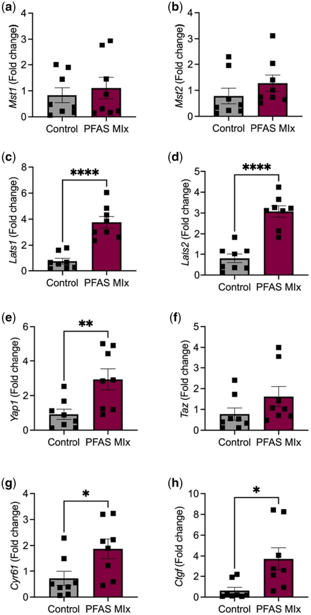
Impact of PFAS mixture on gene expression of Hippo pathway components. Ovarian mRNA transcripts for (a) Mst1; (b) Mst2; (c) Lats1; (d) Lats2; (e) Yap1; (f) Taz; (g) Cyr61; (h) Ctgf were measured by quantitative real-time PCR. Results are presented as relative fold-change means±SEM; n = 8 per treatment. *P ≤ 0.05; **P ≤ 0.01; ****P ≤ 0.0001.
Fig. 10.

Effect of PFAS on Hippo pathway core kinase protein abundance. (a) Representative western blot of Hippo pathway kinases in vehicle control and PFAS mixture-treated mice. Quantification of protein abundance for (b) pLATS1/LATS1 protein ratio; (c) total LATS1; (d) pYAP1/YAP1 protein ratio; (e) total YAP1; (f) CTGF. Results are presented as relative fold-change means±SEM; n = 8 per treatment; #=P ≤ 0.07; **P ≤ 0.01; ***P ≤ 0.001.
Discussion
The ovary regulates endocrine and reproductive function in females, with the follicle serving as the functional unit of the ovary. Exposure to chemicals throughout the lifespan can have detrimental impacts on fertility by disrupting normal ovarian function. Adolescents are at heightened risk of susceptibility to environmental chemicals due to the rapid growth and developmental changes occurring during this period. In rodent studies, subacute exposure to a single PFAS such as PFOA or PFOS has been shown to reduce follicle numbers and the number of corpora lutea present in adult female mice and rats (Chen et al. 2017; Du et al. 2019; Yang et al. 2022). The current study revealed that adolescent PFAS exposure resulted in a loss of primordial and primary follicles, whereas secondary and antral follicle numbers were unaffected. The lack of apoptosis and overall healthy-looking histology of the ovaries from the PFAS-treated mice suggest that accelerated activation and maturation of follicles contribute to the loss of primordial and primary follicles versus direct toxicity to the follicle pool. The 6-wk recovery time that the mice received after the PFAS exposure could be considered as a limiting factor in determining the exact outcome, as direct toxicity may have occurred early during the exposure and the atretic follicles may have already been cleared by the end of the experimental period. Based on previous work this seems unlikely, as our group and others have evaluated the follicular pool directly after PFOA exposure and observed several growing follicles and increased proliferation in the granulosa cells of activated follicles with no changes in apoptosis (Clark and Davis 2022; Clark et al. 2022a; Yang et al. 2022). In vitro work using a singular PFAS, PFOA, induced cell proliferation with no changes in apoptosis in a human granulosa cell line (Clark et al. 2022a). Similarly, a mixture of PFAS (PFOA/PFOS/perfluorohexanesulfonic acid [PFHxS]) induced proliferation and altered the cell cycle primary human granulosa cells (Clark et al. 2024). Taken together, these data support a role for PFAS-induced cell proliferation in granulosa cells, presumptively resulting in follicle activation and growth. Future studies are needed to address this gap in the knowledge on the exact timeline and mechanism of follicle activation.
Another interesting dynamic of the histological examination of the ovaries from the mice exposed to the PFAS mixture was the increase in collagen throughout the ovarian stroma. The ovary undergoes cyclic remodeling with each reproductive cycle, with constant connective tissue remodeling and wound healing, thus the accelerated activation of follicles could contribute to the increased synthesis deposition of collagen/extracellular matrix (ECM) components. One of the early hallmarks of ovarian aging is excess accumulation of ECM, or fibrosis (Briley et al. 2016; Amargant et al. 2020). Additionally, injury or chronic inflammation can lead to fibrosis (Mack 2018). We observed higher levels of circulating TNFα in PFAS-exposed mice relative to vehicle controls, thus the increased inflammation present could also contribute to the increase in fibrosis. These findings, taken together with the slight decrease in ovarian weight and disturbance of estrous cyclicity in the PFAS-treated mice, suggest that early life PFAS exposure may contribute to the initiation of premature ovarian aging.
Endocrine-disrupting chemicals can interfere with the normal actions of endogenous hormones such as their synthesis, secretion, transport, metabolism, binding, and elimination in various organ systems including the ovary (Land et al. 2022). Female steroid hormones are crucial in regulating many physiological processes including pubertal development, reproduction, and metabolism. Increasing evidence indicates that endocrine disruption as a mechanism of action of PFAS in the ovary (Shi et al. 2009; Feng et al. 2015, 2017; Chen et al. 2017; Du et al. 2019; Cao et al. 2020; Yang et al. 2022; Dangudubiyyam et al. 2023), though all of these studies were completed with a singular PFAS and most utilized higher dosages. In the present study, we did not observe alterations in circulating steroid hormones including estradiol, progesterone, or testosterone after exposure to a mixture of PFAS. Additionally, we did not observe changes in serum levels of peptide hormones AMH and FSH. These results are comparable to rodent studies that used lower doses of PFAS (<2.5 mg/kg/d) that did not see any alterations in steroid or gonadotropin hormones (Chen et al. 2017; González-Alvarez et al. 2022; Yang et al. 2022; Dangudubiyyam et al. 2023). Conversely, a subacute study utilizing PFOA or PFOS (0.1, 1 mg/kg) in neonatal (PND 1 to 5) and juvenile rats (PND 26 to 30) demonstrated increases in estradiol (Du et al. 2019), whereas chronic exposure (6 mo) of PFOS (0.1 mg/kg) to mice significantly decreased estradiol and progesterone secretion (Feng et al. 2015). Additionally, a 10-d study of PFOA (1 mg/kg) in adult female mice demonstrated a decrease in progesterone and an increase testosterone (Yang et al. 2022). Interestingly, the gonadotropin hormone LH was increased in our mice that were exposed to the PFAS mixture relative to their control counterparts. A similar elevation in LH has also been observed in a chronic exposure study (6 mo) to PFOS (0.1 mg/kg) in female mice (Feng et al. 2015). Increased levels of LH are a hallmark of reproductive aging or reproductive disorders such as PCOS (Klein et al. 1996; Tock et al. 2014). We did not observe any PCOS-like phenotype/histology in our model, however, the increase in LH accompanied with the increased fibrosis of the ovary and depletion of the follicle pool offers additional support that adolescent exposure to PFAS can alter ovarian homeostasis.
To further investigate the impact of PFAS on steroidogenesis in the ovary, we measured mRNA and protein of enzymes related to steroid production and fatty acid/cholesterol synthesis. In the ovaries from the mice exposed to the PFAS mixture, there were increases in transcript abundance of Cyp11a1, Cyp17a1, Cyp19a1, Hsd3b1, and Hsd17b1. Significant changes in CYP17A1 and CYP19A1 protein abundance were also observed in ovaries from the PFAS-treated mice. Given the increase in serum LH after PFAS treatment, the increases in Cyp17a1 are expected as Cyp17a1 regulates the biosynthesis of androgens (Burris-Hiday and Scott 2021). We also observed increases in Hmgcr, Scarb1, and Srebp1, genes that are involved in cholesterol and fatty acid synthesis/uptake, key steps in the biosynthesis of steroid hormones in the ovary (Huang et al. 2019; Strauss 2019; Zheng et al. 2023). However, the increases in other steroidogenic enzymes were not associated with increases in serum steroid hormones. These results were unexpected and differ from previous studies similar in the manner discussed above. In pregnant mice exposed to PFOA (gestational days 1 to 20, 2.5 mg/kg), mRNA transcripts for Star, Cyp11a1, and Hsd3b1 were decreased in the ovary (Chen et al. 2017). Female mice exposed to PFOS (0.1 mg/kg) for 6 mo also demonstrated a decrease in Star mRNA abundance, though no changes in other steroidogenic enzymes were observed (Feng et al. 2015). A similar decrease in Cyp19a1 was observed in ovaries from rats treated with PFOA during pregnancy (gestational days 4 to 20, ∼1 mg/kg) (Dangudubiyyam et al. 2023). Conversely, adult female mice exposed to PFOA (1 mg/kg) for 10 d had increased levels of Cyp19a1 gene expression (Yang et al. 2022). As we utilized mRNA and protein from whole ovary samples, the present results encompass a variety of cell types, including stromal (smooth muscle and fibroblast), follicular (granulosa, theca, oocyte), immune, endothelial, epithelial, and perivascular. Recent evidence suggests that the ovarian stroma contains steroidogenic cells (Jabara et al. 2003; Wagner et al. 2020; Wang et al. 2020a), though their definitive characterization and steroidogenic capacity remain to be defined. Further, as the ovary ages, the ovarian stroma may become a source of androgen production (Candelaria et al. 2019; Kinnear et al. 2020). Although our PFAS-exposed mouse is still relatively young and the exposure period brief, the increase of steroidogenic enzymes and androgen production, specifically LH, observed at this early time point (even after the cessation of PFAS intake) is a cause for concern. With sustained PFAS exposure, the high androgen environment could potentially lead to PCOS or other hyperandrogenism-related disorders. Observational studies in women are suggestive of a relationship between detectable levels of serum PFAS and PCOS (Wang et al. 2019; Hammarstrand et al. 2021; Zhan et al. 2023). More research is warranted to identify the specific cell types that PFAS target in the ovary, and if/how these cell types respond in a steroidogenic/endocrine capacity.
The Hippo pathway has emerged as a key governor of ovarian homeostasis, regulating several biological processes such as cell proliferation, migration, differentiation, and cell fate determination (Clark et al. 2022b). The major components of the Hippo signaling cascade (MST1/2, LATS1/2, YAP1, TAZ) are present in all follicular stages (primordial, primary, secondary, antral) within the oocyte, granulosa cells, theca cells, and in the corpus luteum following ovulation (Xiang et al. 2015; De Roo et al. 2017; Ji et al. 2017; Hu et al. 2019; Lv et al. 2019; Plewes et al. 2019; Masciangelo et al. 2020; Sun et al. 2021; Dos Santos et al. 2022). Previous work in an immortalized human granulosa cell line HGrC1, a noncancerous human granulosa cell line that mimics granulosa cell behavior in earlier stages of development, revealed increases in protein expression of YAP1 and its downstream target CTGF after PFOA (1 and 10 µm) exposure (Clark et al. 2022a). These observations of increased YAP1 and CTGF protein expression coincided with increases in cell proliferation and migration, whereas inhibition of Yap1 repressed PFOA-induced cell proliferation (Clark et al. 2022a). In another study, PFOA (50 µm) stimulated increases in Mst1, Mst2, Lats1, Yap1, and Taz transcripts in cultured neonatal mouse ovaries (Clark and Davis 2022). Additionally, downstream Hippo pathway targets Areg, Amotl2, and Cyr61 transcripts were also upregulated after PFOA exposure (Clark and Davis 2022). Together with these alterations in the Hippo pathway, it was observed that PFOA increased the number of secondary follicles present in the cultured neonatal mouse ovary, with inhibition of the Hippo pathway using YAP1 inhibitor Verteporfin halting PFOA-induced follicle activation (Clark and Davis 2022). In this study, we observe similar findings in which the loss of primordial and primary follicle numbers is indicative of PFAS-induced follicle activation, paired with the increases in Yap1 and Ctgf mRNA and protein abundance, provides additional in vivo evidence that the Hippo pathway is a potential target/mechanism of action for PFAS in the ovary. Also consistent with previous reports of Hippo pathway modulation after PFAS exposure is the upregulation of the intermediate protein kinase Lats1/2, indicative of a possible feedback loop due to sustained activation of Yap1 (He et al. 2019; Sun et al. 2021; Clark and Davis 2022). Further, ECM stiffening/fibrosis can regulate the Hippo pathway, driving the nuclear localization of Yap and Taz into fibroblasts and promotion of profibrotic ECM synthesis (Liu et al. 2015). The increase of Yap1 and Ctgf in parallel with the increase in ovarian fibrosis suggests another possible mechanism that the Hippo pathway contributes to PFAS-induced pathophysiology in the ovary. Our data also suggest another possible mechanism for PFAS-induced Hippo-Yap1 signaling via regulation of ovarian lipid homeostasis. In the ovaries from the PFAS mixture-treated mice, we observe increased transcripts of Hmgcr, Scarb1, and Srepb1. In the mouse liver, Yap1 has been shown to directly interact with Srepb1 and can directly bind with the Hmgcr promoter, leading to stimulation of cholesterol synthesis (Shu et al. 2019). Additionally, LH can stimulate expression of SREBP, resulting in de novo cholesterol synthesis (Nakanishi et al. 2021). The dysregulation of energy metabolism in the ovary due to PFAS may have potential to negatively impact fertility. The mechanisms through which the Hippo pathway engages with different binding partners to regulate cellular behavior following chemical exposures are still unclear and warrant further investigation.
The discrepancies of these results can be attributed to the differences in life stage, dosage, exposure time, route of exposure, and species. Further, differences in bioaccumulation and elimination of certain PFAS chemicals are sex and species-dependent (Lau et al. 2007; Fenton et al. 2021). For example, the half-life of the PFAS utilized in our study has been reported to be ∼16 d for PFOA, ∼30 to 38 d for PFOS, ∼18 h for GenX/HFPO-DA, and ∼4.5 h for PFBS in female mice (Fenton et al. 2021). In humans, the half-life of these same PFAS compounds is significantly longer, ∼2.1 to 3.8 yr for PFOA, ∼3.4 to 5.0 yr for PFOS, and 28 d for PFBS (Fenton et al. 2021). There is currently no data on the clearance rate of GenX/HFPO-DA in humans. Animal studies provide valuable initial insights into how humans might respond to environmental exposures. Although exact clearance rates may differ, the overall biological effects and mechanisms of toxicity can often be similar.
Conclusion
PFOA, PFOS, GenX/HFPO-DA, PFBS, and mixtures of these compounds are present in many drinking water supplies and food sources globally, making these exposures highly relevant to humans and animals. A significant innovation of this study was the examination of a PFAS mixture, mimicking real-life exposure scenarios to mixtures of chemicals rather than single substances. To our knowledge, this is the first study that evaluates the effects of a PFAS mixture on the ovary and ovarian function. The impact of environmental toxicant exposures on female reproductive function is primarily influenced by the developmental stage during exposure, as well as the exposure’s intensity and duration. This study demonstrates significant changes in adult ovarian function after an adolescent exposure to a PFAS mixture and supports previous studies suggesting PFAS exposure activates Hippo/Yap1 signaling. Taken together, these findings suggest that PFAS exposure may impact folliculogenesis via the Hippo pathway and provides a potential mechanism in which PFAS exposure may be detrimental for fertility and reproductive health. Although it is nearly impossible for humans to avoid exposure to PFAS, it is critical to understand what the potential effects are on female reproductive health, particularly across various life stages.
Supplementary Material
Contributor Information
Kendra L Clark, Department of Obstetrics and Gynecology, Olson Center for Women’s Health, University of Nebraska Medical Center, Omaha, NE 68198, United States; Department of Environmental, Agricultural, and Occupational Health, University of Nebraska Medical Center, Omaha, NE 68198, United States; Veterans Affairs Nebraska-Western Iowa Health Care System, Omaha, NE, United States.
Jitu W George, Department of Obstetrics and Gynecology, Olson Center for Women’s Health, University of Nebraska Medical Center, Omaha, NE 68198, United States.
John S Davis, Department of Obstetrics and Gynecology, Olson Center for Women’s Health, University of Nebraska Medical Center, Omaha, NE 68198, United States; Veterans Affairs Nebraska-Western Iowa Health Care System, Omaha, NE, United States.
Supplementary material
Supplementary material is available at Toxicological Sciences online.
Funding
This work was supported by NIH F32HD106722 (KLC), VA IK2BX006457 (KLC), NE DHHS LB506 26770 (JWG), VA I01BX004272 (JSD), NIH P01AG029531 (JSD), and the Olson Center for Women’s Health (JSD). JSD is the recipient of a VA Senior Research Career Scientist Award (IK6BX005797). We acknowledge the use of the University of Nebraska Medical Center—UNMC Advanced Microscopy Core Facility, RRID: SCR_022467, P20 GM103427 (NIGMS, NE-INEBRE), P30 GM106397 (NIGMS, NCS), P20 GM130447 (NIGMS, CoNDA), P30 CA036727 (NCI, Buffet Cancer Center), S10RR02730 (NIH), S10OD030486 (NIH), Nebraska Research Initiative, UNMC Vice Chancellor for Research Office.
Conflicts of interest. None declared.
Data availability
The datasets used and/or analyzed in the current study are available from the corresponding author upon reasonable request.
References
- Amargant F, Manuel SL, Tu Q, Parkes WS, Rivas F, Zhou LT, Rowley JE, Villanueva CE, Hornick JE, Shekhawat GS, et al. 2020. Ovarian stiffness increases with age in the mammalian ovary and depends on collagen and hyaluronan matrices. Aging Cell. 19(11):e13259. [DOI] [PMC free article] [PubMed] [Google Scholar]
- Barrett ES, Chen C, Thurston SW, Haug LS, Sabaredzovic A, Fjeldheim FN, Frydenberg H, Lipson SF, Ellison PT, Thune I.. 2015. Perfluoroalkyl substances and ovarian hormone concentrations in naturally cycling women. Fertil Steril. 103(5):1261–1270.e3. [DOI] [PMC free article] [PubMed] [Google Scholar]
- Blake BE, Miller CN, Nguyen H, Chappell VA, Phan TP, Phadke DP, Balik-Meisner MR, Mav D, Shah RR, Fenton SE.. 2022. Transcriptional pathways linked to fetal and maternal hepatic dysfunction caused by gestational exposure to perfluorooctanoic acid (PFOA) or hexafluoropropylene oxide-dimer acid (HFPO-DA or GenX) in CD-1 mice. Ecotoxicol Environ Saf. 248:114314. [DOI] [PMC free article] [PubMed] [Google Scholar]
- Brennan NM, Evans AT, Fritz MK, Peak SA, von Holst HE.. 2021. Trends in the regulation of per- and polyfluoroalkyl substances (PFAS): a scoping review. Int J Environ Res Public Health. 18(20):10900. [DOI] [PMC free article] [PubMed] [Google Scholar]
- Briley SM, Jasti S, McCracken JM, Hornick JE, Fegley B, Pritchard MT, Duncan FE.. 2016. Reproductive age-associated fibrosis in the stroma of the mammalian ovary. Reproduction. 152(3):245–260. [DOI] [PMC free article] [PubMed] [Google Scholar]
- Burris-Hiday SD, Scott EE.. 2021. Steroidogenic cytochrome P450 17A1 structure and function. Mol Cell Endocrinol. 528:111261. [DOI] [PMC free article] [PubMed] [Google Scholar]
- Byers SL, Wiles MV, Dunn SL, Taft RA.. 2012. Mouse estrous cycle identification tool and images. PLoS One. 7(4):e35538. [DOI] [PMC free article] [PubMed] [Google Scholar]
- Candelaria NR, Padmanabhan A, Stossi F, Ljungberg MC, Shelly KE, Pew BK, Solis M, Rossano AM, McAllister JM, Wu S, et al. 2019. VCAM1 is induced in ovarian theca and stromal cells in a mouse model of androgen excess. Endocrinology. 160(6):1377–1393. [DOI] [PMC free article] [PubMed] [Google Scholar]
- Cao XY, Liu J, Zhang YJ, Wang Y, Xiong JW, Wu J, Chen L.. 2020. Exposure of adult mice to perfluorobutanesulfonate impacts ovarian functions through hypothyroxinemia leading to down-regulation of AKT-mTOR signaling. Chemosphere. 244:125497. [DOI] [PubMed] [Google Scholar]
- CDC. 2022. National report on human exposure to environmentalchemicals. Atlanta (GA): U.S. Department of Health and Human Services, Centers for Disease Control and Prevention. 10.15620/cdc:133100 [DOI] [Google Scholar]
- Chen Y, Zhou L, Xu J, Zhang L, Li M, Xie X, Xie Y, Luo D, Zhang D, Yu X, et al. 2017. Maternal exposure to perfluorooctanoic acid inhibits luteal function via oxidative stress and apoptosis in pregnant mice. Reprod Toxicol. 69:159–166. [DOI] [PubMed] [Google Scholar]
- Clark KL, Davis JS.. 2022. Perfluorooctanoic acid (PFOA) promotes follicular growth and alters expression of genes that regulate the cell cycle and the Hippo pathway in cultured neonatal mouse ovaries. Toxicol Appl Pharmacol. 454:116253. [DOI] [PMC free article] [PubMed] [Google Scholar]
- Clark KL, George JW, Hua G, Davis JS.. 2022a. Perfluorooctanoic acid promotes proliferation of the human granulosa cell line HGrC1 and alters expression of cell cycle genes and hippo pathway effector YAP1. Reprod Toxicol. 110:49–59. [DOI] [PMC free article] [PubMed] [Google Scholar]
- Clark KL, George JW, Przygrodzka E, Plewes MR, Hua G, Wang C, Davis JS.. 2022b. Hippo signaling in the ovary: emerging roles in development, fertility, and disease. Endocr Rev. 43(6):1074–1096. [DOI] [PMC free article] [PubMed] [Google Scholar]
- Clark KL, Shukla M, George JW, Gustin S, Rowley MJ, Davis JS.. 2024. An environmentally relevant mixture of perfluoroalkyl substances (PFAS) impacts proliferation, steroid hormone synthesis, and gene transcription in primary human granulosa cells. Toxicol Sci. 200(1):57–69. [DOI] [PMC free article] [PubMed] [Google Scholar]
- Clark KL, Talton OO, Ganesan S, Schulz LC, Keating AF.. 2019. Developmental origins of ovarian disorder: impact of maternal lean gestational diabetes on the offspring ovarian proteome in mice†. Biol Reprod. 101(4):771–781. [DOI] [PubMed] [Google Scholar]
- Conley JM, Lambright CS, Evans N, Farraj AK, Smoot J, Grindstaff RD, Hill D, McCord J, Medlock-Kakaley E, Dixon A, et al. 2023. Dose additive maternal and offspring effects of oral maternal exposure to a mixture of three PFAS (HFPO-DA, NBP2, PFOS) during pregnancy in the Sprague-Dawley rat. Sci Total Environ. 892:164609. [DOI] [PMC free article] [PubMed] [Google Scholar]
- Dangudubiyyam SV, Mishra JS, Kumar S.. 2023. Perfluorooctane sulfonic acid modulates expression of placental steroidogenesis-associated genes and hormone levels in pregnant rats. Reprod Toxicol. 118:108390. [DOI] [PMC free article] [PubMed] [Google Scholar]
- De Roo C, Lierman S, Tilleman K, Peynshaert K, Braeckmans K, Caanen M, Lambalk CB, Weyers S, T’Sjoen G, Cornelissen R, et al. 2017. Ovarian tissue cryopreservation in female-to-male transgender people: insights into ovarian histology and physiology after prolonged androgen treatment. Reprod Biomed Online. 34(6):557–566. [DOI] [PubMed] [Google Scholar]
- Ding N, Harlow SD, Randolph JF, Calafat AM, Mukherjee B, Batterman S, Gold EB, Park SK.. 2020. Associations of perfluoroalkyl substances with incident natural menopause: the study of women’s health across the nation. J Clin Endocrinol Metab. 105(9):e3169–e3182. [DOI] [PMC free article] [PubMed] [Google Scholar]
- Ding N, Harlow SD, Randolph JF, Mukherjee B, Batterman S, Gold EB, Park SK.. 2022. Perfluoroalkyl substances and incident natural menopause in midlife women: the mediating role of sex hormones. Am J Epidemiol. 191(7):1212–1223. [DOI] [PMC free article] [PubMed] [Google Scholar]
- Dos Santos EC, Lalonde-Larue A, Antoniazzi AQ, Barreta MH, Price CA, Dias Gonçalves PB, Portela VM, Zamberlam G.. 2022. Yap signaling in preovulatory granulosa cells is critical for the functioning of the EGF network during ovulation. Mol Cell Endocrinol. 541:111524. [DOI] [PubMed] [Google Scholar]
- Du G, Hu J, Huang Z, Yu M, Lu C, Wang X, Wu D.. 2019. Neonatal and juvenile exposure to perfluorooctanoate (PFOA) and perfluorooctane sulfonate (PFOS): advance puberty onset and kisspeptin system disturbance in female rats. Ecotoxicol Environ Saf. 167:412–421. [DOI] [PubMed] [Google Scholar]
- Feng X, Cao X, Zhao S, Wang X, Hua X, Chen L, Chen L.. 2017. Exposure of pregnant mice to perfluorobutanesulfonate causes hypothyroxinemia and developmental abnormalities in female offspring. Toxicol Sci. 155(2):409–419. [DOI] [PubMed] [Google Scholar]
- Feng X, Wang X, Cao X, Xia Y, Zhou R, Chen L.. 2015. Chronic exposure of female mice to an environmental level of perfluorooctane sulfonate suppresses estrogen synthesis through reduced histone H3K14 acetylation of the star promoter leading to deficits in follicular development and ovulation. Toxicol Sci. 148(2):368–379. [DOI] [PubMed] [Google Scholar]
- Fenton SE, Ducatman A, Boobis A, DeWitt JC, Lau C, Ng C, Smith JS, Roberts SM.. 2021. Per- and polyfluoroalkyl substance toxicity and human health review: current state of knowledge and strategies for informing future research. Environ Toxicol Chem. 40(3):606–630. [DOI] [PMC free article] [PubMed] [Google Scholar]
- Glüge J, Scheringer M, Cousins IT, DeWitt JC, Goldenman G, Herzke D, Lohmann R, Ng CA, Trier X, Wang Z.. 2020. An overview of the uses of per- and polyfluoroalkyl substances (PFAS). Environ Sci Process Impacts. 22(12):2345–2373. [DOI] [PMC free article] [PubMed] [Google Scholar]
- González-Alvarez EM, Severin A, Sayadi M, Keating AF.. 2022. PFOA-induced ovotoxicity differs between lean and obese mice with impacts on ovarian reproductive and DNA damage sensing and repair proteins. Toxicol Sci. 190(2):173–188. [DOI] [PMC free article] [PubMed] [Google Scholar]
- Guo M, Yu Y, Liu H, Zhu C.. 2023. Associations between exposure to a mixture of perfluoroalkyl and polyfluoroalkyl substances and age at menarche in adolescent girls utilizing three statistical models. Chemosphere. 335:139054. [DOI] [PubMed] [Google Scholar]
- Hammarstrand S, Jakobsson K, Andersson E, Xu Y, Li Y, Olovsson M, Andersson EM.. 2021. Perfluoroalkyl substances (PFAS) in drinking water and risk for polycystic ovarian syndrome, uterine leiomyoma, and endometriosis: a Swedish Cohort Study. Environ Int. 157:106819. [DOI] [PubMed] [Google Scholar]
- Harlow SD, Hood MM, Ding N, Mukherjee B, Calafat AM, Randolph JF, Gold EB, Park SK.. 2021. Per- and polyfluoroalkyl substances and hormone levels during the menopausal transition. J Clin Endocrinol Metab. 106(11):e4427–e4437. [DOI] [PMC free article] [PubMed] [Google Scholar]
- He C, Lv X, Huang C, Hua G, Ma B, Chen X, Angeletti PC, Dong J, Zhou J, Wang Z, et al. 2019. YAP1-LATS2 feedback loop dictates senescent or malignant cell fate to maintain tissue homeostasis. EMBO Rep. 20(3):e44948. [DOI] [PMC free article] [PubMed] [Google Scholar]
- Hirshfield AN. 1991. Development of follicles in the mammalian ovary. Int Rev Cytol. 124:43–101. [DOI] [PubMed] [Google Scholar]
- Hoyer PB, Keating AF.. 2014. Xenobiotic effects in the ovary: temporary versus permanent infertility. Expert Opin Drug Metab Toxicol. 10(4):511–523. [DOI] [PubMed] [Google Scholar]
- Hsueh AJ, Kawamura K, Cheng Y, Fauser BC.. 2015. Intraovarian control of early folliculogenesis. Endocr Rev. 36(1):1–24. [DOI] [PMC free article] [PubMed] [Google Scholar]
- Hu LL, Su T, Luo RC, Zheng YH, Huang J, Zhong ZS, Nie J, Zheng LP.. 2019. Hippo pathway functions as a downstream effector of Akt signaling to regulate the activation of primordial follicles in mice. J Cell Physiol. 234(2):1578–1587. [DOI] [PubMed] [Google Scholar]
- Huang Q, Liu Y, Yang Z, Xie Y, Mo Z.. 2019. The effects of cholesterol metabolism on follicular development and ovarian function. Curr Mol Med. 19(10):719–730. [DOI] [PubMed] [Google Scholar]
- Jabara S, Christenson LK, Wang CY, McAllister JM, Javitt NB, Dunaif A, Strauss JF III. 2003. Stromal cells of the human postmenopausal ovary display a distinctive biochemical and molecular phenotype. J Clin Endocrinol Metab. 88(1):484–492. [DOI] [PubMed] [Google Scholar]
- Ji SY, Liu XM, Li BT, Zhang YL, Liu HB, Zhang YC, Chen ZJ, Liu J, Fan HY.. 2017. The polycystic ovary syndrome-associated gene YAP1 is regulated by gonadotropins and sex steroid hormones in hyperandrogenism-induced oligo-ovulation in mouse. Mol Hum Reprod. 23(10):698–707. [DOI] [PubMed] [Google Scholar]
- Khosla S, Monroe DG.. 2018. Regulation of bone metabolism by sex steroids. Cold Spring Harb Perspect Med. 8(1):a031211. [DOI] [PMC free article] [PubMed] [Google Scholar]
- Kinnear HM, Tomaszewski CE, Chang AL, Moravek MB, Xu M, Padmanabhan V, Shikanov A.. 2020. The ovarian stroma as a new frontier. Reproduction. 160(3):R25–R39. [DOI] [PMC free article] [PubMed] [Google Scholar]
- Klein NA, Battaglia DE, Fujimoto VY, Davis GS, Bremner WJ, Soules MR.. 1996. Reproductive aging: accelerated ovarian follicular development associated with a monotropic follicle-stimulating hormone rise in normal older women. J Clin Endocrinol Metab. 81(3):1038–1045. [DOI] [PubMed] [Google Scholar]
- Knox SS, Jackson T, Javins B, Frisbee SJ, Shankar A, Ducatman AM.. 2011. Implications of early menopause in women exposed to perfluorocarbons. J Clin Endocrinol Metab. 96(6):1747–1753. [DOI] [PMC free article] [PubMed] [Google Scholar]
- Kristensen SL, Ramlau-Hansen CH, Ernst E, Olsen SF, Bonde JP, Vested A, Halldorsson TI, Becher G, Haug LS, Toft G.. 2013. Long-term effects of prenatal exposure to perfluoroalkyl substances on female reproduction. Hum Reprod. 28(12):3337–3348. [DOI] [PubMed] [Google Scholar]
- Land KL, Miller FG, Fugate AC, Hannon PR.. 2022. The effects of endocrine-disrupting chemicals on ovarian- and ovulation-related fertility outcomes. Mol Reprod Dev. 89(12):608–631. [DOI] [PMC free article] [PubMed] [Google Scholar]
- Lau C, Anitole K, Hodes C, Lai D, Pfahles-Hutchens A, Seed J.. 2007. Perfluoroalkyl acids: a review of monitoring and toxicological findings. Toxicol Sci. 99(2):366–394. [DOI] [PubMed] [Google Scholar]
- Liu F, Lagares D, Choi KM, Stopfer L, Marinković A, Vrbanac V, Probst CK, Hiemer SE, Sisson TH, Horowitz JC, et al. 2015. Mechanosignaling through YAP and TAZ drives fibroblast activation and fibrosis. Am J Physiol Lung Cell Mol Physiol. 308(4):L344–L357. [DOI] [PMC free article] [PubMed] [Google Scholar]
- Liu Y, Calafat AM, Chen A, Lanphear BP, Jones NY, Cecil KM, Rose SR, Yolton K, Buckley JP, Braun JM.. 2023. Associations of prenatal and postnatal exposure to perfluoroalkyl substances with pubertal development and reproductive hormones in females and males: the home study. Sci Total Environ. 890:164353. [DOI] [PMC free article] [PubMed] [Google Scholar]
- Livak KJ, Schmittgen TD.. 2001. Analysis of relative gene expression data using real-time quantitative PCR and the 2(-Delta c(t)) method. Methods. 25(4):402–408. [DOI] [PubMed] [Google Scholar]
- Lopez-Espinosa MJ, Fletcher T, Armstrong B, Genser B, Dhatariya K, Mondal D, Ducatman A, Leonardi G.. 2011. Association of perfluorooctanoic acid (PFOA) and perfluorooctane sulfonate (PFOS) with age of puberty among children living near a chemical plant. Environ Sci Technol. 45(19):8160–8166. [DOI] [PubMed] [Google Scholar]
- Lv X, He C, Huang C, Wang H, Hua G, Wang Z, Zhou J, Chen X, Ma B, Timm BK, et al. 2019. Timely expression and activation of YAP1 in granulosa cells is essential for ovarian follicle development. FASEB J. 33(9):10049–10064. [DOI] [PMC free article] [PubMed] [Google Scholar]
- Lyngsø J, Ramlau-Hansen CH, Høyer BB, Støvring H, Bonde JP, Jönsson BA, Lindh CH, Pedersen HS, Ludwicki JK, Zviezdai V, et al. 2014. Menstrual cycle characteristics in fertile women from Greenland, Poland and Ukraine exposed to perfluorinated chemicals: a cross-sectional study. Hum Reprod. 29(2):359–367. [DOI] [PubMed] [Google Scholar]
- Mack M. 2018. Inflammation and fibrosis. Matrix Biol. 68-69:106–121. [DOI] [PubMed] [Google Scholar]
- Masciangelo R, Hossay C, Chiti MC, Manavella DD, Amorim CA, Donnez J, Dolmans MM.. 2020. Role of the PI3K and Hippo pathways in follicle activation after grafting of human ovarian tissue. J Assist Reprod Genet. 37(1):101–108. [DOI] [PMC free article] [PubMed] [Google Scholar]
- Nagle CM, Dixon SC, Jensen A, Kjaer SK, Modugno F, deFazio A, Fereday S, Hung J, Johnatty SE, Fasching PA, et al. ; Ovarian Cancer Association Consortium. 2015. Obesity and survival among women with ovarian cancer: results from the ovarian cancer association consortium. Br J Cancer. 113(5):817–826. [DOI] [PMC free article] [PubMed] [Google Scholar]
- Nakanishi T, Tanaka R, Tonai S, Lee JY, Yamaoka M, Kawai T, Okamoto A, Shimada M, Yamashita Y.. 2021. LH induces de novo cholesterol biosynthesis via SREBP activation in granulosa cells during ovulation in female mice. Endocrinology. 162(11):bqab166. [DOI] [PubMed] [Google Scholar]
- Pérez F, Nadal M, Navarro-Ortega A, Fàbrega F, Domingo JL, Barceló D, Farré M.. 2013. Accumulation of perfluoroalkyl substances in human tissues. Environ Int. 59:354–362. [DOI] [PubMed] [Google Scholar]
- Plewes MR, Hou X, Zhang P, Liang A, Hua G, Wood JR, Cupp AS, Lv X, Wang C, Davis JS.. 2019. Yes-associated protein 1 is required for proliferation and function of bovine granulosa cells in vitro†. Biol Reprod. 101(5):1001–1017. [DOI] [PMC free article] [PubMed] [Google Scholar]
- Rickard BP, Tan X, Fenton SE, Rizvi I.. 2022. Select per- and polyfluoroalkyl substances (PFAS) induce resistance to carboplatin in ovarian cancer cell lines. Int J Mol Sci. 23(9):5176. [DOI] [PMC free article] [PubMed] [Google Scholar]
- Rickard BP, Tan X, Fenton SE, Rizvi I.. 2023. Photodynamic priming overcomes per- and polyfluoroalkyl substance (PFAS)-induced platinum resistance in ovarian cancer(†). Photochem Photobiol. 99(2):793–813. [DOI] [PMC free article] [PubMed] [Google Scholar]
- Robarts DR, Paine-Cabrera D, Kotulkar M, Venneman KK, Gunewardena S, Foquet L, Bial G, Apte U.. 2024. Identifying novel mechanisms of per- and polyfluoroalkyl substance-induced hepatotoxicity using frg humanized mice. Arch Toxicol. 98(9):3063–3075. [DOI] [PubMed] [Google Scholar]
- Rodgers JL, Jones J, Bolleddu SI, Vanthenapalli S, Rodgers LE, Shah K, Karia K, Panguluri SK.. 2019. Cardiovascular risks associated with gender and aging. J Cardiovasc Dev Dis. 6(2):19. [DOI] [PMC free article] [PubMed] [Google Scholar]
- Shi Z, Zhang H, Ding L, Feng Y, Xu M, Dai J.. 2009. The effect of perfluorododecanonic acid on endocrine status, sex hormones and expression of steroidogenic genes in pubertal female rats. Reprod Toxicol. 27(3-4):352–359. [DOI] [PubMed] [Google Scholar]
- Shu Z, Gao Y, Zhang G, Zhou Y, Cao J, Wan D, Zhu X, Xiong W.. 2019. A functional interaction between Hippo-Yap signalling and SREBPs mediates hepatic steatosis in diabetic mice. J Cell Mol Med. 23(5):3616–3628. [DOI] [PMC free article] [PubMed] [Google Scholar]
- Strauss JF. 2019. Organization of ovarian steroidogenic cells and cholesterol metabolism. In: Peter CK, Leung EYA, editors. The ovary. 3rd ed. Cambridge (MA): Academic Press. p. 83–94. [Google Scholar]
- Sullivan SD, Sarrel PM, Nelson LM.. 2016. Hormone replacement therapy in young women with primary ovarian insufficiency and early menopause. Fertil Steril. 106(7):1588–1599. [DOI] [PMC free article] [PubMed] [Google Scholar]
- Sun X, Niu X, Qin N, Shan X, Zhao J, Ma C, Xu R, Mishra B.. 2021. Novel insights into the regulation of LATS2 kinase in prehierarchical follicle development via the Hippo pathway in hen ovary. Poult Sci. 100(12):101454. [DOI] [PMC free article] [PubMed] [Google Scholar]
- Sunderland EM, Hu XC, Dassuncao C, Tokranov AK, Wagner CC, Allen JG.. 2019. A review of the pathways of human exposure to poly- and perfluoroalkyl substances (PFASs) and present understanding of health effects. J Expo Sci Environ Epidemiol. 29(2):131–147. [DOI] [PMC free article] [PubMed] [Google Scholar]
- Taylor KW, Hoffman K, Thayer KA, Daniels JL.. 2014. Polyfluoroalkyl chemicals and menopause among women 20-65 years of age (NHANES). Environ Health Perspect. 122(2):145–150. [DOI] [PMC free article] [PubMed] [Google Scholar]
- Tock L, Carneiro G, Pereira AZ, Tufik S, Zanella MT.. 2014. Adrenocortical production is associated with higher levels of luteinizing hormone in nonobese women with polycystic ovary syndrome. Int J Endocrinol. 2014:620605. [DOI] [PMC free article] [PubMed] [Google Scholar]
- Wagner M, Yoshihara M, Douagi I, Damdimopoulos A, Panula S, Petropoulos S, Lu H, Pettersson K, Palm K, Katayama S, et al. 2020. Single-cell analysis of human ovarian cortex identifies distinct cell populations but no oogonial stem cells. Nat Commun. 11(1):1147. [DOI] [PMC free article] [PubMed] [Google Scholar]
- Wang S, Zheng Y, Li J, Yu Y, Zhang W, Song M, Liu Z, Min Z, Hu H, Jing Y, et al. 2020a. Single-cell transcriptomic atlas of primate ovarian aging. Cell. 180(3):585–600.e19. [DOI] [PubMed] [Google Scholar]
- Wang W, Rhodes G, Ge J, Yu X, Li H.. 2020b. Uptake and accumulation of per- and polyfluoroalkyl substances in plants. Chemosphere. 261:127584. [DOI] [PubMed] [Google Scholar]
- Wang W, Zhou W, Wu S, Liang F, Li Y, Zhang J, Cui L, Feng Y, Wang Y.. 2019. Perfluoroalkyl substances exposure and risk of polycystic ovarian syndrome related infertility in Chinese women. Environ Pollut. 247:824–831. [DOI] [PubMed] [Google Scholar]
- Wang Y, Aimuzi R, Nian M, Zhang Y, Luo K, Zhang J.. 2021. Perfluoroalkyl substances and sex hormones in postmenopausal women: NHANES 2013-2016. Environ Int. 149:106408. [DOI] [PubMed] [Google Scholar]
- Xiang C, Li J, Hu L, Huang J, Luo T, Zhong Z, Zheng Y, Zheng L.. 2015. Hippo signaling pathway reveals a spatio-temporal correlation with the size of primordial follicle Pool in mice. Cell Physiol Biochem. 35(3):957–968. [DOI] [PubMed] [Google Scholar]
- Xing Y, Zhou Y, Zhang X, Lin X, Li J, Liu P, Lee HK, Huang Z.. 2023. The sources and bioaccumulation of per- and polyfluoroalkyl substances in animal-derived foods and the potential risk of dietary intake. Sci Total Environ. 905:167313. [DOI] [PubMed] [Google Scholar]
- Yang M, Lee Y, Gao L, Chiu K, Meling DD, Flaws JA, Warner GR.. 2022. Perfluorooctanoic acid disrupts ovarian steroidogenesis and folliculogenesis in adult mice. Toxicol Sci. 186(2):260–268. [DOI] [PMC free article] [PubMed] [Google Scholar]
- Zhan W, Qiu W, Ao Y, Zhou W, Sun Y, Zhao H, Zhang J.. 2023. Environmental exposure to emerging alternatives of per- and polyfluoroalkyl substances and polycystic ovarian syndrome in women diagnosed with infertility: a mixture analysis. Environ Health Perspect. 131(5):57001. [DOI] [PMC free article] [PubMed] [Google Scholar]
- Zhang S, Tan R, Pan R, Xiong J, Tian Y, Wu J, Chen L.. 2018. Association of perfluoroalkyl and polyfluoroalkyl substances with premature ovarian insufficiency in Chinese women. J Clin Endocrinol Metab. 103(7):2543–2551. [DOI] [PubMed] [Google Scholar]
- Zheng M, Andersen CY, Rasmussen FR, Cadenas J, Christensen ST, Mamsen LS.. 2023. Expression of genes and enzymes involved in ovarian steroidogenesis in relation to human follicular development. Front Endocrinol (Lausanne). 14:1268248. [DOI] [PMC free article] [PubMed] [Google Scholar]
- Zhou W, Zhang L, Tong C, Fang F, Zhao S, Tian Y, Tao Y, Zhang J; Shanghai Birth Cohort Study. 2017. Plasma perfluoroalkyl and polyfluoroalkyl substances concentration and menstrual cycle characteristics in preconception women. Environ Health Perspect. 125(6):067012. [DOI] [PMC free article] [PubMed] [Google Scholar]
Associated Data
This section collects any data citations, data availability statements, or supplementary materials included in this article.
Supplementary Materials
Data Availability Statement
The datasets used and/or analyzed in the current study are available from the corresponding author upon reasonable request.