

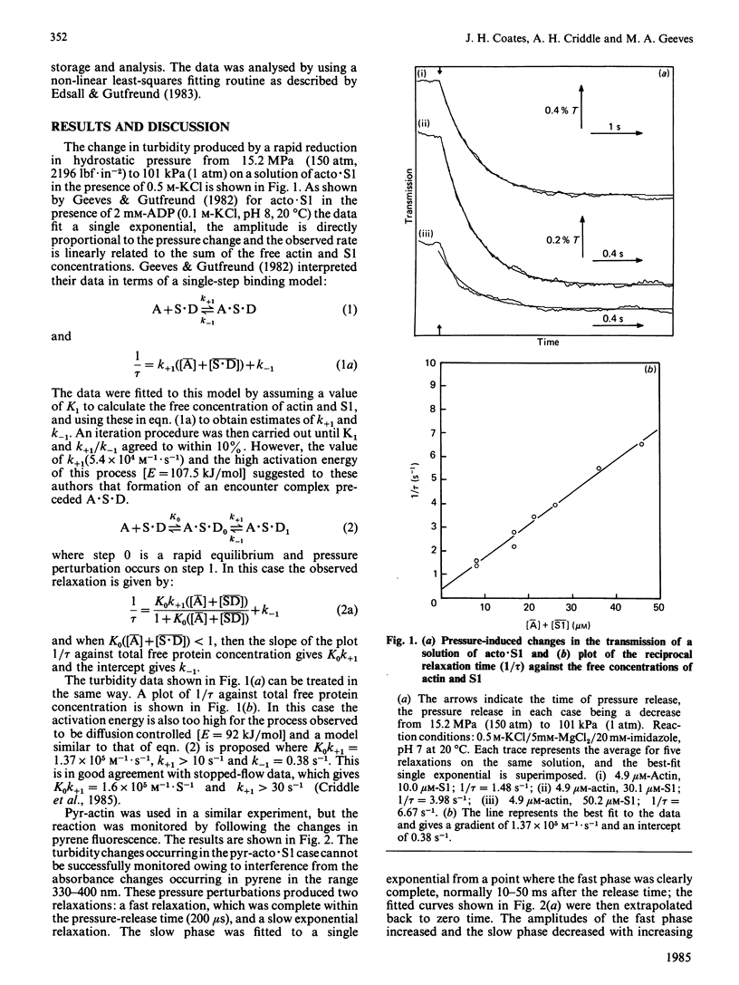
Abstract
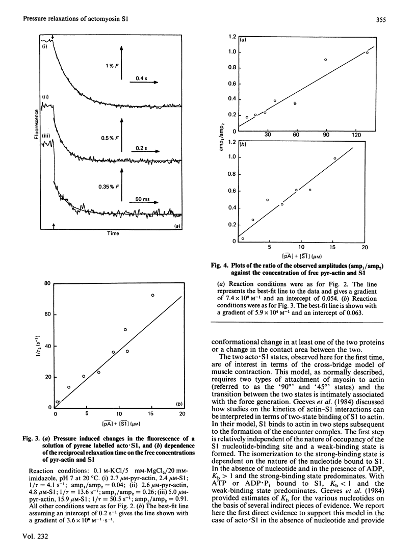
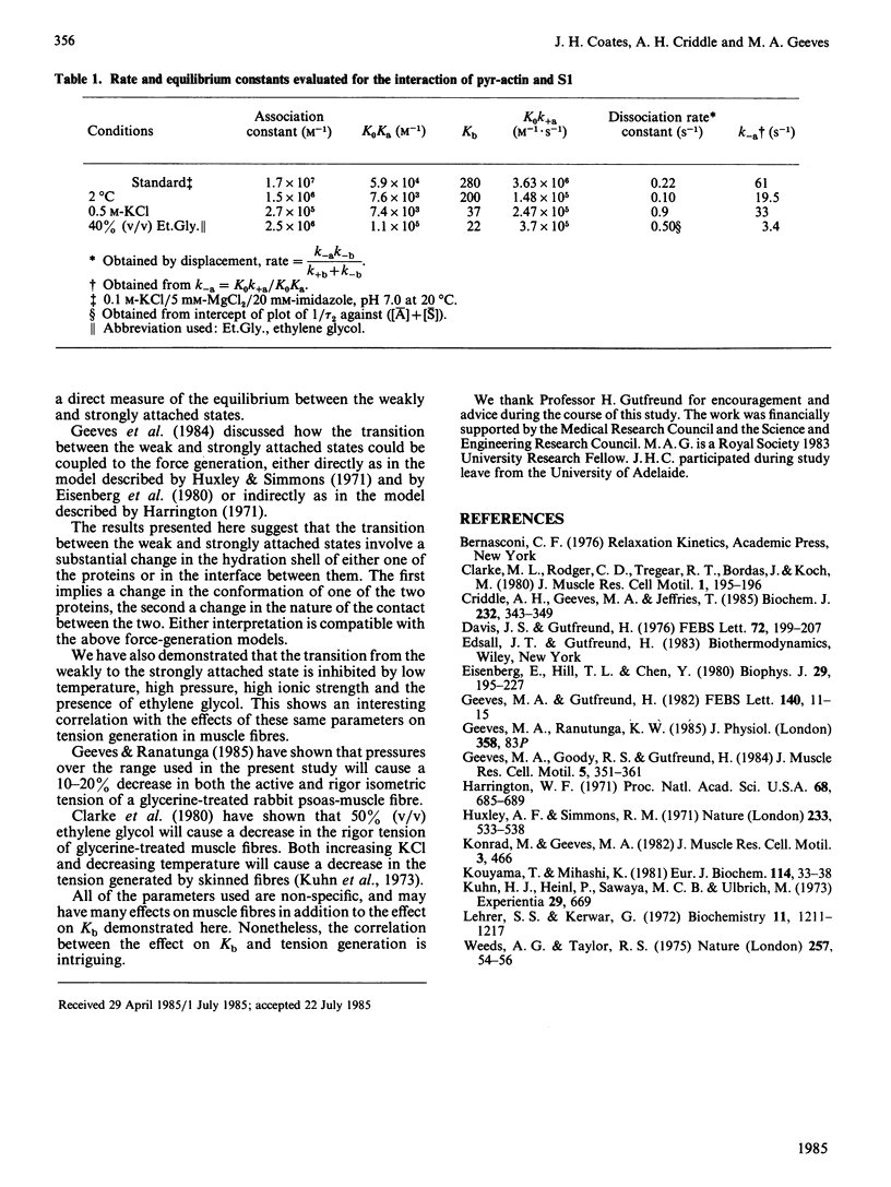
We have used actin labelled at Cys-374 with N-(1-pyrenyl)iodoacetamide [Kouyama & Mihashi (1981) Eur. J. Biochem. 114, 33-38] to monitor pressure-induced relaxations of acto-myosin subfragment 1. This label greatly increases the sensitivity of measurement of dissociated actin and reveals the presence of two relaxations. The experimental data can be fitted by a model in which actin binds subfragment 1 relatively weakly (K = 5.9 X 10(4) M-1) and then isomerizes to a more tightly bound complex (K = 1.7 X 10(7) M-1). This directly observed isomerization supports the model of Geeves, Goody & Gutfreund [(1984) J. Muscle Res. Cell. Motil. 5, 351-361]. The rate of the isomerization is too high to be observed in the pressure-jump apparatus (less than 200 microseconds), but analysis of the amplitudes allows estimation of the equilibrium constant of the isomerization as 280 (20 degrees C, 0.1 M-KCl, pH 7). The equilibrium is sensitive to temperature, pressure, ionic strength and the presence of ethylene glycol. The pressure-sensitivity of the isomerization suggests a significant conformational change of the acto-myosin subfragment 1 complex.
Full text
PDF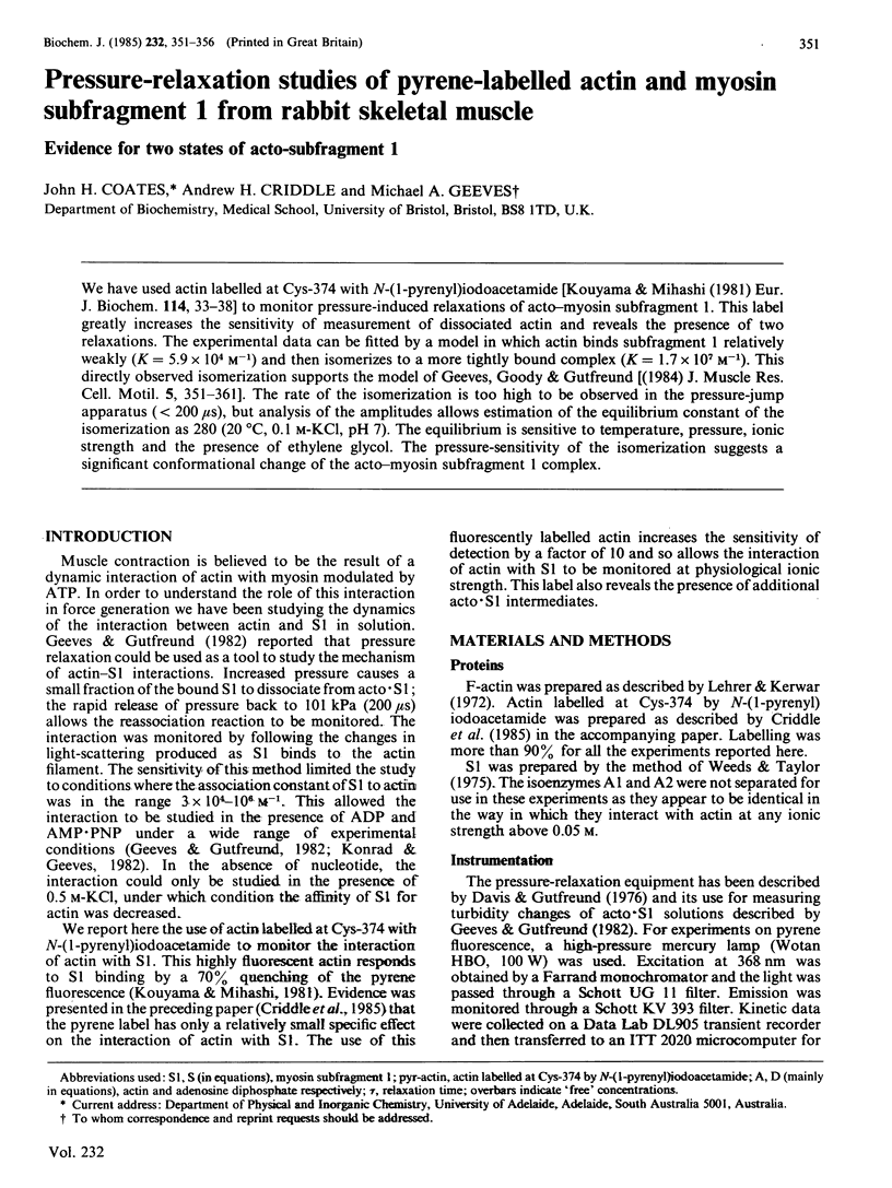
Selected References
These references are in PubMed. This may not be the complete list of references from this article.
- Criddle A. H., Geeves M. A., Jeffries T. The use of actin labelled with N-(1-pyrenyl)iodoacetamide to study the interaction of actin with myosin subfragments and troponin/tropomyosin. Biochem J. 1985 Dec 1;232(2):343–349. doi: 10.1042/bj2320343. [DOI] [PMC free article] [PubMed] [Google Scholar]
- Davis J. S., Gutfreund H. The scope of moderate pressure changes for kinetic and equilibrium studies of biochemical systems. FEBS Lett. 1976 Dec 31;72(2):199–207. doi: 10.1016/0014-5793(76)80971-4. [DOI] [PubMed] [Google Scholar]
- Eisenberg E., Hill T. L., Chen Y. Cross-bridge model of muscle contraction. Quantitative analysis. Biophys J. 1980 Feb;29(2):195–227. doi: 10.1016/S0006-3495(80)85126-5. [DOI] [PMC free article] [PubMed] [Google Scholar]
- Geeves M. A., Goody R. S., Gutfreund H. Kinetics of acto-S1 interaction as a guide to a model for the crossbridge cycle. J Muscle Res Cell Motil. 1984 Aug;5(4):351–361. doi: 10.1007/BF00818255. [DOI] [PubMed] [Google Scholar]
- Geeves M. A., Gutfreund H. The use of pressure perturbations to investigate the interaction of rabbit muscle myosin subfragment 1 with actin in the presence of MgADP. FEBS Lett. 1982 Apr 5;140(1):11–15. doi: 10.1016/0014-5793(82)80509-7. [DOI] [PubMed] [Google Scholar]
- Harrington W. F. A mechanochemical mechanism for muscle contraction. Proc Natl Acad Sci U S A. 1971 Mar;68(3):685–689. doi: 10.1073/pnas.68.3.685. [DOI] [PMC free article] [PubMed] [Google Scholar]
- Huxley A. F., Simmons R. M. Proposed mechanism of force generation in striated muscle. Nature. 1971 Oct 22;233(5321):533–538. doi: 10.1038/233533a0. [DOI] [PubMed] [Google Scholar]
- Kouyama T., Mihashi K. Fluorimetry study of N-(1-pyrenyl)iodoacetamide-labelled F-actin. Local structural change of actin protomer both on polymerization and on binding of heavy meromyosin. Eur J Biochem. 1981;114(1):33–38. [PubMed] [Google Scholar]
- Kuhn H. J., Heinl P., Sawaya M. C., Ulbrich M. The influence of magnesium and calcium ions on the force produced in rigor muscle. Experientia. 1973 Jun 15;29(6):669–671. doi: 10.1007/BF01944763. [DOI] [PubMed] [Google Scholar]
- Lehrer S. S., Kerwar G. Intrinsic fluorescence of actin. Biochemistry. 1972 Mar 28;11(7):1211–1217. doi: 10.1021/bi00757a015. [DOI] [PubMed] [Google Scholar]
- Weeds A. G., Taylor R. S. Separation of subfragment-1 isoenzymes from rabbit skeletal muscle myosin. Nature. 1975 Sep 4;257(5521):54–56. doi: 10.1038/257054a0. [DOI] [PubMed] [Google Scholar]