Abstract
During a survey of microfungi associated with forest plants, four specimens related to Kirschsteiniothelia were collected from decaying wood in Guizhou, Hainan and Yunnan Provinces, China. Kirschsteiniothelia species have sexual and asexual forms. They are commonly found as saprophytes on decaying wood and have been reported as disease-causing pathogens in humans as well. In this study, we introduce three novel Kirschsteiniothelia species (K.bulbosapicalis, K.dendryphioides and K.longirostrata) and describe a new host record for K.atra, based on morphology and multi-gene phylogenetic analyses of a concatenated ITS, LSU and SSU rDNA sequence data. These taxa produced a dendryphiopsis- or sporidesmium-like asexual morph and detailed descriptions and micromorphological illustrations are provided. Furthermore, we provide a checklist for the accepted Kirschsteiniothelia species, including detailed host information, habitat preferences, molecular data, existing morphological type, country of origin and corresponding references.
Key words: Checklist, diversity, Dothideomycetes, Kirschsteiniotheliales, one new host record, taxonomy, three new taxa
Introduction
Kirschsteiniothelia was introduced by Hawksworth (1985) and typified by K.aethiops, based on morphological observation, linking it to its asexual genus, Dendryphiopsis S. Hughes. Later, the type species was reclassified with its asexual morph, K.atra (Hyde et al 2011; Wijayawardene et al. 2014). The connection between the sexual morphs of Kirschsteiniotheliaatra (characterised by cylindrical-clavate, bitunicate, 8-spored or rarely 4-spored asci and ellipsoidal verruculose or smooth ascospores with 1–2 septa, lacking a distinct gelatinous sheath) and the asexual morphs (characterised by macronematous, mononematous and branched conidiophores, monotretic, terminal or intercalary, cylindrical, doliiform conidiogenous cells and acrogenous, solitary, cylindrical, oblong, septate conidia with obtuse ends) were previously established by Hughes (1978). This connection was confirmed from cultures obtained from fragments of the ascomata, based on morphological examination. Schoch et al. (2006) further confirmed the connection between the sexual and asexual morphs of Kirschsteiniothelia, based on both morphology and phylogenetic analysis of SSU, LSU, tef1-α and rpb2. Boonmee et al. (2012) established a novel family, Kirschsteiniotheliaceae, based on the connection between the sexual and asexual morph of Kirschsteiniothelia and multiple gene (LSU, SSU and ITS) phylogeny. Wijayawardene et al. (2014) suggested the use of Kirschsteiniothelia as the updated genus to accommodate Dendryphiopsis species. As a result, Dendryphiopsisatra was re-assigned to Kirschsteiniothelia and synonymised with K.atra, based on morphological and phylogenetic analyses. In the meantime, Wijayawardene et al. (2014) suggested using K.atra to replace K.aethiops as the type species of Kirschsteiniothelia. This recommendation was supported by later studies (Rossman et al. 2015; Su et al. 2016; Bao et al. 2018; Yang et al. 2023; Jin et al. 2024; Sruthi et al. 2024; Tian et al. 2024). Sruthi et al. (2024) legitimately placed five species from Dendryphiopsis under Kirschsteiniothelia, namely, K.arbuscula, K.binsarensis, K.biseptata, K.fascicularis and K.goaensis. Kirschsteiniothelia usually exhibits both sexual and asexual morphs (Hawksworth 1985; Boonmee et al. 2012; Hyde et al. 2013; Sun et al. 2021; Xu et al. 2023). The sexual morph of Kirschsteiniothelia is characterised by superficial, erumpent, papillate, brown or black and hemi-spherical or subglobose ascomata and cylindrical or clavate, bitunicate, pedicellate asci that are usually 8-spored comprising an ocular apical chamber. The ascospores are ellipsoidal, usually asymmetrical, verruculose or smooth and olivaceous to dark brown, comprising 1–2 septa, with a mucilaginous sheath being occasionally present (Chen et al. 2006; Hyde et al. 2018; Meng et al. 2024). Furthermore, ascospores occasionally display longitudinal or sinuate furrows that are visible from the face view (Hawksworth 1985; Boonmee et al. 2012; Hyde et al. 2013; Mehrabi et al. 2017; Yang et al. 2023).
The asexual morph is further categorised into two types, namely the dendryphiopsis- and sporidesmium-like morphs. The dendryphiopsis-like morph was described by Hughes (1978), who found that the ascomatal fragments of Kirschsteiniotheliaaethiops (≡ Amphisphaeriaincrustans) exhibited agar sporulation and morphological traits similar to those of Dendryphiopsisatra. This was later supported by Hawksworth (1985). Subsequently, Boonmee et al. (2012) supported the connection between the sexual morph of Kirschsteiniothelia and the asexual dendryphiopsis-like morph, based on morphological and phylogenetic analyses. The dendryphiopsis-like morph is characterised by macronematous, septate, cylindrical conidiophores that are irregularly or subscorpioidly branched at the apex. Their conidiogenous cells are mono- to polytretic, integrated, terminal or lateral, doliiform or lageniform. Moreover, the conidia are holoblastic, acrogenous, obclavate, rostrate, obovoid to broadly obovoid, solitary or branched in acropetal chains, exhibiting rounded ends. Taxa exhibiting the dendryphiopsis-like characteristics are K.atra, K.arbuscula, K.binsarensis, K.biseptata, K.dendryphioides, K.ebriosa, K.emarceis, K.fascicularis, K.goaensis, K.inthanonensis, K.lignicola, K.longisporum, K.nabanheensis, K.ramus, K.recessa, K.saprophytica, K.septemseptatum, K.shimlaensis, K.vinigena and K.zizyphifolii (Hughes 1978; Boonmee et al. 2012; de Farias et al. 2024; Tian et al. 2024; this study).
The sporidesmium-like asexual morph was described by Su et al. (2016), based on morphological and phylogenetic evidence. Despite having different morphological characteristics from other Kirschsteiniothelia species, the sporidesmium-like morphs fits into the generic concept of Kirschsteiniothelia as they display similar morphologies including unbranched, slender conidiophores that are straight or slightly curved, multi-septate and brown to pale brown, usually truncate at the base and rounded at the apex, producing small conidia. The sporidesmium-like morph is depicted by macronematous, mononematous, unbranched, multi-septate, cylindrical conidiophores, holoblastic, integrated, terminal, determinate, percurrent, cylindrical and caliciform conidiogenous cells and acrogenous, multi-septate, obclavate to obspathulate, rostrate and fusiform conidia that are swollen at the tips or middle of the beak, with or without a conspicuous, gelatinous, hyaline sheath around the tip or middle of the beak. The presence of the sporidesmium-like asexual morph of Kirschsteiniothelia was further supported by subsequent research (Sun et al. 2021; Jayawardena et al. 2022; Xu et al. 2023; Yang et al. 2023). Species exhibiting the sporidesmium-like features are K.acutispora, K.agumbensis, K.aquatica, K.bulbosapicalis, K.cangshanensis, K.crustacea, K.dujuanhuensis, K.dushanensis, K.extensum, K.fluminicola, K.guangdongensis, K.longirostrata, K.pini, K.puerensis, K.rostrata, K.sichuanensis, K.spatiosum, K.submersa, K.tectonae, K.thailandica and K.xishuangbannaensis (Su et al. 2016; Jayawardena et al. 2022; Xu et al. 2023; Yang et al. 2023; Jin et al. 2024; Sruthi et al. 2024; this study).
Although Kirschsteiniothelia comprises numerous species, there are likely to be more undescribed species in this genus as predicted by Bhunjun et al. (2022). Most species have been reported as saprobes inhabiting terrestrial and freshwater environments in tropical and subtropical regions. However, K.ebriosa and K.vinigena have been identified from cork taint of sparkling wine (Bao et al. 2018; Rodríguez-Andrade et al. 2020; Sun et al. 2021; Jayawardena et al. 2022). Moreover, a report indicates the presence of an unidentified Kirschsteiniothelia species that is pathogenic to humans, causing infection superimposed on pre-existing non-infectious bursitis of the ankle. This identification was based on the examination of the strain’s cultural colony and ITS gene fragment (Nishi et al. 2018). To date, there are 59 species of Kirschsteiniothelia, amongst which 18 have been reported only in their sexual morph, 32 reported in their asexual morph and six species documented in both morphs (Boonmee et al. 2012; Su et al. 2016; Sun et al. 2021; Xu et al. 2023; Zhang et al. 2023; de Farias et al. 2024; Sruthi et al. 2024; this study). Amongst the two asexual morphs that have been described so far, only the dendryphiopsis-like morph is linked to the sexual morph, while the sporidesmium-like state has not been associated with the sexual morph (Hawksworth 1985; Wang et al. 2004; Mulenko et al. 2008; Boonmee et al. 2012; Su et al. 2016; Sun et al. 2021; Xu et al.2023; de Farias et al. 2024).
In this study, we aimed to isolate microfungi from unidentified decaying wood collected in Hainan and Yunnan Provinces, China, as well as from Edgeworthiachrysantha, collected in Guizhou Province, China. This study has the following objectives: 1) to describe novel species associated with decaying wood through comprehensive morphological examinations and phylogenetic analyses of ITS, LSU and SSU rDNA sequence data; 2) to provide a checklist that includes host information, habitat preferences, availability of molecular data, morphological characteristics and country of origin.
Materials and methods
Sample collection, isolation and morphological studies
Decaying wood materials of Edgeworthiachrysantha and unidentified plants were collected from Zunyi City in Guizhou Province, Jianfengling National Forest Park, situated at the confluence of Ledong Li Autonomous County and Dongfang City in Hainan Province and Lushui City in Yunnan Province, China. These specimens were initially stored in Ziploc bags and observed using a stereomicroscope (Motic SMZ-171). The collection, observation and isolation were conducted following the methods outlined in Senanayake et al. (2020) and Tang et al. (2022). The observed features were measured using Tarosoft (R) Image Frame Work (version IFW 0.97) and photoplates were constructed using Adobe Photoshop 2019 (Adobe Systems, USA).
Specimens were deposited at the herbaria of the Kunming Institute of Botany, Chinese Academy of Sciences (HKAS), located in Kunming, China and the Guizhou Academy of Agriculture Sciences (GZAAS), situated in Guiyang, China. In addition, ex-type living cultures were preserved at the Kunming Institute of Botany Culture Collection (KUMCC) and the Guizhou Culture Collection (GZCC). Faces of Fungi and Fungal name numbers were obtained following the guidelines in Jayasiri et al. (2015), Wang et al. (2023) and Fungal names (2024). Species identification and establishment were determined following the guidelines outlined by Jeewon and Hyde (2016), Maharachchikumbura et al. (2021) and Pem et al. (2021).
DNA extraction, PCR amplification and sequencing
Freshly scraped mycelia from the pure cultures obtained by single spore isolation were transferred to 1.5 ml microcentrifuge tubes and stored in the refrigerator at -20 °C. Genomic DNA extraction was carried out using DNA extraction kits provided by Sangon Biotech (Shanghai) Co. Ltd., China. Polymerase Chain Reaction (PCR) was employed for DNA template amplification, using the following primer pairs: ITS5/ITS4 for ITS, NS1/NS4 for SSU (White et al. 1990) and LR0R/LR5 for LSU (Vilgalys and Hester 1990; Cubeta et al. 1991). Further details regarding DNA extraction, PCR amplification, sequencing and phylogenetic analyses are given in Tang et al. (2022, 2023).
In PCR amplification, the total volume of the PCR mixture was 50 μl, comprising the DNA template (2 μl), forward primer (2 μl), reverse primer (2 μl), 2 × Taq PCR Master Mix (25 μl) and 19 μl of double-distilled water. The PCR profiles consisted of 35 cycles, with annealing temperatures set at 52 °C for 1 minute and extension for 90 seconds at 72 °C for ITS, LSU and SSU loci. PCR products were verified on 1% agarose gel prior to submission to Sangon Biotech (Shanghai) Co., Ltd., China, for sequencing.
Phylogenetic analyses
Sequences obtained were subjected to a BLAST search in the NCBI database (https://blast.ncbi.nlm.nih.gov/Blast.cgi). Forward and reverse sequences were assembled using the Contig Express version 3.0.0 application. The ITS, LSU and SSU sequence data of Kirschsteiniothelia species were retrieved and downloaded from GenBank (Table 1). Individual sequences were aligned using MAFFT version 7 (https://mafft.cbrc.jp/alignment/server/index.html) with the “auto” option (Katoh et al. 2017). The aligned sequences were trimmed using trimAl version 1.2 with the ‘-gt 0.6’ command (Capella-Gutiérrez et al. 2009) and multiple genes were assembled using SequenceMatrix (Vaidya et al. 2011).
Table 1.
Taxa used in this study and their respective GenBank accession numbers.
| Taxon | Strain number | ITS | LSU | SSU |
|---|---|---|---|---|
| Kirschsteiniotheliaacutispora | MFLU 21-0127T | OP120780 | ON980758 | ON980754 |
| K.atra | CBS 109.53 | – | AY016361 | AY016344 |
| K.atra | MFLUCC 15-0424 | KU500571 | KU500578 | KU500585 |
| K.atra | MFLUCC 16-1104 | MH182583 | MH182589 | MH182615 |
| K.atra | S-783 | MH182586 | MH182595 | MH182617 |
| K.atra | GZCC 23-0731 | PQ248940 | PQ248936 | PQ248932 |
| K.atra | DENT | MG602687 | – | – |
| K.aquatica | MFLUCC 16-1685T | MH182587 | MH182594 | MH182618 |
| K.arasbaranica | IRAN 2509C | KX621986 | KX621987 | KX621988 |
| K.arasbaranica | IRAN 2508CT | KX621983 | KX621984 | KX621985 |
| K.agumbensis | NFCCI 5714T | PP029048 | – | PP029049 |
| K.bulbosapicalis | GZCC 23-0732T | PQ248937 | PQ248933 | PQ248929 |
| K.cangshanensis | GZCC19-0515 | – | MW133829 | MW134609 |
| K.cangshanensis | MFLUCC 16-1350T | MH182584 | MH182592 | – |
| K.chiangmaiensis | MFLU 23-0358T | OR575473 | OR575474 | OR575475 |
| K.crustacea | MFLU 21-0129T | MW851849 | MW851854 | |
| K.dendryphioides | KUNCC 10431T | OP626354 | PQ248935 | PQ248931 |
| K.dendryphioides | KUNCC 10499 | PQ248938 | – | – |
| K.dujuanhuensis | KUNCC 22-12671 | OQ874971 | OQ732682 | |
| K.dushanensis | GZCC 19-0415T | OP377845 | MW133830 | MW134610 |
| K.ebriosa | CBS H–23379T | – | LT985885 | – |
| K.emarceis | MFLU 10-0037T | NR_138375 | NG_059454 | – |
| K.esperanzae | T. Raymundo 6581T | OQ877253 | OQ880482 | – |
| K.extensum | MFLU 21-0130T | MW851850 | MW851855 | – |
| K.fluminicola | MFLUCC 16-1263T | MH182582 | MH182588 | – |
| K.guangdongensis | ZHKUCC 22-0233T | OR164946 | OR164974 | – |
| K.inthanonensis | MFLUCC 23-0277T | OR762773 | OR762781 | OR764784 |
| K.laojunensis | KUN-L 88727T | PP081651 | PP081658 | – |
| K.lignicola | MFLUCC 10-0036T | HQ441567 | HQ441568 | HQ441569 |
| K.longirostrata | GZCC 23-0733T | PQ248939 | PQ248934 | PQ248930 |
| K.longisporum | UESTCC 24.0190T | PQ038266 | PQ038273 | PQ046108 |
| K.nabanheensis | HJAUP C2006 | OQ023274 | OQ023275 | OQ023037 |
| K.nabanheensis | HJAUP C2004T | OQ023197 | OQ023273 | OQ023038 |
| K.phoenicis | MFLU 18-0153 | NR_158532 | NG_064508 | – |
| K.phoenicis | MFLUCC 18-0216T | MG859978 | MG860484 | MG859979 |
| K.pini | UESTCC 24.0131T | PP835321 | PP835315 | PP835318 |
| K.puerensis | ZHKUCC 21-0271T | OP450977 | OP451017 | OP451020 |
| K.puerensis | ZHKUCC 22-0272 | OP450978 | OP451018 | OP451021 |
| K.ramus | GZCC 23-0596T | OR098711 | OR091333 | – |
| K.rostrata | MFLUCC15-0619 | KY697280 | KY697276 | NG_063633 |
| K.rostrata | MFLU 15-1154 | NR_156318 | NG_059790 | KY697278 |
| K.rostrata | MFLUCC 16-1124 | – | MH182590 | – |
| K.saprophytica | MFLUCC 23-0276 | OR762775 | OR762782 | – |
| K.saprophytica | MFLUCC 23-0275T | OR762774 | OR762783 | – |
| K.septemseptatum | MFLU 21-0126T | OP120779 | ON980757 | ON980752 |
| K.sichuanensis | UESTCC 24.0127T | PP785368 | PP784322 | – |
| Kirschsteiniothelia sp. | KUNCC 23-13755 | OR589301 | – | – |
| Kirschsteiniothelia sp. | KUNCC 23-14559 | OR589302 | – | – |
| Kirschsteiniothelia sp. | KUNCC 23-13756 | OR589303 | – | – |
| Kirschsteiniothelia sp. | E38 | MN912317 | MN912273 | – |
| Kirschsteiniothelia sp. | CSN602 | MT813880 | – | – |
| Kirschsteiniothelia sp. | CSN604 | MT813881 | – | – |
| Kirschsteiniothelia sp. | UTHSCSA D122 44 | – | ON191450 | – |
| Kirschsteiniothelia sp. | UTHSCSA D122 45 | – | ON191449 | – |
| Kirschsteiniothelia sp. | 7020611638 | – | MZ380317 | – |
| K.spatiosum | MFLU 21-0128T | OP077294 | – | ON980753 |
| K.submersa | S-601 | MH182585 | MH182593 | – |
| K.submersa | S-481 | – | MH182591 | MH182616 |
| K.submersa | MFLUCC 15-0427T | KU500570 | KU500577 | KU500584 |
| K.tectonae | MFLUCC 12-0050T | KU144916 | KU764707 | – |
| K.tectonae | MFLUCC 13-0470 | KU144924 | – | – |
| K.thailandica | MFLUCC 20-0116T | MT985633 | MT984443 | MT984280 |
| K.thujina | JF13210 | KM982716 | KM982718 | KM982717 |
| K.vinigena | CBS H-23378T | – | NG_075229 | – |
| K.xishuangbannaensis | ZHKUCC 22-0221 | OP289563 | OP303182 | OP289565 |
| K.xishuangbannaensis | ZHKUCC 22-0220T | OP289566 | OP303181 | OP289564 |
| K.zizyphifolii | MFLUCC 23-0270T | OR762768 | OR762776 | OR764779 |
| Strigulaguangxiensis | HMAS-L0138040 | NR146255 | MK206256 | – |
| S.nemathora | MPN 72 | – | JN887405 | JN887389 |
Notes: Ex-type strains are indicated by “T” in superscript and newly-generated sequences are in red. Abbreviations: CBS: Westerdijk Fungal Biodiversity Institute, Utrecht, Netherlands; CSN: collection of Chris Spies at ARC-Nietvoorbij, Stellenbosch, South Africa; GZCC: Guizhou Culture Collection, Guizhou, China; HJAUP: Herbarium of Jiangxi Agricultural University, Plant Pathology; HMAS-L: Fungarium of the Institute of Microbiology, Chinese Academy of Sciences; IRAN: Iranian Fungal Culture Collection, Iranian Research Institute of Plant Protection, Tehran, Iran; JF: Jacques Fournier; KUNCC: Kunming Institute of Botany Culture Collection; KUN-L: Lichen Herbarium of Kunming Institute of Botany, Chinese Academy of Science, Yunnan, China; MFLUCC: Mae Fah Luang University Culture Collection, Chiang Rai, Thailand; MFLU: Mae Fah Luang University Herbarium Collection; NFCCI: National Fungal Culture Collection of India NFCCI-A National Facility; ZHKUCC: Zhongkai University of Agriculture and Engineering Culture Collection, Guangzhou, China. K.: Kirschsteiniothelia; S.: Strigula; “–”: Data not available.
The phylogenetic analyses of the concatenated ITS, LSU and SSU sequences were conducted using Maximum Likelihood (ML) and Bayesian Inference (BI). Maximum Likelihood analysis was conducted using the IQ tree web server (http://iqtree.cibiv.univie.ac.at) and BI was carried out in the CIPRES web portal (Miller et al. 2010). The BI was performed using the tool “MrBayes on XSEDE” (Huelsenbeck and Ronquist 2001; Swofford 2002; Stamatakis et al. 2008; Ronquist et al. 2012). Prior to conducting BI, the model of evolution for each gene region was estimated using MrModelTest version 2 (Tang et al. 2023). The aligned Fasta file was converted into a Nexus format for subsequent Bayesian analysis using AliView version 1.27 (Daniel et al. 2010). Phylograms were visualised using FigTree version 1.4.0 and edited in the Adobe Photoshop 2019 programme (Adobe Systems, USA) and Adobe Illustrator version 51.1052.0.0 (Adobe Inc., San Jose, California, USA).
Results
Phylogenetic analyses
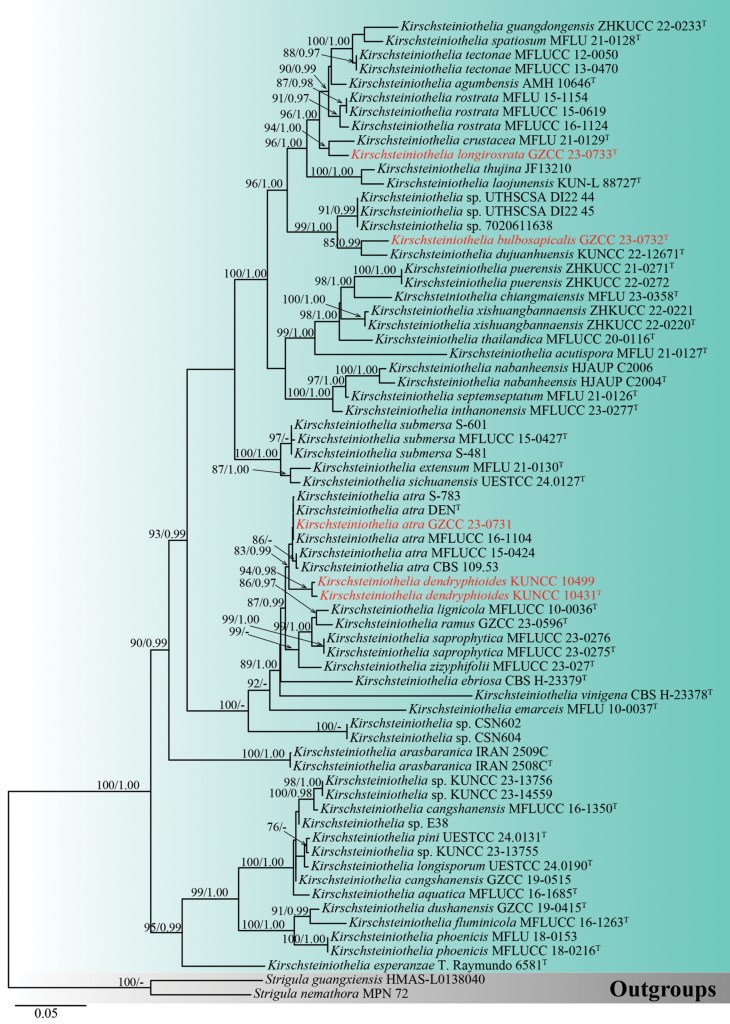
According to the analysis of the concatenated ITS, LSU and SSU rDNA sequence data, all isolates collected in this study cluster within Kirschsteiniothelia. The dataset with 67 strains of Kirschsteiniothelia, including gaps, comprises 2290 characters (ITS: 1–506 base pairs (bp), LSU: 507–1330 bp and SSU: 1331–2283 bp). The highest-scoring RAxML tree is presented in Fig. 1, with a final ML optimisation likelihood value of -16683.670 (ln). The best-fit model for the BI analysis was GTR+I+G for ITS, LSU and SSU. Bayesian posterior probabilities (PP) from MCMC were analysed, achieving a final average standard deviation of split frequencies of 0.009914.
Figure 1.
Phylogram of Kirschsteiniothelia taxa, based on the RAxML analysis of the combined ITS, LSU and SSU rDNA sequence dataset. Bootstrap support values for Maximum Likelihood (ML) equal to or greater than 75% and Bayesian posterior probabilities (PP) equal to or greater than 0.95 are shown above the nodes. The tree is rooted with Strigulaguangxiensis (HMAS-L0138040) and S.nemathora (MPN 72). Newly-generated strains are denoted in red and type strains are indicated with a superscript “T”.
Taxonomy
. Kirschsteiniothelia atra
(Corda) D. Hawksw., Fungal Diversity 69: 37 (2014)
A2C30658-7CE2-56C4-A2C1-734E7B9B1346
Fungal Names number: FN 104401
Facesoffungi number: FoF01738
Figure 2.
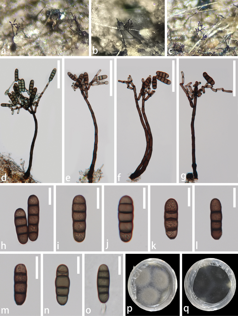
Kirschsteiniotheliaatra (GZAAS 23-0807, new host record) a–c colonies on natural substrate d–g conidiophores and conidiogenous cells bearing conidia h–n conidia o a germinated conidium p upper surface view of culture q lower surface view of culture. Scale bars: 100 μm (d–g); 20 μm (h–o).
≡ Amphisphaeriaaethiops Sacc., Syll. fung. (Abellini) 1: 722 (1882)
= Dendryphiopsisatra (Corda) S. Hughes, Can. J. Bot. 31: 655 (1953)
≡ Dendryphionatrum Corda, Icon. fung. (Prague) 4: 33 (1840)
≡ Kirschsteiniotheliaaethiops (Sacc.) D. Hawksw., J. Linn. Soc., Bot. 91(1–2): 185 (1985)
Description.
Saprobic on decaying wood of Edgeworthiachrysantha. Sexual morph: see Hawksworth (1985). Asexual morph: Colonies on the natural substrate superficial, effuse, gregarious, dark brown to black, glistening. Mycelium immersed, composed of branched, septate, thin-walled, smooth, brown hyphae. Conidiophores 253–396 × 8–15.5 µm (x̄= 334.6 × 11.7 µm, n = 20), macronematous, mononematous, erect, straight or flexuous, cylindrical, septate, smooth, brown to dark brown, becoming paler towards the apex and comprising numerous short branches. Conidiogenous cells 14.5–29 × 5–10 µm (x̄= 20.6 × 6.8 µm, n = 30), tretic, integrated, sometimes percurrent, terminal, doliiform or lageniform, subhyaline to pale brown, with new cells developing from the apical or subapical part of the subtending cells. Conidia 32–56.5 × 11–19.5 µm (x̄ = 42.3 × 14.5 µm, n = 30), solitary, acrogenous, cylindrical, sometimes clavate, 3–4-septate, constricted and darker at the septa, smooth, brown and rounded at the apex.
Culture characteristics.
Conidia germinating on Potato Dextrose Agar (PDA) within 24 h, and producing germ tubes either from the apex or base. Colonies circular, flat, dense, radial sulcate, edge entire, pearl-gray on the surface, dark brown on the reverse and becoming grey-white along the margin.
Material examined.
China • Guizhou Province, Zunyi City, Suiyang County, saprobic on decaying branches of Edgeworthiachrysantha, 13 February 2023, Xue-Mei Chen, SY12 (GZAAS 23-0807), living culture GZCC 23-0731.
Known distribution (based on molecular data).
China (Su et al. 2016; this study).
Known hosts (based on molecular data).
Edgeworthiachrysantha (This study), Unidentified decaying wood (Su et al. 2016).
Note.
Morphologically, our collection matches the characteristics of Kirschsteiniotheliaatra, including macronematous, mononematous conidiophores with numerous short branches; tretic, doliiform, or lageniform conidiogenous cells that develop new cells from the apical or subapical part of the subtending cells; and cylindrical, occasionally clavate conidia that are 3–4-septate, constricted and darker at the septa, which are rounded at the apex (Su et al. 2016). In the phylogenetic analyses, our collection (GZCC 23-0731) clusters with Kirschsteiniotheliaatra (CBS 109.53, DEN, MFLUCC 15-0424, MFLUCC 16-1104 and S–783) (Fig. 1). Excluding gaps, no difference was observed in the comparison of nucleotides across the ITS (491 bp), LSU (788 bp) and SSU (844 bp) regions between our collection and Kirschsteiniotheliaatra (MFLUCC 16-1104). Based on these findings, we identify our isolate as Kirschsteiniotheliaatra, following the guidelines established by Jeewon and Hyde (2016) and Maharachchikumbura et al. (2021). This is the first time Kirschsteiniotheliaatra has been reported from Edgeworthiachrysantha.
. Kirschsteiniothelia bulbosapicalis
X. Tang, K.D. Hyde, Jayaward. & J.C. Kang, sp. nov.
7F6A1323-2181-545A-9A9D-5A8F92D695F1
Fungal Names number: FN 572044
Facesoffungi number: FoF16485
Figure 3.
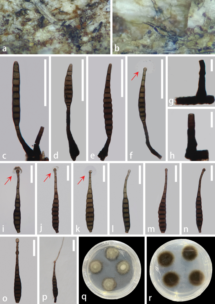
Kirschsteiniotheliabulbosapicalis (GZCC 23-0732, holotype) a, b colonies natural substrate c–f conidiophores, conidiogenous cells bearing conidia (red arrows indicate mucilaginous sheaths) g, h conidiophores i–o conidia (red arrows indicate mucilaginous sheaths) p a germinated conidium q upper surface view of culture r lower surface view of culture. Scale bars: 100 μm (c–f); 20 μm (g, h); 50 μm (i–p).
Etymology.
The specific epithet ‘bulbosapicalis’ refers to the bulbous area of the conidia at the apex.
Holotype.
GZAAS 23-0808.
Description.
Saprobic on unidentified decaying wood. Sexual morph: Undetermined. Asexual morph: Colonies on the natural substrate superficial, effuse, gregarious, hairy, black, glistening. Mycelium semi-immersed, on the substrate, pale brown to dark brown. Conidiophores (–47)58–128(–199) μm × 7.5–12.5(–16.5) μm (x̄ = 86.7 × 10.6 μm, n = 15), macronematous, mononematous, solitary, straight or slightly flexuous, cylindrical, unbranched, septate, smooth, brown to dark brown, truncate at the apex and wider at the base. Conidiogenous cells 6–17 μm × 7–10.5 μm (x̄ = 10.6 × 8.6 μm, n = 15), monoblastic, holoblastic, terminal, determinate, proliferating, cylindrical, brown to dark brown. Conidia 118–236.5 μm × 15–27 μm (x̄ = 174.8 × 21 μm, n = 30), solitary, acrogenous, cylindrical, ovoid to obclavate, rostrate, smooth, straight or slightly curved, 8–13-septate, slightly constricted at the septa, olivaceous to reddish-brown to dark brown, bulbous at the apex and/or third or fourth cell, truncate at the base, with a spherical hyaline mucilaginous sheath.
Culture characteristics.
Conidia germinating on PDA within 24 hours, producing germ tubes from the apex. Colonies displayed a circular morphology with an umbonate elevation, dense growth and a filiform margin. The surface appeared greyish-green, occasionally exhibiting paler mycelium in the bulge region. The reverse colonies exhibited a circular shape with a filiform margin, displaying a dark brown colour, becoming olivaceous towards the periphery.
Material examined.
China • Hainan Province, Jianfengling National Forest Park, saprobic on unidentified decaying wood, 23 August 2021, Zili Li, JBT04 (GZAAS 23-0808, holotype), ex-type living culture GZCC 23-0732.
Note.
Kirschsteiniotheliabulbosapicalis exhibits sporidesmium-like characteristics and shares similar morphologies with other Kirschsteiniothelia species. However, K.bulbosapicalis can be distinguished from other Kirschsteiniothelia species in having different sizes of conidiophores, conidiogenous cells and the unique feature of its conidia, which comprises one or two bulbous structures at or near the apex, with a spherical hyaline mucilaginous sheath. Phylogenetically, K.bulbosapicalis is sister to K.dujuanhuensis (KUNCC 22-12671) with 85% ML and 0.99 PP support (Fig. 1). Similar to our new species, K.dujuanhuensis also comprises a spherical hyaline mucilaginous sheath. Kirschsteiniotheliabulbosapicalis is characterised by larger conidiophores [(–47)58.5–128(–199) μm × 7.5–12.5(–16.5) μm, L/W ratio = 8.2] compared to K.dujuanhuensis [29–74(–119) × 9–11 μm, L/W ratio = 5.1] and larger conidia (118–236.5 μm × 15–27 μm, L/W ratio = 8.3) compared to K.dujuanhuensis [(114–)122–155(–170) × 10–13(–16) μm, L/W ratio = 11.5]. In addition, K.bulbosapicalis exhibits cylindrical to ovoid or obclavate conidia with 8–13 septa and often consist of bulbous structures at the apex and/or the third or fourth cell, as well as a spherical hyaline mucilaginous sheath. In contrast, K.dujuanhuensis typically contains obclavate to subcylindrical conidia that are 6–15 septate.
In addition, the comparison of the nucleotides between the sequences of K.bulbosapicalis and K.dujuanhuensis showed differences of 9% (47/512 bp) across ITS, 1% (8/812 bp) across LSU and 0.1% (2/1003 bp) across SSU, excluding gaps. Based on these findings, we introduce K.bulbosapicalis as a novel species, in accordance with the guidelines established by Jeewon and Hyde (2016) and Maharachchikumbura et al. (2021).
. Kirschsteiniothelia dendryphioides
X. Tang, K.D. Hyde, Jayaward. & J.C. Kang, sp. nov.
A19BE87A-BA0F-5DD7-BF33-60B1C765E8A2
Fungal Names number: FN 572046
Facesoffungi number: FoF16486
Figure 4.
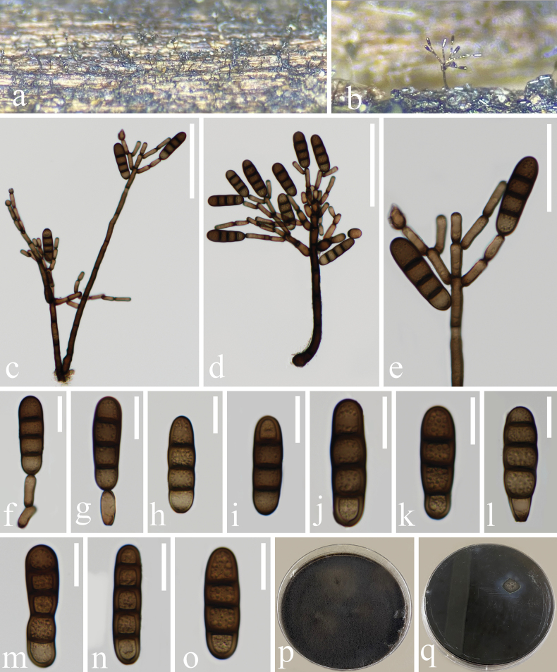
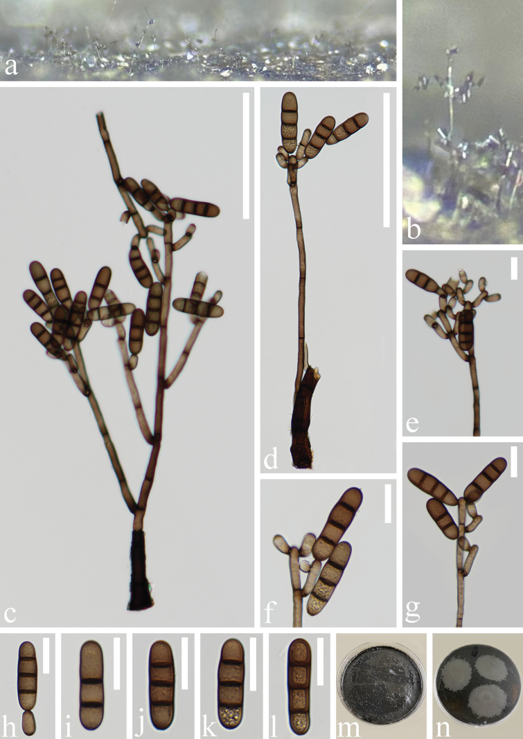
Kirschsteiniotheliadendryphioides (HKAS 136930, holotype) a, b colonies on natural substrate c, d conidiophores, conidiogenous cells bearing conidia e–g conidiogenous cells bearing conidia h–o conidia p upper surface view of culture q lower surface view of culture; Scale bars: 100 μm (c, d); 50 μm (e); 20 μm (f–o).
Figure 5.
Kirschsteiniotheliadendryphioides (HKAS 135651, paratype) a, b colonies on natural substrate c, d conidiophores, conidiogenous cells bearing conidia e–g conidiogenous cells bearing conidia h–l conidia m upper surface view of culture n lower surface view of culture. Scale bars: 100 μm (c, d); 20 μm (e–l).
Etymology.
The specific epithet “dendryphioides” is derived from the resemblance to the dendryphiopsis-like features.
Holotype.
HKAS 136930.
Description.
Saprobic on an unidentified decaying wood. Sexual morph: Undetermined. Asexual morph: Colonies on the natural substrate superficial, effuse, scattered, hairy, black, glistening. Mycelium partly immersed, on the substrate, pale brown to dark brown. Conidiophores 179–467 × 4.5–8 μm (x̄ = 318.2 × 6.1 μm, n = 10), macronematous, mononematous, erect, subscorpioid branched, straight or flexuous, cylindrical, septate, smooth, brown to dark brown, becoming paler towards the apex. Conidiogenous cells 9–19 × 4–8 μm (x̄ = 13.3 × 6.1 μm, n = 30), monotretic, terminal or intercalary, integrated, sometimes percurrent, cylindrical, doliiform, mostly discrete, determinate, smooth, pale brown to brown, both ends appearing darker, with new cells developing from the apical or subapical part of the subtending cells. Conidia 30–55 × 9–13.5 μm (x̄ = 40 × 11.1 µm, n = 30), solitary, acrogenous, cylindrical, oblong and occasionally clavate, smooth, guttulate, 2–4-septate, slightly or deeply constricted and darker at the septa, brown, rounded at the apex and sometimes truncate at the base, exhibiting obtuse ends.
Culture characteristics.
Conidia germinating on PDA within 24 hours. Colonies circular, characterised by dense, flat, spreading and fluffy growth, with an entire margin. The surface displayed a dark brown hue, while the reverse colonies exhibited a circular shape with an entire margin, also appearing dark brown.
Material examined.
China • Yunnan Province, Lushui City, Sanhe Village, Gaoligong Mountain, saprobic on decaying wood in a freshwater stream, 5 May 2021, Rong-ju Xu, XS17 (HKAS 136930, holotype), ex-type living culture, KUNCC 10431; ibid. • saprobic on submerged decaying wood in freshwater habitats, 22 August 2021, Rong-ju Xu, SYC-05 (HKAS 135651, paratype), living culture, KUNCC 10499.
Notes.
Kirschsteiniotheliadendryphioides exhibits dendryphiopsis-like characteristics and shares similar morphologies with other Kirschsteiniothelia species. However, K.dendryphioides differs from other species in the size of its conidiophores, conidiogenous cells and conidia. Kirschsteiniotheliadendryphioides is distinct from K.atra in having larger conidiophores (179–467 × 4.5–8 μm, L/W ratio = 52.2 vs. 148–228 µm × 6–8 μm, L/W ratio = 27), shorter conidiogenous cells (9–19 × 4–8 μm, L/W ratio = 2.2 vs. 25–33 µm × 5–7 μm, L/W ratio = 4.8) and smaller conidia (30–55 × 9–13.5 μm, L/W ratio = 3.6 vs. 54–63 ×14–18 μm, L/W ratio = 3.4).
The establishment of Kirschsteiniotheliadendryphioides as a new species is further supported by molecular data. Based on our phylogenetic analyses, K.dendryphioides strains (KUNCC 10431 and KUNCC 10499) form a subclade sister to the strains of Kirschsteiniotheliaatra (CBS 109.53, DEN, MFLUCC 15-0424, MFLUCC 16-1104 and S-783) with 83% ML and 0.99 PP support (Fig. 1). The comparison of the nucleotides between the sequences of K.dendryphioides and K.atra (MFLUCC 16-1104) shows a difference of 1.9% (9/481 bp) across ITS and 2.4% (11/458 bp) across SSU, but no difference was observed across LSU (777 bp), excluding gaps. Based on these findings, we introduce Kirschsteiniotheliadendryphioides as a novel species, following guidelines outlined in Jeewon and Hyde (2016) and Maharachchikumbura et al. (2021). We were unable to compare the nucleotide differences across LSU and SSU of KUNCC 10499 as it lacks sequence data for these loci.
. Kirschsteiniothelia longirostrata
X. Tang, K.D. Hyde, Jayaward. & J.C. Kang, sp. nov.
22B93B83-4A45-56CA-9239-434CB037CD93
Fungal Names number: FN 572045
Facesoffungi number: FoF16487
Figure 6.
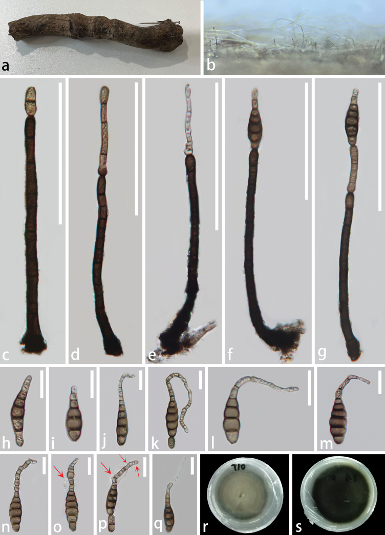
Kirschsteiniothelialongirosrata (GZCC 23-0733, holotype) a unidentified submerged wood b colonies on natural substrate c, d conidiophores, conidiogenous cells e–g conidiophores, conidiogenous cells bearing conidia h–p conidia (red arrows indicate mucilaginous sheaths) q a germinated conidium; r Upper surface view of culture s lower surface view of culture. Scale bars: 100 μm (c–g); 20 μm (h–q).
Etymology.
The specific epithet ‘longirostrata’ refers to the conidia containing a long rostrate.
Holotype.
GZAAS 23-0809.
Description.
Saprobic on an unidentified submerged decaying wood. Sexual morph: Undetermined. Asexual morph: Colonies on the natural substrate superficial, effuse, gregarious, hairy, black, glistening. Mycelium partly immersed on the substrate, composed of branched, septate, smooth-walled hyphae, pale to dark brown. Conidiophores 80–252 × 4.5–9.5 μm (x̄ = 161.3 × 6.8 μm, n = 20), macronematous, mononematous, solitary, cylindrical, straight, or slightly flexuous, unbranched, percurrent, smooth, guttulate, 4–13-septate, sometimes slightly constricted at the septa, brown to dark brown tapering towards the apex and wider at the base. Conidiogenous cells 6.5–16 × 5–9 μm (x̄ = 13× 7 μm, n = 20), monoblastic, terminal or indeterminate, percurrently proliferating, cylindrical, pale brown to brown. Conidia 36.5–109(–160) × 8–16 μm (x̄ = 71× 12 μm, n = 30), solitary, acrogenous, cylindrical, obpyriform to obclavate, rostrate 15–100(–120) × 2.5–6 μm (x̄ = 48 × 4.3 μm, n = 30), smooth, straight or curved, guttulate, 6–18-septate, slightly constricted and darker at the septa, proliferating, pale brown to brown, becoming paler towards the apex, with a truncate base and a mucilaginous sheath surrounding the upper part of the apex.
Culture characteristics.
Conidia germinating on PDA within 24 hours, producing germ tubes from the apex. Colonies displayed a circular morphology with dense, flat, spreading and fluffy growth, with an entire margin. The surface exhibited an olivaceous-green hue with a darker edge, while the reverse colonies displayed a circular shape with an entire margin, appearing blackish-green.
Material examined.
China • Hainan Province, Jianfengling National Forest Park, saprobic on submerged unidentified decaying wood, 23 August 2021, Zili Li, T10 (GZAAS 23-0809, holotype) ex-type living culture GZCC 23-0733.
Notes.
Kirschsteiniothelialongirostrata exhibits sporidesmium-like characteristics and shares similar features with other Kirschsteiniothelia species. Kirschsteiniothelialongirostrata can be distinguished from other Kirschsteiniothelia species in having different sizes and shapes of conidiophores, conidiogenous cells and unique features of conidia, such as obpyriform to obclavate, long rostrate, proliferating, with a mucilaginous sheath surrounding the upper part of the apex. Unlike K.crustacea, K.longirostrata has cylindrical, proliferating conidiogenous cells and obpyriform to obclavate conidia, with longer (15–100(–120) × 2.5–6 μm), guttulate, proliferating rostrate structures and a mucilaginous sheath surrounding the upper part of the apex.
Molecular data further supports the establishment of Kirschsteiniothelialongirostrata as a novel taxon. Based on our phylogenetic analyses, K.longirostrata is sister to K.crustacea (MFLU 21-0129) with 94% ML and 1.00 PP support (Fig. 1). The comparison of the nucleotides between the sequences of K.longirostrata (GZCC 23-0733) and K.crustacea (MFLU 21-0129) shows differences of 8.4% (39/467 bp) across ITS and 0.7% (5/718 bp) across LSU, excluding gaps. However, we were unable to compare the nucleotide differences across SSU as K.crustacea lacks sequence data for this locus. Based on these findings, we introduce Kirschsteiniothelialongirostrata as a novel species, following guidelines outlined in Jeewon and Hyde (2016) and Maharachchikumbura et al. (2021).
Discussion
During surveys on saprobic fungi associated with woody plants in the subtropical and tropical forests of the Guizhou, Hainan and Yunnan Provinces in China, we discovered three previously undocumented taxa and one known species, which belong to Kirschsteiniothelia. They were found on decaying wood, including some unidentified hosts and Edgeworthiachrysantha. All these species have been reported with their asexual morph, either exhibiting the dendryphiopsis-like or sporidesmium-like morphologies.
Our newly-described taxa include two sporidesmium-like species, namely Kirschsteiniotheliabulbosapicalis and K.longirostrata and one dendryphiopsis-like taxon, K.dendryphioides. Both Kirschsteiniotheliabulbosapicalis and K.longirostrata exhibit distinct characteristics from other species of Kirschsteiniothelia. Kirschsteiniotheliabulbosapicalis is characterised by acrogenous, cylindrical, ovoid, obclavate, rostrate, straight or slightly curved conidia with 8–13 septa, often bulbous at the apex and/or third or fourth cell, with a spherical hyaline mucilaginous sheath. Kirschsteiniothelialongirostrata displays solitary, acrogenous, cylindrical, obpyriform to obclavate, rostrate, smooth, straight or curved and guttulate conidia that are paler towards the apex, consisting of 6–18 septa, slightly constricted and darker at the septa, with a mucilaginous sheath surrounding the tail-like upper part of the apex. Kirschsteiniothelialongirostrata has the longest tail amongst all current Kirschsteiniothelia species, which proliferates from the apex of the conidium. Our phylogenetic analyses reveal that our new species belong to Kirschsteiniothelia with stable support values and, in particular, is closely related to K.crustacea.
Kirschsteiniotheliadendryphioides displays solitary, acrogenous, cylindrical, oblong and occasionally clavate, smooth, guttulate, 2–4-septate conidia with slightly or deeply constricted and darker at the septa, rounded at the apex and sometimes truncate at the base, exhibiting obtuse ends. However, Kirschsteiniotheliadendryphioides is characterised by larger conidiophores, shorter conidiogenous cells and smaller conidia when compared to K.atra. Although they are morphologically similar, there are also sufficient dissimilarities in the DNA sequence data.
The new host record for Kirschsteiniotheliaatra shows characteristics of solitary, acrogenous, cylindrical, sometimes clavate conidia that are 3–4-septate, constricted and darker at the septa and smooth and rounded at the apex. Based on a comparison of morphological and phylogenetic analyses, no significant differences were observed in the DNA base pairs and the morphological variations fall within the range of intraspecific diversity. Therefore, we identified our new collection (GZCC 23–0731) as the known species K.atra. Furthermore, this report extends the known host range of K.atra (Table 2).
Table 2.
Kirschsteiniothelia species and data related to their host, habitat, country and reported morph.
| Taxa | Host | Habitat | Morphological character | Asexual Morph character | Country | Molecular data | References |
|---|---|---|---|---|---|---|---|
| Kirschsteiniotheliaabietina | Tsugacanadian | Terrestrial | Sexual | N/A | USA | N/A | Fairman (1905); Wang et al. (2004) |
| K.acerina | On absorbing mycorrhizal rootlets of Acersaccharum | Terrestrial | Sexual | N/A | USA | N/A | Hawksworth (1985) |
| K.acutispora | Unidentified decaying wood | Terrestrial | Asexual | Sporidesmium-like | Thailand | A | Jayawardena et al. (2022) |
| K.agumbensis | On decaying wood of Garcinia sp. | Terrestrial | Asexual | Sporidesmium-like | India | A | Sruthi et al. (2024) |
| K.atra | Abiesbalsamea, Acernegundo, Acer sp., Agathisaustralis, Alnusglutinosa, A.incana, Alstonia sp., Betulapapyrifera, Brachyglottisrepanda, Bursera sp., Carpinusbetulus, Carpinus sp., Celtis sp., Clematis sp., Coprosmaaustralis, Corylusavellana, Cupressusmacrocarpa, Cupressus sp., Drypetesalba, Edgeworthiachrysantha, Fraxinuspennsylvanica, Fraxinus sp., Fuchsiaexcorticata, Hederahelix, Juglans sp., Knightiaexcelsa, Leptospermumscoparium, Loniceracoerulea, Machaerocereus sp., Macropiperexcelsum, Nothofagustruncata, Phoenixdactylifera, Pinusbanksiana, Populusangustifolia, P.balsamifera, P.tremuloides, Prunus sp., Quercusrobur, Quercus sp., Rhopalostylis sp., Salix sp., Tiliaamericana, Tsugacanadensis, Unidentified decaying wood | Freshwater, terrestrial | Sexual and Asexual | Dendryphiopsis-like | Australia, Belgium, China, Czech Republic, France, Germany, Mexico, New Zealand, Poland, Russian Federation, Sweden, Unite Kingdom, USA | A | Aptroot (1995, 1997); Cannon et al. (1985); Chlebicki and Chmiel (2006); Conners (1967); Cooke (1985); Eriksson (1992, 2014); Ginns (1986); Hughes (1978); Hyde (1993); Kobayashi (2007); Matsushima (1971, 1975); McKenzie et al. (2000, 2004); Minter et al. (2001); Mulenko et al. (2008); Nattrass (1961); Nordén et al. (1997); Popov et al. (2008); Rao and Varghese (1981); Réblová and Svrcek (1997); Schmid-Heckel (1988); Sieber et al. (1995); Sierra (1984); Su et al. (2016); Sutton (1973); This study; Wang and He (2007); Wang (2010); Wang et al. (2004). |
| K.aquatica | Unidentified decaying wood | Freshwater | Asexual | Sporidesmium-like | China | A | Bao et al. (2018) |
| K.arasbaranica | Dead branches of Quercuspetraea | Terrestrial | Sexual | N/A | Iran | A | Mehrabi et al. (2017) |
| K.arbuscula | On bark of Acer, Rhuscopallinum, Carya, Magnoliaglauca, and Acerrubrum | Terrestrial | Asexual | Dendryphiopsis-like | USA | N/A | Berkeley (1875); Ellis (1976); Pratibha et al. (2010); Sruthi et al. (2024) |
| K.atkinsonii | Freycinetiaarnotti | Terrestrial | Sexual | N/A | Greece | N/A | Stevens (1925) |
| K.binsarensis | On dead twig | Terrestrial | Asexual | Dendryphiopsis-like | India | N/A | Subramanian and Srivastava (1994); Sruthi et al. (2024) |
| K.biseptata | On dead wood | Terrestrial | Asexual | Dendryphiopsis-like | South Africa | N/A | Morgan-Jones et al. (1983); Sruthi et al. (2024) |
| K.bulbosapicalis | Unidentified decaying wood | Terrestrial | Asexual | Sporidesmium-like | China | A | This study |
| K.cangshanensis | Unidentified decaying wood | Freshwater | Asexual | Sporidesmium-like | China | A | Bao et al. (2018) |
| K.chiangmaiensis | Unidentified decaying wood | Terrestrial | Sexual | N/A | Thailand | A | Louangphan et al. (2024) |
| K.crustacea | Bamboo | Terrestrial | Asexual | Sporidesmium-like | Thailand | A | Jayawardena et al. (2022) |
| K.dendryphioides | Unidentified decaying wood | Freshwater | Asexual | Dendryphiopsis-like | China | A | This study |
| K.dolioloides | Ramulisdecorticatispineis | Terrestrial | Sexual | N/A | Switzerland | N/A | Wegelin (1894); Wang et al. (2004) |
| K.dujuanhuensis | Unidentified submerged wood | Freshwater | Asexual | Sporidesmium-like | China | A | Unpublished |
| K.dushanensis | Unidentified decaying wood | Freshwater | Asexual | Sporidesmium-like | China | A | Yang et al. (2023) |
| K.ebriosa | Sparkling wine | N/A | Asexual | Dendryphiopsis-like | Spain | A | Rodríguez-Andrade et al. (2020) |
| K.emarceis | Unidentified decaying wood | Terrestrial | Sexual and asexual | Dendryphiopsis-like | Thailand | A | Boonmee et al. (2012) |
| K.esperanzae | Unidentified decaying wood | Terrestrial | Sexual | N/A | Mexico | N/A | Raymundo et al. (2023) |
| K.extensum | Unidentified decaying wood | Terrestrial | Asexual | Sporidesmium-like | Thailand | A | Jayawardena et al. (2022) |
| K.fascicularis | On bark of Liquidambar sp. | Terrestrial | Asexual | Dendryphiopsis-like | USA | N/A | Berkeley (1875); Hughes (1958); Sruthi et al. (2024) |
| K.fluminicola | Unidentified decaying wood | Freshwater | Asexual | Sporidesmium-like | China | A | Bao et al. (2018) |
| K.goaensis | On dead and decaying bark of tree | Terrestrial | Asexual | Dendryphiopsis-like | India | N/A | Pratibha et al. (2010); Sruthi et al. (2024) |
| K.guangdongensis | Dead branches of unidentified plant | Terrestrial | Asexual | Sporidesmium-like | China | A | Senanayake et al. (2023) |
| K.inthanonensis | Unidentified decaying wood | Terrestrial | Asexual | Dendryphiopsis-like | Thailand | A | de Farias et al. (2024) |
| K.laojunensis | Bark of Abiesfabri | Terrestrial | sexual | N/A | China | A | Meng et al. (2024) |
| K.lignicola | Unidentified decaying wood | Terrestrial | Sexual and Asexual | Dendryphiopsis-like | Thailand | A | Boonmee et al. (2012) |
| K.longirostrata | Unidentified submerged decaying wood | Freshwater | Asexual | Sporidesmium-like | China | A | This study |
| K.longisporum | Dead branches of Pinustaeda | Terrestrial | Asexual | Dendryphiopsis-like | China | A | Tian et al. (2024) |
| K.nabanheensis | Unidentified broadleaf tree | Terrestrial | Asexual | Dendryphiopsis-like | China | A | Liu et al. (2023b) |
| K.phileura | Tiliaamericana | Terrestrial | Sexual | N/A | USA | N/A | Cooke and Ellis (1876); Saccardo (1882) |
| K.phoenicis | Phoenixpaludosa | Marine | Sexual | N/A | Thailand | A | Hyde et al. (2018) |
| K.pini | On decaying branches of a Pinus sp. | Terrestrial | Asexual | Sporidesmium-like | China | A | Jin et al. (2024) |
| K.populi | Populusangustifolia | Terrestrial | Sexual | N/A | USA | N/A | Greene (1901); Wang et al. (2004) |
| K.proteae | Proteacynaroides | N/A | Sexual | N/A | South Africa | N/A | Marincowitz et al. (2008) |
| K.puerensis | Coffee wood | Terrestrial | Asexual | Sporidesmium-like | China | A | Hyde et al. (2023) |
| K.ramus | Unidentified decaying wood | Freshwater | Asexual | Dendryphiopsis-like | China | A | Zhang et al. (2023) |
| K.recessa | Unidentified decaying wood, Acerrubrum, Alnusrubra, Pyrus sp., rotten wood | Terrestrial | Sexual and asexual | Dendryphiopsis-like | Canada, Italy, USA, | N/A | Hawksworth (1985); Barr et al. (1986); Aptroot (1995); Wang et al. (2004) |
| K.reticulata | Unidentified twigs | Terrestrial | Sexual | N/A | China | N/A | Chen et al. (2006) |
| K.rostrata | Unidentified decaying wood | Freshwater | Asexual | Sporidesmium-like | China | A | Bao et al. (2018) |
| K.saprophytica | Unidentified decaying wood | Terrestrial | Sexual and asexual | Dendryphiopsis-like | Thailand | A | de Farias et al. (2024) |
| K.septemseptatum | Unidentified decaying wood | Terrestrial | Asexual | Dendryphiopsis-like | Thailand | A | Jayawardena et al. (2022) |
| K.sichuanensis | On decaying branches of an unidentified woody plant | Terrestrial | Asexual | Sporidesmium-like | China | A | Jin et al. (2024) |
| K.shimlaensis | Cedrusdeodara | Terrestrial | Asexual | Dendryphiopsis-like | India | N/A | Verma et al. (2021) |
| K.smilacis | Smilax sp. | Terrestrial | Sexual | N/A | China | N/A | Chen et al. (2006) |
| K.spatiosum | Unidentified decaying wood | Terrestrial | Asexual | Sporidesmium-like | Thailand | A | Jayawardena et al. (2022) |
| K.striatispora | Juniperuscommunis | Terrestrial | Sexual | N/A | China, Switzerland | N/A | Hawksworth (1985) |
| K.submersa | Unidentified decaying wood | Freshwater | Asexual | Sporidesmium-like | China | A | Su et al. (2016) |
| K.tectonae | Microcospaniculata, Tectonagrandis | Terrestrial | Asexual | Sporidesmium-like | Thailand | A | Li et al. (2016); de Farias et al. (2024) |
| K.thailandica | Ficusmicrocarpa | Terrestrial | Asexual | Sporidesmium-like | Thailand | A | Sun et al. (2021) |
| K.thujina | Abiesbalsamea, Thujaoccidentalis | Terrestrial | Sexual | N/A | Canada, USA | A | Saccardo (1882); Hawksworth (1985) |
| K.umbrinoidea | Aesculushippocastanum | Terrestrial | Sexual | N/A | Italy | N/A | Passerini (1887); Wang et al. (2004) |
| K.vinigena | Cork stopper, sparkling wine | N/A | Asexual | Dendryphiopsis-like | Spain | A | Rodríguez-Andrade et al. (2020) |
| K.xera | Prunus sp. | Terrestrial | Sexual | N/A | USA | N/A | Fairman (1910); Wang et al. (2004) |
| K.xishuangbannaensis | Heveabrasiliensis | Terrestrial | Asexual | Sporidesmium-like | China | A | Xu et al. (2023) |
| K.zizyphifolii | Nayariophytonzizyphifolium | Terrestrial | Sexual and asexual | Dendryphiopsis-like | Thailand | A | de Farias et al. (2024) |
N/A: data not available; A: data available.
Whether we can use sporidesmium-like and dendryphiopsis-like morphs to differentiate species is currently obscure. Based on our phylogeny, species with sporidesmium-like and dendryphiopsis-like morphs do constitute distinct clades. There are three dendryphiopsis-like species (K.inthanonensis, K.nabanheensis and K.septemseptatum) that constitute a strongly-supported subclade, but they are nested within sporidesmium-like species. Therefore, segregating species based on this aspect should be dealt with caution. As with other asexual fungi, some species of Kirschsteiniothelia occur solely in the sexual morph and, hence, we are unable to compare their morphologies with other asexual species.
Besides establishing three novel Kirschsteiniothelia species and a new host record, we provide a checklist of all Kirschsteiniothelia taxa (Table 2), which incorporate 41 asexual morph species, amongst which 21 exhibit the sporidesmium-like morph, while 20 display the dendryphiopsis-like features (Hawksworth 1985; Boonmee et al. 2012; Su et al. 2016; Sun et al. 2021; Xu et al. 2023; de Farias et al. 2024; Jin et al. 2024; Tian et al. 2024). This checklist includes data on the host, habitat preferences, reported morphology, country of origin and availability of molecular data for all species of Kirschsteiniothelia. The checklist also provides the latest ecological information of species in the genus.
From the checklist (Table 2), we decipher that most Kirschsteiniothelia species have been reported from China (23 species), followed by Thailand (14 species), with a few distributed across different countries including Australia, Belgium, Canada, Czechia, France, Germany, Greece, Italy, India, Iran, Mexico, New Zealand, Poland, Russian Federation, Sweden, Switzerland, South Africa, Spain, United Kingdom and USA (Boonmee et al. 2012; Mehrabi et al. 2017; Bao et al. 2018; Rodríguez-Andrade et al. 2020; Jayawardena et al. 2022; Senanayake et al. 2023; Liu et al. 2023b; Yang et al. 2023; de Farias et al. 2024; Louangphan et al. 2024). As noted in Table 2, the proportion of new species of Kirschsteiniothelia discovery in China and Thailand reaches 63%, while other countries and regions have only sporadically discovered one or two species. Hence, we presume that Kirschsteiniothelia is highly diverse with many potentially more unknown species and other tropical and subtropical regions that should be explored. In addition, most new species in China and Thailand were primarily found in tropical and subtropical regions, with nearly all species growing on decayed wood (Bao et al. 2018; Rodríguez-Andrade et al. 2020; Sun et al. 2021; Jayawardena et al. 2022; Senanayake et al. 2023; Liu et al. 2023b; Yang et al. 2023; de Farias et al. 2024; Louangphan et al. 2024). We speculate that the high proportion of new species discovered in China and Thailand can be attributed to the following reasons: 1) The tropical/subtropical climatic conditions in China and Thailand are suitable for the growth of these species, with optimal temperature and humidity levels that favour spore germination and colony growth; 2) Samples of Kirschsteiniothelia are easily observed on natural substrates and easy to collect; 3) They proliferate quite easily and are potentially good decomposers of substrates and they grow well on most common culture media without requiring specific cultivation conditions; 4) There are many mycologists in China and Thailand who are actively involved in fungal taxonomy and are more likely to discover more new species.
Most species of Kirschsteiniothelia are saprobes occurring mainly in terrestrial and followed by freshwater habitats, with only a few taxa reported from environments, such as cork stoppers and as a pathogen that infects human beings (Hawksworth 1985; Boonmee et al. 2012; Su et al. 2016; Nishi et al. 2018; Rodríguez-Andrade et al. 2020; Sun et al. 2021; Xu et al. 2023; de Farias et al. 2024). According to our results, species of Kirschsteiniothelia are also present in various habitats, albeit in small numbers. That may be because most studies have overlooked these more specific habitats. We recommend that future research should focus on exploring this genus in diverse environments to potentially discover additional species. In addition, most of the newly-discovered species are asexual on decayed wood samples. The latter provides ample organic nutrients which favour the emergence of the asexual morph and allows them to colonise new areas and propagate (Zalamea et al. 2016; Liu et al. 2023a).
From a morphological perspective, the sporidesmium-like species are more diverse compared to the dendryphiopsis-like taxa. Interspecies differences are mainly attributed to the number and size of the conidiophores, conidiogenous cells and septa in the conidia. However, relying only on these features for species delineation is challenging and insufficient. Prior to the incorporation of molecular data, the taxonomy of Kirschsteiniothelia was challenging, resulting in controversial classifications. This genus was initially accommodated in Pleosporaceae by Hawksworth (1985) and Barr (1987), but later transferred to Pleomassariaceae by Barr (1993), based on morphological data. Based on molecular data, Schoch et al. (2006) suggested that Kirschsteiniothelia does not belong to any family within Pleosporales, but would rather be in a new family. Subsequently, Boonmee et al. (2012) introduced Kirschsteiniotheliaceae to accommodate Kirschsteiniothelia species, based on morphology and phylogenetic analyses. Hernández-Restrepo et al. (2017) established a novel order, Kirschsteiniotheliales, to accommodate the Kirschsteiniotheliaceae taxa. Subsequent studies have followed this classification, using ITS, LSU and SSU rDNA sequence data in their phylogenies (Sun et al. 2021; Xu et al. 2023; de Farias et al. 2024).
At present, most studies use LSU, ITS and SSU rDNA genes for inferring phylogeny relationships amongst Kirschsteiniothelia species. Despite close morphological similarities and overlap amongst species, we noted that there are rather unexpected sequence dissimilarities across the different genes analysed here. We presume that, despite high morphological similarities, these asexual species are characterised by high genetic diversity. This difference in genetic trait presumably enables them to adapt and flourish in different environments, gives them better chances of survival and drives speciation. So far, the ITS, LSU and SSU rDNA genes have most commonly been used to identify species within this genus, but as the number of species increases, we recommend incorporating protein genes like tub, tef1-α and rpb2 (Badotti et al. 2017).
Supplementary Material
Acknowledgements
The author would like to express gratitude to Shaun Pennycook for his valuable suggestions on naming the new fungus. Xia Tang also wishes to thank Mae Fah Luang University for the Thesis Writing Grant and for awarding me a tuition scholarship for my Ph.D. studies at Mae Fah Luang University, Thailand. Rajesh Jeewon thanks the University of Mauritius in Reduit, Mauritius.
Citation
Tang X, Jeewon R, Jayawardena RS, Gomdola D, Lu Y-Z, Xu R-J, Alrefaei AF, Alotibi F, Hyde KD, Kang J-C (2024) Additions to the genus Kirschsteiniothelia (Dothideomycetes); Three novel species and a new host record, based on morphology and phylogeny. MycoKeys 110: 35–66. https://doi.org/10.3897/mycokeys.110.133450
Contributor Information
Kevin D. Hyde, Email: kdhyde3@gmail.com.
Ji-Chuan Kang, Email: jckang@gzu.edu.cn.
Additional information
Conflict of interest
The authors have declared that no competing interests exist.
Ethical statement
No ethical statement was reported.
Funding
This research was supported by grants from the National Natural Science Foundation of China (NSFC Grants Nos. 32170019 & 31670027 & 31460011) and the Open Fund Program of Engineering Research Centre of Southwest Bio-Pharmaceutical Resources, Ministry of Education, Guizhou University (No. GZUKEY20160702) and the Thailand Research Fund grant “Impact of climate change on fungal diversity and biogeography in the Greater Mekong Sub-region” (RDG6130001). K.D Hyde and Fatimah AI-Otibi were funded by the Distinguished Scientist Fellowship Program (DSFP), King Saud University, Kingdom of Saudi Arabia.
Author contributions
Xia Tang conducted the experiments, analysed the data, and wrote the first draft of the manuscript. Rajesh Jeewon, Yong-Zhong Lu, Ruvishika S. Jayawardena, Kevin D. Hyde and Ji-Chuan Kang planned the experiments. Xia Tang, Deecksha Gomdola and Rong-Ju Xu analysed the data. Xia Tang conducted the experiments. Rajesh Jeewon, Deecksha Gomdola, Yong-Zhong Lu, Ruvishika S. Jayawardena, Fatimah Alotibi, Abdulwahed Fahad Alrefaei, Kevin D. Hyde and Ji-Chuan Kang corrected and revised the manuscript. Kevin D. Hyde and Ji-Chuan Kang funded the experiments. All authors revised and agreed to the published version of the manuscript.
Author ORCIDs
Xia Tang https://orcid.org/0000-0003-2705-604X
Rajesh Jeewon https://orcid.org/0000-0002-8563-957X
Ruvishika S. Jayawardena https://orcid.org/0000-0001-7702-4885
Deecksha Gomdola https://orcid.org/0000-0002-0817-1555
Yong-Zhong Lu https://orcid.org/0000-0002-1033-5782
Rong-Ju Xu https://orcid.org/0000-0002-3968-8442
Abdulwahed Fahad Alrefaei https://orcid.org/0000-0002-3761-6656
Fatimah Alotibi https://orcid.org/0000-0003-3629-5755
Kevin D. Hyde https://orcid.org/0000-0002-2191-0762
Ji-Chuan Kang https://orcid.org/0000-0002-6294-5793
Data availability
All the data that support the findings of this study are available in the main text. DNA sequences generated have been submitted to GenBank.
References
- Aptroot A. (1995) Redisposition of some species excluded from Didymosphaeria (Ascomycotina). Nova Hedwigia 60: 325–379. [Google Scholar]
- Aptroot A. (1997) Corticolouspyrenocarpous ascomycetes (lichenized and non-lichenized) from the Sonoran Desert (Arizona and Mexico). Nova Hedwigia 64(1–2): 169–176. 10.1127/nova.hedwigia/64/1997/169 [DOI] [Google Scholar]
- Badotti F, de Oliveira FS, Garcia CF, Vaz ABM, Fonseca PLC, Nahum LA, Oliveira G, Góes-Neto A. (2017) Effectiveness of ITS and sub-regions as DNA barcode markers for the identification of Basidiomycota (Fungi). BMC Microbiology 17(1): 1–12. 10.1186/s12866-017-0958-x [DOI] [PMC free article] [PubMed] [Google Scholar]
- Bao DF, Luo ZL, Liu JK, Bhat DJ, Sarunya N, Li WL, Su HY, Hyde KD. (2018) Lignicolous freshwater fungi in China III: New species and record of Kirschsteiniothelia from northwestern Yunnan Province. Mycosphere: Journal of Fungal Biology 9(4): 755–768. 10.5943/mycosphere/9/4/4 [DOI] [Google Scholar]
- Barr ME. (1987) Prodomus to class Loculoascomycetes. Publ. by the author, Amherst.
- Barr ME. (1993) Notes on the Pleomassariaceae. Mycotaxon 49: 129–142. 10.1001/archderm.1993.01680230018001 [DOI] [Google Scholar]
- Barr ME, Rogerson CT, Smith SJ, Haines JH. (1986) An annotated catalog of the pyrenomycetes described by Charles H. Bulletin – New York State Museum 459: 1–74. 10.5962/bhl.title.135547 [DOI] [Google Scholar]
- Berkeley MJ. (1875) Notices of North American fungi. Grevillea 3: 97–112. [Google Scholar]
- Bhunjun CS, Niskanen T, Suwannarach N, Wannathes N, Chen YJ, McKenzie EH, Maharachchikumbura SSN, Buyck B, Zhao CL, Fan YG, Zhang JY, Dissanayake AJ, Marasinghe DS, Jayawardena RS, Kumla J, Padamsee M, Chen YY, Liimatainen K, Ammirati JF, Phukhamsakda C, Liu JK, Phonrob W, Randrianjohany É, Hongsanan S, Cheewangkoon R, Bundhun D, Khuna S, Yu WJ, Deng LS, Lu YZ, Hyde KD, Lumyong S. (2022) The numbers of fungi: Are the most speciose genera truly diverse? Fungal Diversity 114(1): 387–462. 10.1007/s13225-022-00501-4 [DOI]
- Boonmee S, Ko TWK, Chukeatirote E, Hyde KD, Chen H, Cai L, McKenzie EHC, Jones EBG, Kodsueb R, Hassan BA. (2012) Two new Kirschsteiniothelia species with Dendryphiopsis anamorphs cluster in Kirschsteiniotheliaceae fam. nov. Mycologia 104(3): 698–714. 10.3852/11-089 [DOI] [PubMed] [Google Scholar]
- Cannon PF, Hawksworth DL, Sherwood-Pike MA. (1985) The British Ascomycotina. An Annotated Checklist. Commonwealth Mycological Institute, Kew, Surrey, England, 302.
- Capella-Gutiérrez S, Silla-Martínez JM, Gabaldón T. (2009) trimAl: A tool for automated alignment trimming in large-scale phylogenetic analyses. Bioinformatics (Oxford, England) 25(15): 1972–1973. 10.1093/bioinformatics/btp348 [DOI] [PMC free article] [PubMed] [Google Scholar]
- Chen CY, Wang CL, Huang JW. (2006) Two new species of Kirschsteiniothelia from Taiwan. Mycotaxon 98: 153–158. [Google Scholar]
- Chlebicki A, Chmiel MA. (2006) Microfungi of Carpinusbetulus from Poland I. Annotated list of microfungi. Acta Mycologica 41(2): 253–278. 10.5586/am.2006.027 [DOI] [Google Scholar]
- Conners IL. (1967) An Annotated Index of Plant Diseases in Canada and Fungi Recorded on Plants in Alaska, Canada and Greenland. Publ. Res. Br. Canada Dept Agric 1251: 1–381. [Google Scholar]
- Cooke WB. (1985) Fungi of Lassen Volcanic National Park. Cooperative National Park Resources Studies Unit, University of California at Davis, Institute of Ecology 21: 251.
- Cooke MC, Ellis JB. (1876) New Jersey fungi (cont.). Grevillea 5(34): 49–55. [Google Scholar]
- Cubeta MA, Echandi E, Abernethy T, Vilgalys R. (1991) Characterization of anastomosis groups of binucleate Rhizoctonia species using restriction analysis of an amplified ribosomal RNA gene. Phytopathology 81(11): 1395–1400. 10.1094/Phyto-81-1395 [DOI] [Google Scholar]
- Daniel GP, Daniel GB, Miguel RJ, Florentino FR, David P. (2010) ALTER: Program-oriented conversion of DNA and protein alignments. Nucleic Acids Research 38: W14–W18. 10.1093/nar/gkq321 [DOI] [PMC free article] [PubMed]
- de Farias ARG, Afshari N, Silva VSH, Louangphan J, Karimi O, Boonmee S. (2024) Three novel species and new records of Kirschsteiniothelia (Kirschsteiniotheliales) from northern Thailand. MycoKeys 101: 347. [DOI] [PMC free article] [PubMed]
- Ellis MB. (1976) More dematiaceous hyphomycetes. Commonwealth Mycological Institute, Kew, Surrey. 10.1079/9780851983653.0000 [DOI]
- Eriksson OE. (1992) The non-lichenized pyrenomycetes of Sweden. Btjtryck, Lund, Sweden 208.
- Eriksson OE. (2014) Checklist of the non-lichenized ascomycetes of Sweden. Acta Universitatis Upsaliensis 36: 499.
- Fairman CE. (1905) New or rare Pyrenomycetaceae from western New York. Proceedings of the Rochester Academy of Science 4: 215–224. [Google Scholar]
- Fairman CE. (1910) Fungi Lyndonville nses novi vel minus cogniti. Annales Mycologici 8(3): 322–332. [Google Scholar]
- Fungal names (2024) The Fungal names database. https://nmdc.cn/fungalnames/ [Accessed May 24, 2024]
- Ginns JH. (1986) Compendium of plant disease and decay fungi in Canada 1960–1980. Research Branch, Agriculture Canada 1813: 416. 10.5962/bhl.title.58888 [DOI]
- Greene EL. (1901) PlantaeBakerianae. Catholic University of America 1(1): 1–53. 10.5962/bhl.title.24815 [DOI] [Google Scholar]
- Hawksworth DL. (1985) Kirschsteiniothelia, a new genus for the Microtheliaincrustans-group (Dothideales). Botanical Journal of the Linnean Society 91(1–2): 181–202. 10.1111/j.1095-8339.1985.tb01144.x [DOI] [Google Scholar]
- Hernández-Restrepo M, Gené J, Castañeda-Ruiz RF, Mena-Portales J, Crous PW, Guarro J. (2017) Phylogeny of saprobic microfungi from Southern Europe. Studies in Mycology 86: 53–97. 10.1016/j.simyco.2017.05.002 [DOI] [PMC free article] [PubMed] [Google Scholar]
- Huelsenbeck JP, Ronquist F. (2001) MRBAYES: Bayesian inference of phylogenetic trees. Bioinformatics (Oxford, England) 17(8): 754–589. 10.1093/bioinformatics/17.8.754 [DOI] [PubMed] [Google Scholar]
- Hughes SJ. (1958) Revisiones hyphomycetum aliquot cum appendice de nominibus rejiciendis. Canadian Journal of Botany 36(6): 727–836. 10.1139/b58-067 [DOI] [Google Scholar]
- Hughes SJ. (1978) New Zealand Fungi. 25. Miscellaneous species. New Zealand Journal of Botany 16(3): 311–370. 10.1080/0028825X.1978.10425143 [DOI] [Google Scholar]
- Hyde KD. (1993) Fungi from palms. II. Kirschsteiniotheliaaethiops from the date palm, Phoenixdactylifera. Sydowia 45: 1–4. [Google Scholar]
- Hyde KD, McKenzie EHC, Koko TW. (2011) Towards incorporating anamorphic fungi in a natural classification – checklist and notes for 2010. Mycosphere 2(1): 1–88. [Google Scholar]
- Hyde KD, Jones G, Liu JK, Ariyawansa H, Boehm E, Boonmee S, Braun U, Chomnunti P, Crous PW, Dai DQ, Diederich P, Dissanayake A, Doilom M, Doveri F, Hongsanan S, Jayawardena R, Lawrey JD, Li YM, Liu YX, Lücking R, Monkai J, Muggia L, Nelsen MP, Pang KL, Phookamsak R, Senanayake IC, Shearer CA, Suetrong S, Tanaka K, Thambugala KM, Wijayawardene NN, Wikee S, Wu HX, Zhang Y, Aguirre-Hudson B, Alias SA, Aptroot A, Bahkali AH, Bezerra JL, Bhat DJ, Camporesi E, Chukeatirote E, Gueidan C, Hawksworth DL, Hirayama K, Hoog SD, Kang JC, Knudsen K, Li WJ, Li XH, Liu ZY, Mapook A, McKenzie EHC, Miller AN, Mortimer PE, Phillips AJL, Raja HA, Scheuer C, Schumm F, Taylor JE, Tian Q, Tibpromma S, Wanasinghe DN, Wang Y, Xu JC, Yacharoen S, Yan JY, Zhang M. (2013) Families of Dothideomycetes. Fungal Diversity 63(1): 1–313. 10.1007/s13225-013-0263-4 [DOI] [Google Scholar]
- Hyde KD, Chaiwan N, Norphanphoun C, Boonmee S, Camporesi E, Chethana KWT, Dayarathne MC, de Silva NI, Dissanayake AJ, Ekanayaka AH, Hongsanan S, Huang SK, Jayasiri SC, Jayawardena RS, Jiang HB, Karunarathna A, Lin CG, Liu JK, Liu NG, Lu YZ, Luo ZL, Maharachchimbura SSN, Manawasinghe IS, Pem D, Perera RH, Phukhamsakda C, Samarakoon MC, Senwanna C, Shang QJ, Tennakoon DS, Thambugala KM, Tibpromma S, Wanasinghe DN, Xiao YP, Yang J, Zeng XY, Zhang JF, Zhang SN, Bulgakov TS, Bhat DJ, Cheewangkoon R, Goh TK, Jones EBG, Kang JC, Jeewon R, Liu ZY, Lumyong S, Kuo CH, McKenzie EHC, Wen TC, Yan JY, Zhao Q. (2018) Mycosphere notes 169–224. Mycosphere: Journal of Fungal Biology 9(2): 271–430. 10.5943/mycosphere/9/2/8 [DOI] [Google Scholar]
- Hyde KD, Norphanphoun C, Ma J, Yang HD, Zhang JY, Du TY, Gao Y, Gomes de Farias AR, Gui H, He SC, He YK, Li CJY, Liu XF, Lu L, Su HL, Tang X, Tian XG, Wang SY, Wei DP, Xu RF, Xu RJ, Yang Q, Yang YY, Zhang F, Zhang Q, Bahkali AH, Boonmee S, Chethana KWT, Jayawardena RS, Lu YZ, Karunarathna SC, Tibpromma S, Wang Y, Zhao Q. (2023) Mycosphere notes 387–412 – novel species of fungal taxa from around the world. Mycosphere: Journal of Fungal Biology 14(1): 663–744. 10.5943/mycosphere/14/1/8 [DOI] [Google Scholar]
- Jayasiri SC, Hyde KD, Ariyawansa HA, Bhat J, Buyck B, Cai L, Dai YC, Abd-Elsalam KA, Ertz D, Hidayat I, Jeewon R, Jones EBG, Bahkali AH, Karunarathna SC, Liu JK, Luangsa-ard JJ, Lumbsch HT, Maharachchikumbura SSN, McKenzie EHC, Moncalvo JM, Ghobad-Nejhad M, Nilsson H, Pang KL, Pereira OL, Phillips AJL, Raspé O, Rollins AW, Romero AI, Etayo J, Selçuk F, Stephenson SL, Suetrong S, Taylor JE, Tsui CKM, Vizzini A, Abdel-Wahab MA, Wen TC, Boonmee S, Dai DQ, Daranagama DA, Dissanayake AJ, Ekanayaka AH, Fryar SC, Hongsanan S, Jayawardena RS, Li WJ, Perera RH, Phookamsak R, de Silva NI, Thambugala KM, Tian Q, Wijayawardene NN, Zhao RL, Zhao Q, Kang JC, Promputtha I. (2015) The Faces of Fungi database: Fungal names linked with morphology, phylogeny and human impacts. Fungal Diversity 74(1): 3–18. 10.1007/s13225-015-0351-8 [DOI] [Google Scholar]
- Jayawardena RS, Hyde KD, Wang S, Sun YR, Suwannarach N, Sysouphanthong P, Abdel-Wahab MA, Abdel-Aziz FA, Abeywickrama PD, Abreu VP, Armand A, Aptroot A, Bao DF, Begerow D, Bellanger JM, Bezerra JDP, Bundhun D, Calabon MS, Cao T, Cantillo T, Carvalho JLVR, Chaiwan N, Chen CC, Courtecuisse R, Cui BK, Damm U, Denchev CM, Denchev TT, Deng CY, Devadatha B, de Silva NI, Santos LA, Dubey NK, Dumez S, Ferdinandez HS, Firmino AL, Gafforov Y, Gajanayake AJ, Gomdola D, Gunaseelan S, He SC, Htet ZH, Kaliyaperumal M, Kemler M, Kezo K, Kularathnage ND, Leonardi M, Li JP, Liao CF, Liu S, Loizides M, Luangharn T, Ma J, Madrid H, Mahadevakumar S, Maharachchikumbura SSN, Manamgoda DS, Martín MP, Mekala N, Moreau P-A, Mu YH, Pahoua P, Pem D, Pereira OL, Phonrob W, Phukhamsakda C, Raza M, Ren GC, Rinaldi AC, Rossi W, Samarakoon BC, Samarakoon MC, Sarma VV, Senanayake IC, Singh A, Souza MF, Souza-Motta CM, Spielmann AA, Su WX, Tang X, Tian XG, Thambugala KM, Thongklang N, Tennakoon DS, Wannathes N, Wei DP, Welti S, Wijesinghe SN, Yang HD, Yang YH, Yuan HS, Zhang H, Zhang JY, Balasuriya A, Bhunjun CS, Bulgakov TS, Cai L, Camporesi E, Chomnunti P, Deepika YS, Doilom M, Duan WJ, Han SL, Huanraluek N, Gareth Jones EB, Lakshmidevi N, Li Y, Lumyong S, Luo ZL, Khuna S, Kumla J, Manawasinghe IS, Mapook A, Punyaboon W, Tibpromma S, Lu YZ, Yan JY, Wang Y. (2022) Fungal diversity notes 1512–1610: Taxonomic and phylogenetic contributions on genera and species of fungal taxa. Fungal Diversity 117(1): 1–272. 10.1007/s13225-022-00513-0 [DOI] [PMC free article] [PubMed] [Google Scholar]
- Jeewon R, Hyde KD. (2016) Establishing species boundaries and new taxa among fungi: Recommendations to resolve taxonomic ambiguities. Mycosphere: Journal of Fungal Biology 7(11): 1669–1677. 10.5943/mycosphere/7/11/4 [DOI] [Google Scholar]
- Jin Y, Chen Y, Tian W, Liu JK, Maharachchikumbura SS. (2024) Two novel species of Kirschsteiniothelia from Sichuan Province, China. Phytotaxa 664(2): 98–110. 10.11646/phytotaxa.664.2.2 [DOI] [Google Scholar]
- Katoh K, Rozewicki J, Yamada KD. (2017) MAFFT online service: Multiple sequence alignment, interactive sequence choice and visualization. Briefings in Bioinformatics 20(4): 1160–1166. 10.1093/bib/bbx108 [DOI] [PMC free article] [PubMed] [Google Scholar]
- Kobayashi T. (2007) Index of fungi inhabiting woody plants in Japan. Host, Distribution and Literature. Index of Fungi Inhabiting Woody Plants in Japan: Host, Distribution and Liter.
- Li GJ, Hyde KD, Zhao RL, Hongsanan S, Abdel-Aziz FA, Abdel-Wahab MA, Alvarado P, Alves-Silva G, Ammirati JF, Ariyawansa HA, Baghela A, Bahkali AH, Beug M, Bhat DJ, Bojantchev D, Boonpratuang T, Bulgakov TS, Camporesi E, Boro MC, Ceska O, Chakraborty D, Chen JJ, Chethana KWT, Chomnunti P, Consiglio G, Cui BK, Dai DQ, Dai YC, Daranagama DA, Das K, Dayarathne MC, De Crop E, De Oliveira RJV, de Souza CAF, de Souza JI, Dentinger BTM, Dissanayake AJ, Doilom M, Drechsler-Santos ER, Ghobad-Nejhad M, Gilmore SP, Góes-Neto A, Gorczak M, Haitjema CH, Hapuarachchi KK, Hashimoto A, He MQ, Henske JK, Hirayama K, Iribarren MJ, Jayasiri SC, Jayawardena RS, Jeon SJ, Jerônimo GH, Jesus AL, Jones EBG, Kang JC, Karunarathna SC, Kirk PM, Konta S, Kuhnert E, Langer E, Lee HS, Lee HB, Li WJ, Li XH, Liimatainen K, Lima DX, Lin CG, Liu JK, Liu XZ, Liu ZY, Luangsa-ard JJ, Lücking R, Lumbsch HT, Lumyong S, Leaño EM, Marano AV, Matsumura M, McKenzie EHC, Mongkolsamrit S, Mortimer PE, Nguyen TTT, Niskanen T, Norphanphoun C, O’Malley MA, Parnmen S, Pawłowska J, Perera RH, Phookamsak R, Phukhamsakda C, Pires-Zottarelli CLA, Raspé O, Reck MA, Rocha SCO, de Santiago ALCMA, Senanayake IC, Setti L, Shang QJ, Singh SK, Sir EB, Solomon KV, Song J, Srikitikulchai P, Stadler M, Suetrong S, Takahashi H, Takahashi T, Tanaka K, Tang LP, Thambugala KM, Thanakitpipattana D, Theodorou MK, Thongbai B, Thummarukcharoen T, Tian Q, Tibpromma S, Verbeken A, Vizzini A, Vlasák J, Voigt K, Wanasinghe DN, Wang Y, Weerakoon G, Wen HA, Wen TC, Wijayawardene NN, Wongkanoun S, Wrzosek M, Xiao YP, Xu JC, Yan JY, Yang J, Da Yang S, Hu Y, Zhang JF, Zhao J, Zhou LW, Peršoh D, Phillips AJL, Maharachchikumbura SSN. (2016) Fungal diversity notes 253–366: Taxonomic and phylogenetic contributions to fungal taxa. Fungal Diversity 78(1): 1–237. 10.1007/s13225-016-0366-9 [DOI] [Google Scholar]
- Liu L, Yang J, Zhou S, Gu X, Gou J, Wei Q, Zhang M, Liu Z. (2023a) Novelties in Microthyriaceae (Microthyriales): Two new asexual genera with three new species from freshwater habitats in Guizhou Province, China. Journal of Fungi (Basel, Switzerland) 9(2): 178. 10.3390/jof9020178 [DOI] [PMC free article] [PubMed] [Google Scholar]
- Liu JW, Hu YF, Luo XX, Castañeda-Ruíz RF, Xia JW, Xu ZH, Cui RQ, Shi XG, Zhang LH, Ma J. (2023b) Molecular phylogeny and morphology reveal four novel species of Corynespora and Kirschsteiniothelia (Dothideomycetes, Ascomycota) from China: A Checklist for Corynespora Reported Worldwide. Journal of Fungi (Basel, Switzerland) 9(1): 107. 10.3390/jof9010107 [DOI] [PMC free article] [PubMed] [Google Scholar]
- Louangphan J, Perera RH, De Farias ARG. (2024) A new addition to Kirschsteiniotheliaceae: Kirschsteiniotheliachiangmaiensis sp. nov. from Northern Thailand. Phytotaxa 634(1): 49–62. 10.11646/phytotaxa.634.1.4 [DOI] [Google Scholar]
- Maharachchikumbura SSN, Chen Y, Ariyawansa HA, Hyde KD, Haelewaters D, Perera RH, Samarakoon MC, Wanasinghe DN, Bustamante DE, Liu J, Lawrence DP, Cheewangkoon R, Stadler M. (2021) Integrative approaches for species delimitation in Ascomycota. Fungal Diversity 109(1): 155–179. 10.1007/s13225-021-00486-6 [DOI] [Google Scholar]
- Marincowitz S, Crous PW, Groenewald JZ, Wingfield MJ. (2008) Microfungi occurring on the Proteaceae in the fynbos. CBS Biodiversity Series 7: 1–166. [Google Scholar]
- Matsushima T. (1971) Microfungi of the Solomon Islands and Papua-New Guinea. Nippon Printing Pub-lishing Co., Osaka 78.
- Matsushima T. (1975) Icones Microfungorum a Matsushima Lectorum. Nippon Printing Publishing Co., Osaka.
- McKenzie EHC, Buchanan PK, Johnston PR. (2000) Checklist of fungi on Nothofagus species in New Zealand. New Zealand Journal of Botany 38(4): 635–720. 10.1080/0028825X.2000.9512711 [DOI] [Google Scholar]
- McKenzie EHC, Buchanan PK, Johnston PR. (2004) Checklist of fungi on nikau palm (Rhopalostylissapida and R.bauerivar.chessemanii) in New Zealand. New Zealand Journal of Botany 42(2): 335–355. 10.1080/0028825X.2004.9512908 [DOI] [Google Scholar]
- Mehrabi M, Hemmati R, Asgari B. (2017) Kirschsteiniotheliaarasbaranica sp. nov., and an emendation of the Kirschsteiniotheliaceae. Cryptogamie. Mycologie 38(1): 13–25. 10.7872/crym/v38.iss1.2017.13 [DOI] [Google Scholar]
- Meng QF, Thiyagaraja V, Worthy FR, Ertz D, Wang XY, Jayawardena RS, Fu SB, Saichana N. (2024) Morphological and phylogenetic appraisal of a new Kirschsteiniothelia (Dothideomycetes, Kirschsteiniotheliales) species from Yunnan Province, China. Phytotaxa 661(3): 267–281. 10.11646/phytotaxa.661.3.4 [DOI] [Google Scholar]
- Miller MA, Pfeiffer W, Schwartz T. (2010) Creating the CIPRES Science Gateway for inference of large phylogenetic trees. 2010 Gateway Computing Environments Workshop (GCE). Institute of Electrical and Electronics Engineers, New Orleans, LA 1–8. 10.1109/GCE.2010.5676129 [DOI]
- Minter DW, Rodriguez Hernandez M, Mena Portales J. (2001) Fungi of the Caribbean: an annotated checklist. PDMS Publishing 946.
- Morgan-Jones G, Sinclair RC, Eicker A. (1983) Notes on Hyphomycetes. XLIV. New and rare Dematiaceous species from the Transvaal. Mycotaxon 17: 301–316. [Google Scholar]
- Mulenko W, Majewski T, Ruszkiewicz-Michalska M. (2008) A preliminary checklist of Micromycetes in Poland. W. Szafer Institute of Botany, Polish Academy of Sciences 9: 752.
- Nattrass RM. (1961) Host lists of Kenya fungi and bacteria. Mycological Papers 81: 1–46. [Google Scholar]
- Nishi M, Okano I, Sawada T, Hara Y, Nakamura K, Inagaki K, Yaguchi T. (2018) Chronic Kirschsteiniothelia infection superimposed on a pre-existing non-infectious bursitis of the ankle: The first case report of human infection. BMC Infectious Diseases 18(1): 1–5. 10.1186/s12879-018-3152-3 [DOI] [PMC free article] [PubMed] [Google Scholar]
- Nordén B, Appelquist T, Barck L, Löhmus M. (1997) An ecological field study of wood living pyrenomycetes in a Swedish hardwood forest. Windahlia 22: 57–64. [Google Scholar]
- Passerini G. (1887) Diagnosi di funghi nuovi. Nota II. Atti della Reale Accademia dei Lincei. Rendiconti di classe di Scienze Fisiche, Matematiche e Naturale. Ser. 4 3(1): 89–95.
- Pem D, Jeewon R, Chethana KWT, Hongsanan S, Doilom M, Suwannarach N, Hyde KD. (2021) Species concepts of Dothideomycetes: Classification, phylogenetic inconsistencies and taxonomic standardization. Fungal Diversity 109(1): 283–319. 10.1007/s13225-021-00485-7 [DOI] [Google Scholar]
- Popov ES, Shabunin DA, Melnik VA. (2008) Contributions to the studies of mycobiota in Novgorod and Pskov Regions. Mikologiya i Fitopatologiya 42: 137–151. [Google Scholar]
- Pratibha J, Raghukumar S, Bhat DJ. (2010) New species of Dendryphiopsis and Stauriella from Goa, India. Mycotaxon 113(1): 297–303. 10.5248/113.297 [DOI] [Google Scholar]
- Rao V, Varghese KI. (1981) Some new or interesting Hyphomycetes from India. Proc. Natl. Acad. Sci. India B 51: 240–243. [Google Scholar]
- Raymundo T, Valenzuela R, Martínez-González CR, García-Jiménez J, Cobos-Villagrán A, Sánchez-Flores M, de la Fuente J, Martínez-Pineda M, Pérez-Valdespino A, Ramírez-Martínez JC, Luna-Vega I. (2023) New Ascomycetes from the Mexican Tropical Montane Cloud Forest. Journal of Fungi (Basel, Switzerland) 9(9): 933. 10.3390/jof9090933 [DOI] [PMC free article] [PubMed] [Google Scholar]
- Réblová M, Svrcek M. (1997) New records of Pyrenomycetes from the Czech Republic I. Czech Mycology 49(3–4): 193–206. 10.33585/cmy.49305 [DOI] [Google Scholar]
- Rodríguez-Andrade E, Stchigel AM, Guarro J, Cano-Lira JF. (2020) Fungal Diversity of Deteriorated Sparkling Wine and Cork Stoppers in Catalonia, Spain. Microorganisms 8(1, no. 12): 1–29. 10.3390/microorganisms8010012 [DOI] [PMC free article] [PubMed]
- Ronquist F, Teslenko M, Van Der Mark P, Ayres DL, Darling A, Höhna S, Larget B, Liu L, Suchard MA, Huelsenbeck JP. (2012) MrBayes 3.2: Efficient Bayesian Phylogenetic Inference and model choice across a large model space. Systematic Biology 61(3): 539–542. 10.1093/sysbio/sys029 [DOI] [PMC free article] [PubMed] [Google Scholar]
- Rossman AY, Crous PW, Hyde KD, Hawksworth DL, Aptroot A, Bezerra JL, Bhat JD, Boehm E, Braun U, Boonmee S, Camporesi E, Chomnunti P, Dai DQ, D’souza MJ, Dissanayake A, Jones EBG, Groenewald JZ, Hernández-Restrepo M, Hongsanan S, Jaklitsch WM, Jayawardena RS, Jing LW, Kirk PM, Lawrey JD, Mapook A, McKenzie EHC, Monkai J, Phillips AJL, Phookamsak R, Raja HA, Seifert KA, Senanayake I, Slippers B, Suetrong S, Tanaka K, Taylor JE, Thambugala KM, Tian Q, Tibpromma S, Wanasinghe DN, Wijayawardene NN, Wikee S, Woudenberg JHC, Wu HX, Yan JY, Yang T, Zhang Y. (2015) Recommended names for pleomorphic genera in Dothideomycetes. IMA Fungus 6(2): 507–523. 10.5598/imafungus.2015.06.02.14 [DOI] [PMC free article] [PubMed] [Google Scholar]
- Saccardo PA. (1882) Sylloge Pyrenomycetum, Vol. I. Sylloge Fungorum 1: 1–768. [Google Scholar]
- Schmid-Heckel H. (1988) Pilze in den Berchtesgadener Alpen. Forschungsberichte Nationalpark Berchtes-gaden 15: 1–136. [Google Scholar]
- Schoch CL, Shoemaker RA, Seifert KA, Hambleton S, Spatafora JW, Crous PW. (2006) A multigene phylogeny of the Dothideomycetes using four nuclear loci. Mycologia 98(6): 1041–1052. 10.1080/15572536.2006.11832632 [DOI] [PubMed] [Google Scholar]
- Senanayake IC, Rathnayaka AR, Marasinghe DS, Calabon MS, Gentekaki E, Lee HB, Hurdeal VG, Pem D, Dissanayake LS, Wijesinghe SN, Bundhun D, Nguyen TT, Goonasekara ID, Abeywickrama PD, Bhunjun CS, Jayawardena RS, Wanasinghe DN, Jeewon R, Bhat DJ, Xiang MM. (2020) Morphological approaches in studying fungi: Collection, examination, isolation, sporulation and preservation. Mycosphere: Journal of Fungal Biology 11(1): 2678–2754. 10.5943/mycosphere/11/1/20 [DOI] [Google Scholar]
- Senanayake C, Rossi W, Leonardi M, Weir A, McHugh MC, Rajeshkumar K, Verma RK, Karunarathna SC, Tibpromma S, Ashtekar N, Ashtamoorthy SK, Raveendran S, Kour G, Singh A, De la Peña-Lastra S, Mateos A, Kolařík M, Antonín V, Ševčíková H, Esteve-Raventós F, Larsson E, Pancorbo F, Moreno G, Altés A, Turégano Y, Du TY, Lu L, Li QR, Kang JC, Gunaseelan S, Kezo K, Kaliyaperumal M, Fu J, Samarakoon MC, Gafforov Y, Teshaboeva S, Kunjan PC, Chamaparambath A, Flakus A, Etayo J, Rodriguez-Flakus P, Zhurbenko MP, de Silva NI, Tennakoon DS, Latha KPD, Manimohan P, Raj KNA, Calabon MS, Ahmadpour A, Heidarian Z, Alavi Z, Alavi F, Ghosta Y, Azizi R, Luo M, Zhao M-P, Kularathnage ND, Hua L, Yang YH, Liao CF, Zhao H-J, Lestari AS, Jayasiri SC, Yu FM, Lei L, Liu JW, Karimi O, Tang SM, Sun YR, Wang Y, Zeng M, Htet ZH, Linaldeddu BT, Alves A, Phillips AJL, Bregant C, Montecchio L, De Kesel A, Hustad VP, Miller AN, Fedosova AG, Kučera V, Raza M, Hussain M, Chen YP, Thiyagaraja V, Gomdola D, Rathnayaka AR, Dissanayake AJ, Suwannarach N, Hongsanan S, Maharachchikumbura SSN, Dissanayake LS, Wijayawardene NN, Phookamsak R, Lumyong S, Jones EBG, Yapa N, Wanasinghe DN, Xie N, Doilom M, Manawasinghe IS, Liu JK, Zhao Q, Xu B, Hyde KD, Song J. (2023) Fungal diversity notes 1611–1716: Taxonomic and phylogenetic contributions on fungal genera and species emphasis in south China. Fungal Diversity 122(1): 161–403. 10.1007/s13225-023-00523-6 [DOI] [Google Scholar]
- Sieber TN, Kowalski T, Holdenrieder O. (1995) Fungal assemblages in stem and twig lesions of Quercusrobur in Switzerland. Mycological Research 99(5): 534–538. 10.1016/S0953-7562(09)80709-0 [DOI] [Google Scholar]
- Sierra AM. (1984) Hifomicetes Demaciaceos de Sierra del Rosario, Cuba. Editorial Academica, Havana 181.
- Sruthi OP, Rajeshkumar KC, Gowrav SM, Ansil PA, Ashtamoorthy SK. (2024) Morphological and phylogenetic evidence for recognition of a new species of Kirschsteiniothelia, K.agumbensis and validation of five new combinations in Kirschsteiniotheliaceae. Phytotaxa 649(2): 159–181. 10.11646/phytotaxa.649.2.2 [DOI] [Google Scholar]
- Stamatakis A, Hoover P, Rougemont J. (2008) A rapid bootstrap algorithm for the RAxML Web servers. Systematic Biology 57(5): 758–771. 10.1080/10635150802429642 [DOI] [PubMed] [Google Scholar]
- Stevens FL. (1925) Hawaiian fungi. Bulletin of the Bernice Bishop Museum 19: 1–189. 10.5962/bhl.title.58561 [DOI] [Google Scholar]
- Su HY, Hyde KD, Maharachchikumbura SSN, Ariyawansa HA, Luo ZL, Promputtha I, Tian Q, Lin CG, Shang QJ, Zhao YC, Chai HM, Liu XY, Bahkali AH, Bhat JD, McKenzie EHC, Zhou DQ. (2016) The families Distoseptisporaceae fam. nov., Kirschsteiniotheliaceae, Sporormiaceae and Torulaceae, with new species from freshwater in Yunnan Province, China. Fungal Diversity 80(1): 375–409. 10.1007/s13225-016-0362-0 [DOI] [Google Scholar]
- Subramanian CV, Srivastava V. (1994) Two new hyphomycetes from Kumaon Himalayas. Proceedings of the Indian National Sciences Academy 60: 167–171. [Google Scholar]
- Sun YR, Jayawardena RS, Hyde KD, Wang Y. (2021) Kirschsteiniotheliathailandica sp. nov. (Kirschsteiniotheliaceae) from Thailand. Phytotaxa 490(2): 172–182. 10.11646/phytotaxa.490.2.3 [DOI] [Google Scholar]
- Sutton BC. (1973) Hyphomycetes from Manitoba and Saskatchewan, Canada. Mycological Papers 132: 1–143. [Google Scholar]
- Swofford DL. (2002) PAUP*: Phylogenetic analysis using parsimony (and other methods), version 4.0 b10. MA: Sinauer Associates, Sunderland, UK.
- Tang X, Jayawardena RS, Stephenson SL, Kang JC. (2022) A new species Pseudoplagiostomadipterocarpicola (Pseudoplagiostomataceae, Diaporthales) found in northern Thailand on members of the Dipterocarpaceae. Phytotaxa 543(4): 233–243. 10.11646/phytotaxa.543.4.3 [DOI] [Google Scholar]
- Tang X, Jeewon R, Lu YZ, Alrefaei AF, Jayawardena RS, Xu RJ, Ma J, Chen XM, Kang JC. (2023) Morphophylogenetic evidence reveals four new fungal species within Tetraplosphaeriaceae (Pleosporales, Ascomycota) from tropical and subtropical forest in China. MycoKeys 100: 171–204. 10.3897/mycokeys.100.113141 [DOI] [PMC free article] [PubMed] [Google Scholar]
- Tian WH, Jin Y, Liao YC, Faraj TK, Guo XY, Maharachchikumbura SS. (2024) New and Interesting Pine-Associated Hyphomycetes from China. Journal of Fungi (Basel, Switzerland) 10(8): 546. 10.3390/jof10080546 [DOI] [PMC free article] [PubMed] [Google Scholar]
- Vaidya G, Lohman DJ, Meier R. (2011) SequenceMatrix: Concatenation software for the fast assembly of multi-gene datasets with character set and codon information. Cladistics 27(2): 171–180. 10.1111/j.1096-0031.2010.00329.x [DOI] [PubMed] [Google Scholar]
- Verma RK, Prasher IB, Sushma, Gautam AK, Rajeshkumar KC, Castañeda-Ruiz RF. (2021) Kirschsteiniotheliashimlaensis sp. nov. from Himachal Pradesh, India. Mycotaxon 136(2): 401–407. 10.5248/136.401 [DOI] [Google Scholar]
- Vilgalys R, Hester M. (1990) Rapid genetic identification and mapping of enzymatically amplified ribosomal DNA from several Cryptococcus species. Journal of Bacteriology 172(8): 4238–4246. 10.1128/jb.172.8.4238-4246.1990 [DOI] [PMC free article] [PubMed] [Google Scholar]
- Wang CJK. (2010) Wood-Inhabiting Microfungi (Molds) of New York. Published by the author, Amherst MA, 238.
- Wang YZ, He WH. (2007) Notes on Amphisphaeria-like species in HMAS, with a new combination: Phaeodothishainanensis (Pleosporales). Mycotaxon 100: 97–104. [Google Scholar]
- Wang YZ, Aptroot A, Hyde KD. (2004) Revision of the Ascomycete Genus Amphisphaeria. Fungal Diversity Research Series 13: 1–168. [Google Scholar]
- Wang F, Wang K, Cai L, Zhao MJ, Kirk PM, Fan GM, Sun QL, Li B, Wang S, Yu ZF, Han D, Ma J, Wu L, Yao Y. (2023) Fungal names: A comprehensive nomenclatural repository and knowledge base for fungal taxonomy. Nucleic Acids Research 51(D1): D708–D716. 10.1093/nar/gkac926 [DOI] [PMC free article] [PubMed]
- Wegelin H. (1894) Beitrag zur Pyrenomycetenflora der Schweiz. Mitteilungen der Thurgaischen Naturforschenden Gesellschaft 11: 1–12. 10.1111/j.1438-8677.1894.tb04545.x [DOI] [Google Scholar]
- White TJ, Bruns T, Lee SJWT, Taylor J. (1990) Amplification and direct sequencing of fungal ribosomal RNA genes for phylogenetics. PCR protocols: a guide to methods and applications 18: 315–322. 10.1016/B978-0-12-372180-8.50042-1 [DOI] [Google Scholar]
- Wijayawardene NN, Crous PW, Kirk PM, Hawksworth DL, Boonmee S, Braun U, Dai DQ, D’souza MJ, Diederich P, Dissanayake A, Doilom M, Hongsanan S, Jones EBG, Groenewald JZ, Jayawardena R, Lawrey JD, Liu J-K, Luecking R, Madrid H, Manamgoda DS, Muggia L, Nelsen MP, Phookamsak R, Suetrong S, Tanaka K, Thambugala KM, Wanasinghe DN, Wikee S, Zhang Y, Aptroot A, Ariyawansa HA, Bahkali AH, Bhat DJ, Gueidan C, Chomnunti P, De Hoog GS, Knudsen K, Li W-J, McKenzie EHC, Miller AN, Phillips AJL, Piatek M, Raja HA, Shivas RS, Slippers B, Taylor JE, Tian Q, Wang Y, Woudenberg JHC, Cai L, Jaklitsch WM, Hyde KD. (2014) Naming and outline of Dothideomycetes–2014 including proposals for the protection or suppression of generic names. Fungal Diversity 69(1): 1–55. 10.1007/s13225-014-0309-2 [DOI] [PMC free article] [PubMed] [Google Scholar]
- Xu RF, Phukhamsakda C, Dai DQ, Karunarathna SC, Tibpromma S. (2023) Kirschsteiniotheliaxishuangbannaensis sp. nov. from pará rubber (Heveabrasiliensis) in Yunnan, China. Current Research in Environmental & Applied Mycology 13(1): 34–56. 10.5943/cream/13/1/3 [DOI] [Google Scholar]
- Yang J, Liu LL, Jones EG, Hyde KD, Liu ZY, Bao DF, Liu NG, Li WL, Sheng HW, Yu XD, Liu JK. (2023) Freshwater fungi from karst landscapes in China and Thailand. Fungal Diversity 119(1): 1–212. 10.1007/s13225-023-00514-7 [DOI] [Google Scholar]
- Zalamea M, González G, Lodge DJ. (2016) Physical, chemical, and biological properties of soil under decaying wood in a tropical wet forest in Puerto Rico. Forests 7(8): 168. 10.3390/f7080168 [DOI] [Google Scholar]
- Zhang YZ, Chen QL, Ma J, Lu YZ, Chen HB, Liu NG. (2023) Morphological and multi-gene phylogenetic analyses reveal five new hyphomycetes from freshwater habitats. Frontiers in Microbiology 14: 1253239. 10.3389/fmicb.2023.1253239 [DOI] [PMC free article] [PubMed]
Associated Data
This section collects any data citations, data availability statements, or supplementary materials included in this article.
Supplementary Materials
Data Availability Statement
All the data that support the findings of this study are available in the main text. DNA sequences generated have been submitted to GenBank.


