Abstract
Background
This study aims to validate a machine learning algorithm previously developed in a training population on a different randomly chosen population (i.e., test set). The discrimination potential of the palatal intraoral scan-based geometric and superimposition methods was evaluated.
Methods
A total of 23 participants (16 females and seven males) from different countries underwent palatal scans using the Emerald intraoral scanner. Geometric-based identification involved measuring the height, width, and depth of the palatal vault in each scan. These parameters were then input into Fisher’s linear discriminant equations with coefficients determined previously on a training set. Sensitivity and specificity were calculated. For the superimposition method, scan repeatability was compared to between-subjects differences, calculating mean absolute differences (MAD) between aligned scans. Multiple linear regression analysis determined the effects of sex, longitude, and latitude of country of origin on concordance.
Results
The geometric-based method achieved 91.2% sensitivity and 97.1% specificity, consistent with the results from the training set, showing no significant difference. Latitude and longitude did not significantly affect geometric-based matches. In the superimposition method, the between-subjects MAD range (1.068–0.214 mm) and the repeatability range (0.011–0.093 mm) did not overlap. MAD was minimally affected by longitude and not influenced by latitude. The sex determination function recognized females over males with 69.0% sensitivity, similar to the training set. However, the specificity (62.5%) decreased.
Conclusions
The assessment of geometric and superimposition discrimination has unequivocally demonstrated its robust reliability, remaining impervious to population. In contrast, the distinction between sexes carries only moderate reliability. The significant correlation observed among longitude, latitude, and palatal height suggests the feasibility of a comprehensive large-scale study to determine one’s country of origin.
Clinical significance
Portable intraoral scanners can aid forensic investigations as adjunct identification methods by applying the proposed discriminant function to palatal geometry without population restrictions.
Trial registration
The Clinicatrial.gov registration number is NCT05349942 (27/04/2022).
Keywords: Geometry, Palate, Machine learning, Intraoral scanner, Sex, Human identification
Highlights
Digital palatal vault can be used for human identification.
The palatal-based subject discrimination is robust across different populations.
Sex can also be determined, but with moderate success.
Human palate geometry depends on geographic coordinates.
Introduction
In recent research, a groundbreaking discovery showcased the remarkable accuracy of palatal geometry-based human identification through an intraoral scanner (IOS) [1]. A supervised machine learning algorithm, incorporating height, width, and depth as variables, was successfully developed and tested on a Caucasian population from the same country. However, a significant question remains to be addressed: Can this equation deliver equally reliable results in a randomly selected test population? The implications for forensic practice are substantial, as the full potential of this function can only be harnessed if it proves applicable without limitations.
In forensic science, dental records are one of the primary habituated approaches in human postmortem identification [2–4]. IOSs have emerged as a portable and fast alternative for data collection in mass disasters, although their use in the investigation field is not yet widespread. The superimposition of intraoral scans is highly sensitive, specific, and widely used in digital dentistry to measure accuracy [5, 6]. Since the hard palate is shielded by the alveolar arch, teeth, cheeks, and lips, there will be a more negligible risk of deterioration in case of trauma compared to deoxyribonucleic acid (DNA) or fingerprints [7]. Therefore, studies suggested [8–12] using palatal scan superimposition for identification. Previously, we found that it even distinguished identical twin siblings with 99% accuracy [11]. The method remains effective despite variations in IOS hardware and software [13, 14], regardless of changes over 2 years [14]. In contrast, the conventional gypsum cast method’s inferior accuracy restricts its use for identification [14]. Hence, IOSs hold promise for the future of forensic dentistry and 3D comparison [15–17].
However, the alignment algorithm runs long and is impractical for searching an extensive database. Recently, it has been indicated that the alignment of two palatal scans is primarily driven by the geometry of the palatal vault rather than the surface morphology, such as palatal rugae [1]. Geometric data, including the palatal vault’s height, width, and depth, require less storage space, leading to faster database searches and matching processes. Consequently, geometric-based identification could be an alternative to the superimposition method, especially when the rugae pattern deteriorates after death. To support this notion, a linear discriminant function was developed using height, width, and depth as variables to distinguish between individuals and determine sex [1]. The coefficient of the three variables was determined in a Caucasian population (from the same country). The discriminant function resulted in a high success rate of finding an individual’s scan with 91.2% sensitivity and 97.8% specificity. However, the accuracy of a new classification function can be biased and artificially inflated (i.e., overparameterized) if the same training set is used (internal validation) [18]. Therefore, an external validation for a machine learning model is essential before use in a real scenario [19, 20].
Nevertheless, in a forensic investigation, especially in disaster victim identification (DVI), the victims might come from different countries (i.e., different populations) [4, 21]. Consequently, the previous discriminant function developed on a homogenous population might fail in a mixed population due to the difference in dimensional characteristics between training and the test sets.
Sex determination is also crucial for victim identification [22, 23]. However, when using palatal geometry, the accuracy of sex determination was moderate, with 82.2% sensitivity and 89.3% specificity [1]. This method’s effectiveness may be further reduced in mixed populations due to differences in palatal size among different populations.
Thus, this study aimed to perform an external validation of the previously developed A) palatal geometric [1] and (B) superimposition-based [11] discrimination algorithm on a different population. Our first null hypothesis was that the accuracy of either method does not change in the test population. The second null hypothesis was that the accuracy of the sex determination did not change in the test population.
Materials and methods
We conducted a cross-sectional observational study. The study was retrospectively registered in ClinicalTrials.gov, and the registration number was NCT05349942 (27/04/2022). The participants were recruited from postgraduate students who attended a one-week methodological research training at the Semmelweis University and contributed to using their scanned data anonymously. The investigators did not influence the composition of the participants, the countries of origin, or the sex distribution. Upon arrival, no inclusion or exclusion criteria except the consent to participate in the study were applied to avoid any intentional inflation of the accuracy of the discrimination algorithms. The presented participants were considered a unique cohort simulating a real forensic (i.e., disaster) scenario.
Twenty-three subjects (16 females and seven males; 23–35 years) from 11 countries of Asia and Europe were included in the study (Table 1). Written informed consent was obtained from the participants, and the study was carried out according to the Declaration of Helsinki. Ethical approval was granted from the national committee (36699-2/2018/EKU). The method protocol is summarized in a flowchart (Fig. 1). The subjects were scanned three times each. The critical step during scanning was considering a specific pattern (zigzag) for utilizing the Planmeca Emerald IOS (software version 6.2.1.19, Planmeca, Helsinki, Finland) starting from the incisive papilla.
Table 1.
Countries of origin with sample numbers
| Female | Male | Sum | |
|---|---|---|---|
| Belgium | 1 | 0 | 1 |
| Bulgaria | 1 | 0 | 1 |
| China | 0 | 2 | 2 |
| Croatia | 0 | 2 | 2 |
| Germany | 1 | 1 | 2 |
| Greece | 2 | 0 | 2 |
| Hungary | 1 | 0 | 1 |
| Iran | 1 | 0 | 1 |
| Italy | 0 | 1 | 1 |
| Portugal | 2 | 0 | 2 |
| Turkey | 7 | 1 | 8 |
| sum | 16 | 7 | 23 |
Fig. 1.
The protocol is summarized in a flowchart
The global coordinates (longitude and latitude) of the countries for each subject were recorded. The difference between the coordinates of the two subjects was utilized to assess the population diversity.
Measurement of the palatal vault geometry
The height, depth, and width were measured according to Simon et al. [1] in GOM Inspect software (GOM GmbH, Braunschweig, Germany). The palatal width was defined by the distance between the right molar (R.M.) palatal grooves at the level of the gingival margin to left molar ones (L.M.) (Fig. 2A). The depth was measured by projecting a line perpendicular to the width line from the most anterior point of the incisive papilla (I.P.). The I.P., R.M., and L.M. points defined the bottom horizontal plane (BHP). The second horizontal plane parallel to BHP and tangent to the highest point of the palate determined the top horizontal plane (THP). The tangent point was found by Chebyshev’s best-fit algorithm (Fig. 2B). The palatal height was calculated by the distance between the two planes (THP and BHP).
Fig. 2.
The distance between the molars in picture A shows the width of the palate. The distance between the incisive papilla and the C.P. shows the depth of the palate. In picture B, the distance between the top horizontal plane (THP) and the bottom horizontal plane (BHP) shows the height of the palate
Identification by geometry
The difference between the geometry parameters of the two scans was used to recognize the two scans as ‘identity’ or ‘stranger’. One subject had a missing first molar on both sides; one had a missing one on the left and another one on the right side (affects nine scans). Out of the total 60 scans from the remaining 20 subjects, two scans were found to be defective. All scans were paired in MS EXCEL using a customized combination algorithm; the order of the items in the subset did not matter. The combinations of scans resulted in 57 identities and 1 596 stranger pairs. First, the absolute value of the differences between the geometric parameters measured in the current validation data set was calculated and square-rooted to normalize the distribution (sqrd_height, sqrd_depth, and sqrd_width). These three variables (square-rooted parameter differences) were loaded into the following two Fisher’s linear discriminant functions developed in the training set previously [1].
 |
 |
The higher Y score indicates the predicted class, ‘identity’ vs. ‘stranger’.
The coefficients in Fisher’s linear discriminant functions were previously determined using the Linear Discriminant Analysis process in the SPSS statistical package (V28, IBM, USA). The training set was based on a single Caucasian population with 118 subjects (three scan repeats) providing 62,481 combinations of scan pairs [1].
Identification based on scan superimposition
In the first step, the scans were cropped in the GOM software (GOM GmbH, Germany) to keep only the incisive papilla and the area of palatal rugae. In the second step, the scans were superimposed by the pre-alignment procedure with an additional best-fit process using the iterative closest-point algorithm.
Two replicate scans of the same subject were aligned to determine the repeatability. For each subject, three comparisons were made: scan1-scan2, scan1-scan3, and scan2-scan3. In the between-subjects group, one scan of each subject was aligned with the scans of the other 22 subjects. From the 69 scans, one scan in 3 subjects was not successful. Therefore, the number of aligned pairs was 66 for repeatability. In addition, one alignment between two subjects was not successful even though it was manually forced. Thus, 253 pairs were used for the between-subjects group. The alignment algorithm offers three options, which can be selected manually: short, normal, and long. The preference was to have the outcomes in the shortest time. In the cases of repeatability, the alignment process ran generally in the shortest time. On the other hand, in the between-subjects group, the software needed more time to bring the two different scans with different anatomies as close as possible to each other.
The difference between the aligned scans was assessed by calculating the mean absolute distance (MAD), the ratio of the integrated absolute distance between aligned surfaces, and the measured surfaces’ area.
Sex determination by geometry
Fisher’s linear discriminant functions for sex determination were developed previously on a Caucasian twin population [1]. The following two functions calculate discrimination scores for the female and the male.
 |
 |
The highest score predicts the sex. These functions were applied without modification to the current population to evaluate their consistency and generalizability.
Statistical analysis
In the geometric-based identification method, the sensitivity, specificity, and accuracy were calculated by counting the correct and incorrect matches in n = 1653 pairs. The relationship between successful matches (dependent outcome), the longitude and latitude between the country of origin (covariate independent), and sex difference (categorical independent) were explored by logistic regression analysis.
In the superimposition-based identification method, a multiple linear regression analysis was done with the between-subjects MAD (n = 253) as dependent factors and longitude, latitude, and sex as independent factors.
In sex determination, the sensitivity, specificity, and accuracy were calculated considering the correct and incorrect matches in n = 58 pairs.
Multiple linear regression analysis evaluated the correlation between height, width, and depth as dependent factors and longitude, latitude, and sex as independent factors.
The sensitivity, specificity, and accuracy were given with their 95% confidence interval in brackets (95% CI). The results (sensitivity and specificity) of the test set were compared to the results of the training set by chi-square statistics. The accepted level of statistically significant difference was p < 0.05. All statistical analysis was done in SPSS (V28, IBM, USA).
Results
Identification by palatal geometry
The former linear discriminant function with the variables (height, depth, and width) measured in the current population could distinguish between identity and stranger by 91.2% sensitivity (95% CI: 0.800-0.967%) and 97.1% specificity (95% CI: 0.961–0.978%). Based on chi-square statistics, these values were not significantly different from sensitivity (91.2%, 95%CI: 0.877–0.939%, p = 0.99) and specificity (97.8%, 95% CI: 0.976–0.979%, p = 0.058) determined in the homogenous Caucasian population [1]. The overall accuracy was 97.1 (95% CI: 96.12–97.82%).
Logistic regression indicated no effect of latitude (odds ratio = 1.010, p = 0.735) and longitude (odds ratio = 1.000, p = 0.972) on the number of correct matches. However, if the compared palatal pairs belonged to strangers with an opposite sex, the prediction rate increased 5.4 times (odds ratio = 5.4, p < 0.001).
Identification based on scan superimposition
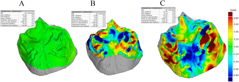
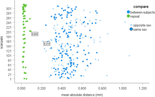
Representative cases for the intra- and between-subject superimposition are illustrated in Fig. 3. The between-subjects variability (1.068–0.214 mm) did not overlap the repeatability range (0.011–0.093 mm) (Table 2; Fig. 4). Sex differences did not significantly affect the MAD in the multiple linear regression analysis (partial correlation = -0.06, p = 0.343). Longitude difference showed a weak negative correlation with MAD (partial correlation = -0.16, p < 0.01), whereas latitude difference had a weak correlation with marginal significance level (partial correlation = -0.12, p = 0.058).
Fig. 3.
Color heat maps of scan superimposition of representative cases. The color code shows the difference between the two scans between − 800 microns and + 800 microns in the positive and negative directions. Green indicates a value close to zero. The deeper in red or blue, the greater the distance is between the two scans in the particular area. Yellow is the intermediary range. The grey area indicates a part that was not included in one of the scans. (A) The color map displays the superimposition of two repeated scans from a female individual of Turkish descent. (B) A superimposed image showcases a Turkish female and a male of Chinese origin. (C) The comparison involves the superimposition of scans from two distinct Turkish female individuals
Table 2.
The intra- (repeatability) and between-subjects mean absolute deviation (in mm)
| Mean | C.I. lower | C.I. upper | Median | Q1 | Q3 | Min | Max | |
|---|---|---|---|---|---|---|---|---|
| Repeatability | 0.029 | 0.025 | 0.033 | 0.025 | 0.010 | 0.040 | 0.011 | 0.093 |
| Between-subjects | 0.476 | 0.460 | 0.493 | 0.457 | 0.309 | 0.605 | 0.214 | 1.068 |
C.I. 95% Confidence interval
Q1, First quartile
Q3, Third quartile
Min, The lowest values
Max, The highest value
Fig. 4.
Scatterplot of the mean absolute distance (MAD) among subjects. Green dots depict the repeatability, and the blue color indicates between-subjects MAD. The blue dot shows the alignment between subjects having the same sex. The blue cross shows the alignment between subjects having the opposite sex. The box label indicates the highest repeatability and the lowest between-subjects MAD
Although ethnicity and sex influenced neither geometric discrimination nor the accuracy of superimposition identification, height significantly correlated with latitude (partial correlation = 0.59, p < 0.001), longitude (partial correlation = -0.46, p < 0.001), and sex (partial correlation = 0.71, p < 0.001). The depth was significantly dependent on longitude (partial correlation = -0.32, p < 0.05) and sex (partial correlation = 0.31, p < 0.05), but not on latitude (partial correlation = -0.13, p = 0.332). The width was significantly influenced by sex (r = 0.38, p < 0.01) but not by latitude (partial correlation = 0.18, p = 0.174) and longitude (partial correlation = 0.21, p = 0.117).
Sex determination by geometry
The former linear discriminative function with the variables measured in the current population could recognize females over males with 69.0% sensitivity (95% CI: 0.53–0.82%), which was not significantly (p = 0.089) lower compared to the homogenous Caucasian (82.2%, 95% CI: 0.72–0.89%) [1]. Contrarily, the specificity (62.5%, 95% CI: 0.36–0.84%) was significantly (p < 0.05) lower than in the homogenous population (89.3%, 95% CI: 0.71–0.97%). The overall accuracy was 67.24 (95% CI: 53.66–78.99%).
Discussion
Identification based on palatal geometry and by superimposition of digital scans
Based on our findings, digital forensic analysis of the palatal vault emerges as an efficient, rapid, and accurate method for human identification, with no limitations imposed by the population. The palatal geometric-based method demonstrated a sensitivity of 91.2% and specificity of 97.1% in the test or validation population from different countries. Incredibly, these results showed remarkable similarity (no overlap in confidence intervals) to the values (91.2% and 97.8%) obtained in the training set using a homogenous Caucasian population [1]. In conclusion, the previously proposed discriminant function could be successfully applied to a different mixed population.
Similarly, the superimposition method exhibited minimal deviations, with 214 μm for the smallest between-subjects deviation and 93 μm for the highest repeatability in the current mixed population. These values were remarkably close to that of the previously investigated Caucasian population, which reported 208 μm and 106 μm, respectively [11]. Notably, the previous study set the lower between-subjects threshold using the difference between mono-zygotic twin siblings, which is expected to be smaller than between two non-relatives.
Hence, the superimposition and geometric identification methods demonstrated consistency across diverse populations, irrespective of race and sex, indicating their generalizability.
Taneva et al. [8] found that both the indirect (gypsum model scan) and direct (intraoral scan) methods can be used to distinguish between subjects based on the superimposition method in the U.S. population. They measured the deviation of the rugae’s endpoint. Depending on the landmarks, the threshold values were set between 23 and 97 μm for identical subjects and 15–248 μm for strangers.
Gibelli et al. [9] used scanned gypsum models to superimpose palates. However, they set a 500 μm threshold value to distinguish between an individual and a stranger. The high threshold value could be due to the indirect digitization method. In line with this, a recent study [14] indicated that the inaccuracy of indirect digitization might limit identification. First, making a palatal impression without flaws and bubbles is challenging. Second, the additional steps, such as creating a gypsum cast and laboratory digitization, increase the overall error. Furthermore, the IOS is quicker and more straightforward at a disaster site and does not tear deteriorated tissue.
In another study [10], the IOS was used to discriminate between strangers and identical scans. The applied threshold for discrimination was lower (300 μm) than that of the study using indirect digitization [9]. Nevertheless, this value was still high compared to the current study (218 μm). Furthermore, in some cases, the deviation between identical twin siblings was as low as 208 μm [11]. The 300 μm value might relate to the older IOS version, the Omnicam (Dentsply Sirona) [10]. However, the graphical illustration in that manuscript indicated that the intra-subject repeat was less than 100 μm. Consequently, their selection of an excessively high cut-off value probably was too conservative.
Sex determination based on palatal geometry and by superimposition of digital scans
The sex difference between subjects did not improve the discriminative potential of the superimposition technique, and the subject’s country of origin had only a minor effect. Therefore, the superimposition method seems unsuitable for sex determination. Nevertheless, the sex discriminative function based on geometry resulted in lower sensitivity and specificity (69.0%, 62.5%) in a population with different countries compared to homogenous ones (82.2% sensitivity and 89.3% specificity) [1]. Interestingly, the male had a lower match (specificity) than the female (sensitivity) in the current data set. However, in the training set, the opposite was found. The male had a higher match (specificity) than the female. The height had the highest discrimination potential in the Fisher’s Linear Discriminant function [1] and was strongly correlated with sex. The standard deviation of height, depth, and width were 1.1, 2.6, and 2.5 mm in females and 3.1, 3.6, and 2.4 mm in males in this mixed ethnical population. As females have three times less variation (standard deviation) in height, it might cause a higher sensitivity to find them. However, in the previous training set, the height standard deviation was slightly different between the sexes. The function was developed in the training set and was applied without any modification to the test set. Therefore, the difference in height standard deviations between populations could cause the weaker and opposite sensitivity of the current results.
Furthermore, Males had a larger palate than females in the Hungarian population [1], in the Lebanese population [24], and in India [25]. However, the height was significantly influenced by the latitude and longitude in our study, suggesting a synergetic effect of sex and population on height. Consequently, the variance between populations could prevent accurate sex determination.
An implication for ethnic differentiation based on palatal geometry
Antemortem and postmortem scan comparisons could be valuable in mass disasters by suggesting the likelihood of ethnic differences. This could help direct further forensic investigation by indicating the country of origin where antemortem data should be explored.
There is no precise definition of race or ethnicity. While the differences between distant populations, such as Chinese and Caucasians, are clear, more subtle—like those between Germans and Greeks or Persians and Iraqis—are less pronounced. We hypothesized that the geographic distance between countries of origin is more reliable (objectively measured on a continuous scale) than subjective classifications such as Black, Caucasian, Arabic, Indian, or Asian.
Our results indicated that the identification based on either method is not affected by the country of origin, suggesting that the particular person primarily determines the palatal vault. However, sex, latitude, and longitude significantly affected height and depth. Thus, northern and western people have a higher palate than southern and eastern people. Westerners also have a slightly deeper palate than Easterners.
The stronger correlation of sex with the geometry compared to the latitude and longitude suggests that ethnicity cannot be reliably predicted without first determining sex. Therefore, a large-scale study, including a well-balanced distribution of males and females across various countries, is necessary. Based on our current findings, such a study would have the potential to provide a generalized discriminant function for ethnic prediction. Our pilot study may facilitate the start of international collaboration for that purpose.
Strengths and limitations
The strength of the study is that the validation of the geometric and superimposition-based identification and sex determination method was done in an unforeseen scenario without the intention to affect the participant’s inclusion or exclusion. As a result, the countries of origin and the sex distribution were coincidental. The uneventful distribution could be the reason that sex determination was significantly lower than previously found in a more significant subset. Consequently, new sex determination classification functions should be developed in an extensive training dataset where the participants are selected consciously from several geographic locations, each including many males and females.
Another limitation may be that only one quadrant is routinely scanned for restorations with only one or three teeth. Therefore, the ante-mortem scan may include only half of the arch, while the post-mortem scan may be damaged on the ipsilateral side, making the matching process difficult. Recently, it has been shown that the palate is quite symmetrical in the sagittal plane [26]. However, the morphology of the rugae and the overall vault are significantly different between the sides [27], which prevents the use of the contralateral side for matching.
The geometric-based identification method performed excellently in unplanned forensic scenarios. However, only 87% of the participants can be analyzed as three subjects had at least one maxillary first molar missing, making distance measurements impossible. During the previous development of the identification algorithm, a slightly higher percentage (16%) should have been excluded due to missing first molars [1]. The inclusion of some older subjects may explain this. The percentage of missing molars in the extensive forensic dental database Odontosearch is similar to our results [3]. Contrarily, the superimposition-based method could be applied accurately in cases where the palatal geometry could not be determined. However, superimposition has the limitation of being slower and more computationally intensive. Without automatized software, the manual alignment limits the evaluation of between-subject comparisons in research as more subjects are included in the sample; the number of pairs or combinations can overgrow, following the quadratic function. Therefore, the researcher usually makes a subset by randomly selecting pairs from all possible combinations [9, 10].
The current limitation of both methods is the missing centralized database for ante-mortem intraoral scans; however, some attempts have been made recently [28].
While dental records, fingerprints, and DNA remain the primary identification methods [29, 30], intraoral scanners have emerged as a potential tool for human identification using dental morphology and the anterior palate [1, 8–10, 15–17, 31, 32]. Digital records offer faster database searches, and a statistically based comparison algorithm not only provides binary match/no-match results but also assigns a probability to each outcome [18], enhancing the reliability of identification. However, this method should not be considered a primary identifier. Instead, it complements existing methods by narrowing down the pool of antemortem data and supporting other identification approaches. Given the clear advantages of intraoral scanners, we suggest they be considered an additional forensic investigation tool, though they are not yet part of standard practice.
Conclusion
Palatal vault geometry is a good candidate for forensic feature comparison because the proposed discriminant function can be used universally, making this identification method independent of population. Rapid pre-screening of palatal geometric data followed by superimposition of the selected scans can increase the accuracy of human identification.
Sex can also be determined from palatal geometry but with moderate accuracy. However, an international study with a large sample size is needed to determine the coefficient of such a discriminant function.
Acknowledgements
The authors would like to thank all participants for their contributions to this investigation.
Abbreviations
- BHP
Bottom horizontal plane
- DNA
Deoxyribonucleic acid
- DVI
Disaster victim identification
- I.P
Incisive papilla
- IOS
Intraoral scanner
- L.M
Left molar
- MAD
Mean absolute differences or distance
- R.M
Right molar
- THP
Top horizontal plane
Author contributions
János Vág: Investigation, Data analysis, Conceptualization, Validation, Visualization, Methodology, Writing – original draft, Writing – review & editing, Supervision, Funding acquisition, Project administration. Aida Roudgari: Data Analysis. Ákos Mikolicz: Investigation, Writing – review & editing. Botond Simon: Investigation, Visualization, Writing – review & editing Arvin Shahbazi: Validation, Writing – original draft, Writing – review & editing, Supervision.
Funding
The study was funded by the Hungarian Scientific Research Fund (K_22, 142142), the Hungarian Human Resources Development Operational Program (EFOP-3.6.2-16-2017-00006), and the American Society of Forensic Odontology Research Grant 2022.
Data availability
The datasets used and/or analyzed during the current study are available from the corresponding author upon reasonable request.
Declarations
Ethics approval and consent to participate
All experimental protocols were approved by the ethics committee of the Hungarian National Public Health and Medical Officer Service within the frames of the Codex of Bioethics of the Medical Research Council (36699-2/2018/EKU). All participants agreed to participate by written informed consent. All methods were carried out in accordance with relevant guidelines and regulations.
Consent for publication
Not applicable.
Previous data presentation
Part of the results of this article were presented in a lecture at the Triennial Congress of the International Organization of Forensic Odonto-Stomatology (IOFOS) in Dubrovnik (09/07/2023).
Competing interests
The authors declare no competing interests.
Footnotes
The original online version of this article was revised: blinded text was replaced.
Publisher’s note
Springer Nature remains neutral with regard to jurisdictional claims in published maps and institutional affiliations.
Ákos Mikolicz and Botond Simon equally contributed to the study; therefore, they should be considered as the first authors.
Change history
12/23/2024
A Correction to this paper has been published: 10.1186/s12903-024-05234-1
References
- 1.Simon B, Aschheim K, Vag J. The discriminative potential of palatal geometric analysis for sex discrimination and human identification. J Forensic Sci. 2022;67(6):2334–42. [DOI] [PMC free article] [PubMed] [Google Scholar]
- 2.Charangowda BK. Dental records: an overview. J Forensic Dent Sci. 2010;2(1):5–10. [DOI] [PMC free article] [PubMed] [Google Scholar]
- 3.Adams BJ, Aschheim KW. Computerized Dental comparison: a critical review of Dental Coding and Ranking Algorithms used in victim identification. J Forensic Sci. 2016;61(1):76–86. [DOI] [PubMed] [Google Scholar]
- 4.Petju M, Suteerayongprasert A, Thongpud R, Hassiri K. Importance of dental records for victim identification following the Indian Ocean tsunami disaster in Thailand. Public Health. 2007;121(4):251–7. [DOI] [PubMed] [Google Scholar]
- 5.Borbola D, Berkei G, Simon B, Romanszky L, Sersli G, DeFee M, Renne W, Mangano F, Vag J. In vitro comparison of five desktop scanners and an industrial scanner in the evaluation of an intraoral scanner accuracy. J Dent. 2023;129:104391. [DOI] [PubMed] [Google Scholar]
- 6.Vitai V, Nemeth A, Solyom E, Czumbel LM, Szabo B, Fazekas R, Gerber G, Hegyi P, Hermann P, Borbely J. Evaluation of the accuracy of intraoral scanners for complete-arch scanning: a systematic review and network meta-analysis. J Dent. 2023;137:104636. [DOI] [PubMed] [Google Scholar]
- 7.Malekzadeh AR, Pakshir HR, Ajami S, Pakshir F. The application of Palatal Rugae for sex discrimination in Forensic Medicine in a selected Iranian Population. Iran J Med Sci. 2018;43(6):612–22. [PMC free article] [PubMed] [Google Scholar]
- 8.Taneva ED, Johnson A, Viana G, Evans CA. 3D evaluation of palatal rugae for human identification using digital study models. J Forensic Dent Sci. 2015;7(3):244–52. [DOI] [PMC free article] [PubMed] [Google Scholar]
- 9.Gibelli D, De Angelis D, Pucciarelli V, Riboli F, Ferrario VF, Dolci C, Sforza C, Cattaneo C. Application of 3D models of palatal rugae to personal identification: hints at identification from 3D-3D superimposition techniques. Int J Legal Med. 2018;132(4):1241–5. [DOI] [PubMed] [Google Scholar]
- 10.Bjelopavlovic M, Degering D, Lehmann KM, Thiem DGE, Hardt J, Petrowski K. Forensic identification: Dental scan data sets of the Palatal fold pairs as an individual feature in a Longitudinal Cohort Study. Int J Environ Res Public Health 2023, 20(3). [DOI] [PMC free article] [PubMed]
- 11.Simon B, Liptak L, Liptak K, Tarnoki AD, Tarnoki DL, Melicher D, Vag J. Application of intraoral scanner to identify monozygotic twins. BMC Oral Health. 2020;20(1):268. [DOI] [PMC free article] [PubMed] [Google Scholar]
- 12.Zhao J, Du S, Liu Y, Saif BS, Hou Y, Guo YC. Evaluation of the stability of the palatal rugae using the three-dimensional superimposition technique following orthodontic treatment. J Dent. 2022;119:104055. [DOI] [PubMed] [Google Scholar]
- 13.Mikolicz Á, Simon B, Lőrincz G, Vág J. Clinical precision of Aoralscan 3 and Emerald S on the palatal and dentition areas: evaluation for forensic applications. J of Dent. 2024:105455. 10.1016/j.jdent.2024.105455. [DOI] [PubMed]
- 14.Mikolicz A, Simon B, Gaspar O, Shahbazi A, Vag J. Reproducibility of the digital palate in forensic investigations: a two-year retrospective cohort study on twins. J Dent. 2023;135:104562. [DOI] [PubMed] [Google Scholar]
- 15.Eggmann F, Blatz MB. Recent advances in intraoral scanners. J Dent Res. 2024. 10.1177/00220345241271937. [DOI] [PMC free article] [PubMed]
- 16.Corte-Real A, Ribeiro R, Almiro PA, Nunes T. Digital Orofacial Identification Technologies in real-world scenarios. Appl Sci-Basel 2024, 14(13).
- 17.Corte-Real A, Ribeiro R, Machado R, Silva AM, Nunes T. Digital intraoral and radiologic records in forensic identification: Match with disruptive technology. Forensic Sci Int. 2024;361:112104. [DOI] [PubMed] [Google Scholar]
- 18.Boedeker P, Kearns NT. Linear Discriminant Analysis for Prediction of Group Membership: a user-friendly primer. Adv Methods Practices Psychol Sci. 2019;2(3):250–63. [Google Scholar]
- 19.Ramspek CL, Jager KJ, Dekker FW, Zoccali C, van Diepen M. External validation of prognostic models: what, why, how, when and where? Clin Kidney J. 2021;14(1):49–58. [DOI] [PMC free article] [PubMed] [Google Scholar]
- 20.Ho SY, Phua K, Wong L, Bin Goh WW. Extensions of the external validation for checking learned Model Interpretability and Generalizability. Patterns (N Y). 2020;1(8):100129. [DOI] [PMC free article] [PubMed] [Google Scholar]
- 21.Morgan OW, Sribanditmongkol P, Perera C, Sulasmi Y, Van Alphen D, Sondorp E. Mass fatality management following the south Asian tsunami disaster: case studies in Thailand, Indonesia, and Sri Lanka. PLoS Med. 2006;3(6):e195. [DOI] [PMC free article] [PubMed] [Google Scholar]
- 22.Nagare SP, Chaudhari RS, Birangane RS, Parkarwar PC. Sex determination in forensic identification, a review. J Forensic Dent Sci. 2018;10(2):61–6. [DOI] [PMC free article] [PubMed] [Google Scholar]
- 23.Santos F, Guyomarc’h P, Bruzek J. Statistical sex determination from craniometrics: comparison of linear discriminant analysis, logistic regression, and support vector machines. Forensic Sci Int. 2014;245:e204201–208. [DOI] [PubMed] [Google Scholar]
- 24.Saadeh M, Ghafari JG, Haddad RV, Ayoub F. Sex prediction from morphometric palatal rugae measures. J Forensic Odontostomatol. 2017;35(1):9–20. [PMC free article] [PubMed] [Google Scholar]
- 25.Mankapure PK, Barpande SR, Bhavthankar JD. Evaluation of sexual dimorphism in arch depth and palatal depth in 500 young adults of Marathwada region, India. J Forensic Dent Sci. 2017;9(3):153–6. [DOI] [PMC free article] [PubMed] [Google Scholar]
- 26.Simon B, Mangano FG, Pal A, Simon I, Pellei D, Shahbazi A, Vag J. Palatal asymmetry assessed by intraoral scans: effects of sex, orthodontic treatment, and twinning. A retrospective cohort study. BMC Oral Health. 2023;23(1):305. [DOI] [PMC free article] [PubMed] [Google Scholar]
- 27.Simon B, Farid AA, Freedman G, Vag J. Digital palate analysis to verify the mirror twin phenomenon. In: Freedman G, editor. Oral Health Group; 2023. https://www.oralhealthgroup.com/features/digital-palate-analysis-to-verify-the-mirror-twin-phenomenon/. [Google Scholar]
- 28.Xiong H, Li K, Tan K, Feng Y, Zhou JT, Hao J, et al. TSegFormer: 3D tooth segmentation in intraoral scans with geometry guided transformer. In: Medical image computing and computer assisted intervention – MICCAI 2023: 2023// 2023. Cham: Springer Nature Switzerland; 2023. pp. 421–432. 10.1007/978-3-031-43987-2_41.
- 29.Forrest A. Forensic odontology in DVI: current practice and recent advances. Forensic Sci Res. 2019;4(4):316–30. [DOI] [PMC free article] [PubMed] [Google Scholar]
- 30.Jayakrishnan JM, Reddy J, Vinod Kumar RB. Role of forensic odontology and anthropology in the identification of human remains. J Oral Maxillofac Pathol. 2021;25(3):543–7. [DOI] [PMC free article] [PubMed] [Google Scholar]
- 31.Mou QN, Ji LL, Liu Y, Zhou PR, Han MQ, Zhao JM, Cui WT, Chen T, Du SY, Hou YX, et al. Three-dimensional superimposition of digital models for individual identification. Forensic Sci Int. 2021;318:110597. [DOI] [PubMed] [Google Scholar]
- 32.Sorrentino R, Ruggiero G, Leone R, Di Mauro MI, Cagidiaco EF, Joda T, Lo Russo L, Zarone F. Influence of different palatal morphologies on the accuracy of intraoral scanning of the edentulous maxilla: a three-dimensional analysis. J Prosthodont Res. 2024;68(4):634–42. [DOI] [PubMed] [Google Scholar]
Associated Data
This section collects any data citations, data availability statements, or supplementary materials included in this article.
Data Availability Statement
The datasets used and/or analyzed during the current study are available from the corresponding author upon reasonable request.


