Abstract
Biocontrol agents play a pivotal role in managing pests and contribute to sustainable agriculture. Recent advancements in genetic engineering can facilitate the development of entomopathogenic fungi with desired traits to enhance biocontrol efficacy. In this study, a CRISPR-Cas9 ribonucleoprotein system was utilized to genetically improve the virulence of Beauveria bassiana, a broad-spectrum insect pathogen used in biocontrol of arthropod pests worldwide. CRISPR-Cas9-based disruption of the transcription factor-encoding gene Bbsmr1 led to derepression of the oosporein biosynthetic gene cluster resulting in overproduction of the red-pigmented dibenzoquinone oosporein involved in host immune evasion, thus increasing fungal virulence. Mutants defective for Bbsmr1 displayed a remarkable enhanced insecticidal activity by reducing lethal times and concentrations, while concomitantly presenting negligible or minor pleiotropic effects. In addition, these mutants displayed faster germination on the insect cuticle which correlated with higher density of free-floating blastospores in the hemolymph and accelerated mortality of the host. These findings emphasize the utility of genetic engineering in developing enhanced fungal biocontrol agents with customized phenotypic traits, and provide an efficient and versatile genetic transformation tool for application in other beneficial entomopathogenic fungi.
Supplementary Information
The online version contains supplementary material available at 10.1186/s40694-024-00190-5.
Keywords: Secondary metabolite, Filamentous fungi, Blastospores, Genome editing, Ribonucleoprotein, Homologous recombination, Geneticin selectable marker
Author Summary
Fungal insecticides, while environmentally friendly, often suffer from slow action and require high doses with repeated applications to achieve effective pest control. To address these limitations, we employed CRISPR-Cas9 genome editing to enhance the virulence of the entomopathogenic fungus Beauveria bassiana. Specifically, we knocked out the Bbsmr1 gene, a transcriptional repressor that downregulates the biosynthesis of oosporein—a secondary metabolite crucial for inhibiting insect immunity and promoting fungal virulence. The resulting knockout strains exhibited significantly higher virulence compared to the wild type. Our approach demonstrates that CRISPR-Cas9 is a simple, efficient, and precise tool for editing the genome of B. bassiana, allowing for the customization of fungal biocontrol strains with desired traits such as enhanced virulence. This method holds great promise for developing more robust and effective mycoinsecticides, contributing to sustainable agriculture by reducing reliance on chemical pesticides.
Introduction
Arthropod pests, such as insects and mites, exert a negative impact on food security and agricultural productivity worldwide, leading to substantial economic losses. Invasive insects alone incur approximately $70 billion in costs to the global economy each year [1]. Over the past five decades, conventional agricultural systems have heavily relied on synthetic chemical pesticides as the primary method to control pests and safeguard crop yields on a global scale. However, this reliance on chemical pesticides has raised concerns due to its adverse effects on the environment and human health, as documented in earlier studies [2, 3]. The escalating expenses associated with the discovery, development, and registration of new synthetic pesticides, coupled with the rapid emergence of pest resistance [4] have fueled interest in non-chemical alternatives. The prevailing chemical paradigm within industrial agriculture relies heavily on insecticides, often causing secondary outbreaks or resurgence of insect pests due to the elimination of natural enemies or development of resistant populations [5]. Consequently, more than 550 arthropod species have developed resistance to at least one insecticide, underscoring the constraints of a chemical control approach [6]. Given this context, biological control strategies play a crucial role in sustainable integrated pest management schemes, reducing the dependence on chemical pesticides, thereby mitigating environmental pollution and the threat of pesticide resistance.
Biological control agents are pivotal players of the recent microbial revolution in agriculture, with a global market share of agricultural biologicals estimated in USD 15.10 billion by 2024 and USD 43.53 billion by 2035 [7]. Alternatively, the widespread adoption of beneficial microorganisms as biocontrol agents, particularly entomopathogenic fungi like Metarhizium spp. (Ascomycota: Clavicipitaceae) and Beauveria spp. (Ascomycota: Cordycipitaceae), form a significant component of biopesticide use across organic and conventional agricultural systems worldwide [8]. Despite their potential, challenges exist in the efficacy of these fungi due to their slow infection kinetics and the high inoculum levels required to effectively control pests [9]. Efforts are underway to develop faster and more efficient methods for selecting suitable microbial strains for biopesticide development; however, these processes are often time-consuming and labor-intensive, with success rates remaining low. Consequently, there is a growing interest in exploring genetic transformation techniques to enhance fungal attributes for biocontrol applications, addressing the pressing needs of the agricultural industry.
Engineered fungi are revolutionizing many applications in pharmaceutical, medical, and agricultural settings by helping to better understand gene functions involved in the interactions of fungal pathogens with their hosts. Particularly, the multifunctional endophytic entomopathogenic Beauveria bassiana and other hypocrealean species are also widely exploited for their secondary metabolites, which are useful in biotechnology and medicine [10]. Many entomopathogenic fungal secondary metabolites have antimicrobial and cytotoxic activities, and B. bassiana synthetizes nonribosomal peptides and polyketides, such as beauvericin and bassianolide (cyclooligomeric nonribosomal peptides), a variety of beauverolides (cyclic peptides), oosporein (dibenzoquinone), bassiatin (diketomorpholine), and tenellin (2-pyridone) [11, 12]. It is thought that these fungi synthesize such compounds in order to kill insects as well as to limit bacterial competition within the host [13], a typical co-evolutionary arms race strategy deployed by pathogen versus host. Despite all the benefits in plant protection and health afforded by entomopathogenic fungi, fungal insecticides, also known as mycoinsecticides, still struggle due to their slow speed of kill and high doses required to cause satisfactory and consistent control of the target insect pests across different environmental conditions [9, 14]. Such fungal phenotypic traits can be improved by genetic engineering of exogenous or endogenous virulence factors [15].
B. bassiana induces innate immune responses in the cuticle and gut to undermine the proliferation of bacterial competitors residing in these tissues during their growth in the insect host [16]. After invading insect hosts, B. bassiana produces a variety of toxins, and genome analysis of B. bassiana revealed its potential to produce putative secondary metabolites. A well annotated genome of B. bassiana ARSEF 2860 shows 41 putative secondary metabolites biosynthetic gene clusters. Expression of biosynthetic gene clusters (BGCs) under suitable conditions leads to production of secondary metabolites such as bassiacridin, beauvericin, bassianin, bassianolide, beauverolides, tenellin, oosporein, and oxalic acid [11, 17, 18]. Among these toxins, some have shown antimicrobial and antifungal activity in vitro and some have been associated with specificity and virulence because they suppress the immune response of the host, i.e. they are immunomodulators [19]. Of particular interest, the red pigment dibenzoquinone known as oosporein acts as an antimicrobial compound post-insect death that inhibits microbial competition and assures nutrients are available for fungal growth and reproduction [13].
Notably, B. bassiana can produce red pigmented compounds during the late stages of growth, and the cluster of six genes responsible for the production of the red pigment oosporein has been functionally characterized in this fungus [13, 20]. Considering that many B. bassiana commercial strains have been widely adopted in pest control given their broad host range and resourceful arsenal of biomolecules, this fungus offers a tractable model to study and expand our knowledge of genetic engineering toward enhancement of virulence in mycoinsecticides through the overexpression of secondary metabolites. In this sense, harnessing the production of secondary metabolites in genetically engineered entomopathogenic fungi may unlock a plethora of bioactive compounds with unique insecticidal activities, thus enhancing the reliability and performance of these fungal biocontrol agents as effective alternatives to chemical pesticides [21].
Blastospores are hydrophilic yeast-like unicellular asexual propagules that grow vegetatively by mitosis, either through budding or cell division, in the host hemocoel that accelerate infection process and host mummification [22]. When in the host hemocoel, blastospore propagation is accompanied by the production of a plethora of secondary metabolites that are key to circumvent the host immune system [11, 17]. B. bassiana and Metarhizium anisopliae blastospores, for instance, absorb nutrients in the hemocoel and produce insecticidal metabolites, such as beauvericin and destruxins, resulting in insect death within a matter of days depending on the fungal strain and host species [10, 16]. Interestingly, blastospores have shown similar or in several cases higher virulence than aerial conidiospores of various hypocrealean entomopathogenic fungi (Akanthomyces muscarium, Metarhizium spp., B. bassiana, Cordyceps javanica) against numerous noxious insects and ectoparasitic ticks [23–28], due to their intrinsic fast germination and penetration ability through the host integument [24, 29]. Therefore, blastospores could be the preferred type of active propagule for infection of arthropod hosts, especially those pests inhabiting the phylloplane of crops.
CRISPR-Cas9 is a powerful genetic tool that enables efficient and precise genome editing in filamentous fungi, surpassing traditional methods like plasmid and Agrobacterium-mediated transformation. Proof-of-concept studies using the CRISPR-Cas9 system on entomopathogenic fungi were previously demonstrated for B. bassiana [30] and Metarhizium brunneum [31, 32], but none of them generated strains with improved ecological fitness or virulence properties to make them better biocontrol agents for pest management. Despite considerable past efforts to develop transgenic fungal strains expressing arachnid (spider or scorpion) or wasp toxins [33, 34], their release into the field is impeded by public, environmental, and safety concerns that are rooted in existing regulatory policies. These concerns have thus far limited their broad utilization as registered biocontrol agents. Alternatively, the genetic manipulation of endogenous genes associated with virulence factors in entomopathogenic fungi using CRISPR-Cas9 emerges as a preferred method for generating markerless mutants with enhanced phenotypic characteristics when compared to wild-type strains [21, 35]. This alternative method holds promise in potentially streamlining the approval process and gaining acceptance from regulatory agencies in both the United States and Brazil in the near future.
In this study, protoplasts of B. bassiana were engineered using CRISPR-Cas9 ribonucleoproteins (RNP) resulting in blastospores with improved insecticidal activity through targeted disruption of the C2H2-type zinc finger protein encoded by Bbsmr1, which acts as a negative regulator of oosporein biosynthesis. The Bbsmr1 negative regulation of the oosporein biosynthetic gene cluster likely acts via regulation of the oosporein transcription factor OpS3, which in turn positively regulates the rest of the OpS gene cluster [13]. In addition, Bbsmr1 was also found to positively regulate the asexual conidiation development and stress response in B. bassiana by binding to the promoter region of the BbbrlA gene [36]. Oosporein by itself cannot directly kill insects and acts as an antimicrobial compound [13], but also promotes infection by evading the insect immunity and facilitating fungal growth [11, 20, 37]. In this context, our main biological hypothesis is that hypervirulent strains with disrupted Bbsmr1 can produce blastospores capable of overexpressing the bioactive molecule oosporein during an earlier stage of the infection process in the insect host, thus presumably resulting in accelerated killing potential and lower inoculum required as compared to its wild-type (hereafter designated WT) strain. This proof-of-concept work broadens our knowledge in gene function in B. bassiana and helps accelerate the pipeline for obtaining genetically engineered strains with improved virulence for use in biological control purposes. Unlike previous efforts in generating transgenic entomopathogenic fungi with heterologous expression of genes encoding for toxins with powerful insecticidal properties derived from other organisms [33, 34, 38, 39], our genetic engineering approach places emphasis on accurately editing endogenous genes via CRISPR-Cas9 in the target fungal genome to unlock its full potential as an effective biocontrol agent.
Our findings reported in this study provide novel insights into a cost-effective genome editing strategy of an endogenous transcription factor associated with secondary metabolism regulation to improve virulence of an entomopathogenic fungus for more effective biocontrol. Targeted gene inactivation using CRISPR-Cas9 may enhance fungal fitness and overall biocontrol performance and can be applied to other entomopathogenic fungi. Therefore, we envision the next generation of mycoinsecticides emerging from gene-edited fungal strains to deliver consistent and highly effective biological solutions that perform reliably across diverse conditions, compete effectively with chemical insecticides, and advance sustainable agriculture while contributing to global food security.
Methods
Microbial cultures, media and growth conditions
The wild-type strain (WT) Beauveria bassiana ARSEF2860 was originally isolated from Schizaphis graminum (Hemiptera: Aphididae) on spring wheat in 1987 in Parma, Idaho, USA and was obtained from the USDA ARSEF culture collection. Its genome is available at Genbank (GCA_000280675.1). Pure cultures were maintained in 20% glycerol stocks at – 80 °C and fresh cultures were grown on a weekly basis on 20 mL of potato dextrose agar (PDA – Difco™, Sparks, MD, USA) medium in 100 × 15-mm Petri dish plates. Fresh fungal cultures retrieved from frozen stocks were grown at 28 °C and in the dark for 14 days prior to using in experiments. Fungal germlings were produced in potato dextrose broth (PDB – Difco™). Transformant selection was carried out in R-Top medium containing geneticin (G418-sulfate, VWR Chemicals, Solon, OH, USA) at 300 µg/mL, poured into sterile Petri dishes (90 × 15 mm) containing on the bottom PDA medium amended with the same concentration of this antibiotic. This geneticin concentration was previously selected according to its ability to impair germination and growth of B. bassiana WT protoplasts.
Bioinformatics
The B. bassiana ARSEF2680 gene Bbsmr1 (1,473 bp ORF, no introns) was identified at GenBank Protein Id: XP_008598185.1 (EJP66373.1) and gene code BBA_04866 according to Xiao et al. [40]. The upstream and downstream regions of Bbsmr1 were identified at MycoCosm database [41]. The protospacer/sgRNA sequence was identified using CCTop [42] (https://cctop.cos.uni-heidelberg.de/), and Clustal Omega – Multiple Sequence Alignment software was applied for DNA alignment [43].
DNA purification, PCR procedures and plasmid construction
Primers used in this study are listed in Table S1. B. bassiana wild-type (WT) ARSEF2860 and mutant genomic DNA were purified using a phenol/chloroform protocol [44].
The donor DNA was constructed by amplification of the geneticin/G418 resistance gene marker (2,816 bp) under the control of a constitutive promoter trpC (GenBank: X02390.1) from the pII99 plasmid (5.3 kb) using the primers combination AZF1_sg8_Gen_KO-F + AZF1_sg8_Gen_KO-R (2,916 bp DNA fragment). Each primer contained 50 nucleotides corresponding to the upstream or downstream homologous flanking regions of the Bbsmr1 gene target site. This repair template cassette was amplified using master mix repliQa® HiFi ToughMix following the program: 10 s at 98 °C and 35 cycles (15 s at 68 °C and 30 s at 68 °C). The generic map of the geneticin cassette, including homology arms, diagnose primer’s locations and insertion site is available in Fig. S1.
DNA fragment amplification for the in vitro Cas9 cleavage assay and diagnostic PCR employed to track positive transformants were produced in a thermal cycler with GoTaq® Green Master Mix (Promega) using the following program: 95 °C for 3 min, 35 cycles of 30 s at 95 °C, 30 s at 61 °C, 2.5 min at 72 °C) and a final extension of 5 min at 72 °C. The DNA fragment used as a template for in vitro Cas9-cleavage assay was amplified using primers Bbsmr1-F and Bbsmr1-R (1,984 bp fragment) (Table S1, Fig. S1). Moreover, diagnostic PCR to detect positive transformants presenting the insert geneticin resistance gene was conducted using the following combinations of primers: Bbsmr1-F + Bbsmr1-R generating a larger fragment of ~4.8 kb long, which contains the geneticin resistance gene; Bbsmr1-F + Gent_R resulting in a fragment of 2,392 bp long; and Gent_F + Bbsmr1-R producing a fragment of 2,843 bp long in ΔBbsmr1 strains (Table S1). These primers were designed to amplify regions upstream and downstream of homology sites, including part of the insert DNA from geneticin resistance gene. This diagnostic strategy allows the confirmation of the specific DNA insertion site in contrast to the WT strain that exhibited a PCR product of ~1.9 kb after amplification with the pair of primers Bbsmr1-F + Bbsmr1-R. In addition, purified DNA fragments were sequenced by Sanger sequencing using the primer Bbsmr1_F (− 501 bp upstream of Bbsmr1 cleavage site).
Cas9 overexpression and protein purification
Transformed E. coli Rosetta™ (DE3) strain containing plasmid with Cas9 cassette was induced to overexpress this protein from plasmid pHis-parallel1-NLSH2BCas9 (Addgene plasmid #112065; http://n2t.net/addgene:112065; RRID: Addgene_112065) [45] after addition of 0.3 mM IPTG for 12 h of growth in 1-L of LB medium at 37 °C and 200 rpm. This protocol was adapted from Pokhrel et al. [46] and its details are described in Fig. S2. A final purified concentration of Cas9 reached 2.0 mg/mL, and then stored at – 80 °C until use.
In vitro Cas9-sgRNA RNP cleavage efficiency assay
In brief, the sgRNA was synthetized using a commercially available kit (EnGen® sgRNA Synthesis Kit, S. pyogenes Protocol NEB #E3322). In the first step, ssDNA target-specific oligonucleotide was designed by selecting 20 nucleotide sequence (not including the PAM [NGG]) of the target gene Bbsmr1 using a forward primer designed (Table S1). PCR cycle for synthesis of ssDNA oligos was set to 3 min at 98 °C, 35 cycles (10 s at 98 °C, 30 s at 60 °C, 30 s at 72 °C) and a final extension at 72 °C for 2 min. Secondly, following the manufacturer’s protocol (https://www.neb.com/en/products/e3322-engen-sgrna-synthesis-kit-s-pyogenes), the target-specific oligos were mixed with the EnGen 2× sgRNA Reaction Mix (NTPs, dNTPs, S. pyogenes Cas9 Scaffold Oligo), 0.1 M DTT and the EnGen sgRNA Enzyme Mix (DNA and RNA polymerases), and all steps occurred in a single reaction during a 30-min incubation at 37 °C. The resulting sgRNA contained the target-specific/crRNA sequence as well as the tracrRNA. For purification of sgRNA to remove proteins, salts and most unincorporated nucleotides, we used spin columns compatible with the size of sgRNA (~ 100 nts) from Zymo™ RNA Clean-Concentrator kit (#ZR1013). sgRNA was stored at – 80 °C until use in the transformation assay.
Previously to the enzymatic reaction, DNA amplicon Bbsmr1 produced, as described previously, was cleaned and concentrated using ice cold 0.1× volume of 3 M sodium acetate (30 µL) plus 2.5× volumes of 100% ethanol (350 µL). DNA was further precipitated by centrifugation at 18,000 rpm, 4 °C for 10 min. DNA pellet was washed twice with ice-cold 80% pure ethanol, centrifuged again (18000 rpm, 4 °C, 10 min) and then air-dried for 10 min prior to re-suspending with 30 µL of 0.1% DEPC water. In that way, it was possible to reach a concentration of donor DNA higher than 300 ng/µL with high quality revealed by 0.8% agarose gel and absorbance ratio (A260/A280) of 1.7-2.0. Four samples of donor DNA were pooled together in a single sample to reach a concentration of 6 µg required for the Cas9-sgRNA RNP transformation assay using fungal protoplasts.
The in vitro reaction for Cas9-sgRNA RNP cleavage efficiency assay consisted in mixing 2 µL (1 µg) Cas9, 2 µL NEB3.1 buffer (10×), 2 µL (200 ng) sgRNA, 2 µL (200 ng or 300 ng) DNA template, and 12 µL of 0.1% DEPC nuclease-free water, totaling 20 µL of final volume. The preformed ribonucleoproteins based on 1 µg Cas9 + 200 ng sgRNA were used to digest 200 ng or 300 ng DNA template of Bbsmr1 at 37 °C for 2 h in the thermocycler (adapted from Wang et al. [45]). After that, the whole digested products (20 µL) were loaded in the agarose gel (1%) and run at 80 V to separate the bands. As for the control, the same amount of DNA template was loaded in the gel to serve as basis of comparison to the digested sample.
Protoplasting and fungal transformation
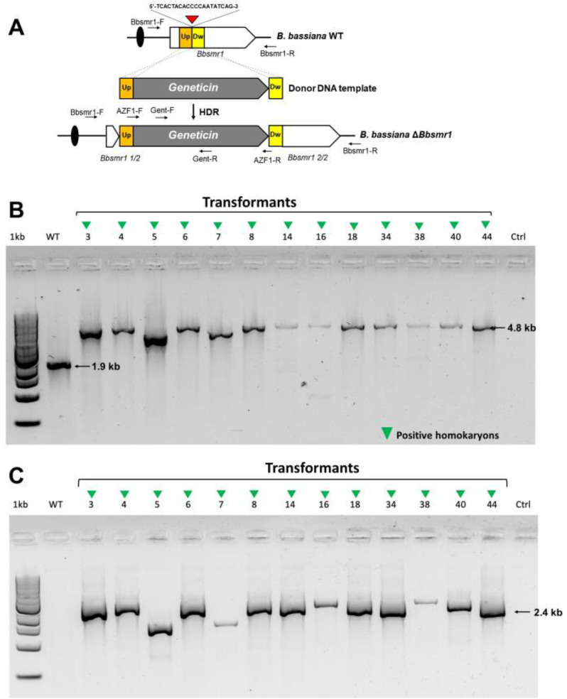
Protoplastation and transformation were previously described in Fusarium oxysporum by Pokhrel et al. [46]. Illustrative and detailed step-by-step transformation protocol for B. bassiana is available in Fig. S2. In addition, Fig. 1A shows the general scheme of gene edition strategy used in this work.
Fig. 1.
Gene editing strategy and construction of B. bassiana (ΔBbsmr1) mutants. A) Gene edition strategy. Red arrow corresponds to the specific Cas9 target site. Black arrows correspond to diagnostic primers. B) Diagnostic PCR for detection of positive mutants (ΔBbsmr1) using specific markers (long read primers: Bbsmr1-F + Bbsmr1-R) before monosporic purification step in 1% agarose gel. The PCR products expected band sizes were 1.9 kb and 4.8 kb for the WT strain and the putative transformants (containing the insert DNA of geneticin resistance gene), respectively. C) Diagnostic PCR for site-specific mutation in Bbsmr1 using specific markers (short read primers: Bbsmr1-F + Gent_R) in 1% agarose gel to confirm the positive transformants. The ~ 2.4 kb band corresponds to a positive ΔBbsmr1 mutant, confirming the integration of geneticin cassette into Bbsmr1 gene. As expected, no band appears in WT due to the absence of the geneticin resistance gene. All homokaryon mutants are highlighted with a green inverted triangle and they show a band in both gels (B, C). Ctrl is the negative control without any DNA, and 1 kb marker was used in the the gels
Relative expression of oosporein-associated key genes by RT-qPCR
Two knockout mutants of Bbsmr1 and the WT strain (as the control treatment) were grown under pH 8.0 in a rotary incubator shaker in PDB with 100 mM Tris buffer at 28 °C and 250 rpm for 3 days to produce mycelium for RNA extraction. Fungal biomass was subsequently collected and harvested using centrifugation. The cell pellets were flash frozen with liquid nitrogen and kept at − 80 °C until RNA extraction. The frozen cell pellets were lyophilized in 1.5 ml tubes and the dried pellet was ground with a small plastic pestle (Kimble Inc). RNA extraction was performed using a Qiagen RNAeasy plant kits (Qiagen Inc, Germantown, MD, USA) with on column DNAase treatment using the manufacturer’s protocol, with the following change in the cell disruption method. The ground cell pellet was resuspended in 1 mL RTL buffer (from the kit) and the cells disrupted by passing the solution through a 22 ga needle 10 times, which was adapted from Cortés-Maldonado et al. [47]. The qPCR primers for target genes and housekeeping gene (BbActin) are listed in Table S1.
To assess the relative expression of Bbsmr1 in the B. bassiana defective mutants (ΔBbsmr1_3 and ΔBbsmr1_38) compared with the WT parent, RNA extraction and purification were performed for 3-day-old liquid grown cultures with pH 8.0 using the protocol detailed above. Reverse transcription was conducted using 300 ng of RNA from each sample via the QuantiTec reverse transcription kit (Qiagen). This protocol included a genomic DNA removal step using gDNA Wipeout Buffer (Qiagen) prior to the reverse transcription step via Quantiscript RT buffer (Qiagen). Gene-specific primers used to detect the Bbsmr1 and BbOsp genes were those designed by Fan et al. [13] (Table S1). Gene expression was evaluated in a 10 µL reaction volume using 1 µL of sample cDNA and the PowerUP SYBR green Master mix qPCR kit (Qiagen) following the recommended qPCR cycling conditions (holding at 95 °C for 10 min, 40 cycles of 15 s at 95 °C, and 1 min at 60 °C), including an endpoint Melt Curve analysis. The qPCR assay was carried out on an Applied Biosystems QuantStudio 6 Flex Real-time PCR system (ThermoFisher Scientific™). Four biological replicates were evaluated for each treatment group, and the expression levels were normalized against the fungal gene Actin (Table S1). The expression data was analyzed using the ∆∆Ct method [48] and their significance evaluated on log-transformed values via one-way ANOVA with Dunnett’s multiple comparison test. The statistical analysis and graphs were generated on Prism 10.0 (GraphPad).
Off-target assessment by genome analysis
The absence of off-target effects in the fungus genome after CRISPR-Cas9-mediated disruption of Bbsmr1 was confirmed by draft genome sequencing of our mutants ΔBbsmr1_3 and ΔBbsmr1_38. DNA was extracted from one-mL samples from a 4 day of cultivation in PDB were collected and harvested by centrifugation. The cell pellet was freeze-dried and DNA extracted using a CTAB based method of Watanabe et al. [49]. Sequencing libraries were constructed using the Illumina DNA prep kit following the manufacturer’s protocol. The libraries were sequenced on an Illumina Novaseq X10 using 2 × 150 bp paired end sequencing. The raw reads were quality trimmed and assembled using CLC Genomic workbench 23.05. All sequencing data is available under GenBank Bioproject PRJNA1093450. To determine possible sequence independent off-target effects, sequencing reads from the mutants were mapped (> 0.5 alignment query and > 0.9 sequence identity) to the wild-type draft genome using CLC Genomic workbench 23.05. Variants were called to detect single and multiple nucleotide variants, insertions, deletions and their combinations (≥ 90% frequency). In addition, their functional consequences of the variants were predicted (amino acid changes), when they fell within a coding region. To determine possible sequence dependent off-target effects, the guide RNA sequence was used to identify regions of the genome with greater than 13 of 20 nucleotides shared. These regions in the mutant genome were manually examined for possible effects.
Phenotypic quantification of oosporein
The standard oosporein (purity > 95% by HPLC, DiagnoCine, Totowa, NJ, USA) was dissolved using 1 mg in 1 mL of DMSO and standard calibration curve was made using a range of 0 to 100 ppm, measured with a spectrophotometer UV-VIS at a wavelength of 430 nm, following the protocol proposed by Lara-Juache et al. [50]. One-mL samples from days 2 to 4 of cultivation in PDB were collected and centrifuged at 25,200×g (= 12000 rpm) for 15 min at 4 °C to pellet down fungal biomass and extract 50-µL aliquot of the supernatant to be used in the oosporein quantification. Oosporein measurements were performed with six biological replicates for each day of growth and each strain. Standard calibration curve of oosporein is given by y = 0.0003x + 0.0022, where x is oosporein concentration in ppm (mg/L) and y is the absorbance measured at 430 nm.
Virulence bioassays
The greater wax moth larvae, Galleria mellonella (Lepidoptera: Pyralidae), were purchased from Waxworms.net (Vanderhorst Wholesale Inc., Saint Marys, Ohio, USA). Larvae from the last instar were used in all bioassays and did not require any diet during the bioassay. All bioassays were performed with at least two independent batches of larvae and blastospores.
Blastospores were produced in liquid medium (adapted from Mascarin et al. [27]) containing free-vitamin basal salts prepared with miliQ-water (amount per L: 4.0 g KH2PO4, 0.8 g CaCl2.2H2O, 0.6 g MgSO4.7H2O, 0.1 g FeSO4.7H2O, 16 mg MnSO4.H2O, and 14 mg ZnSO4.7H2O), 8% (w/v) anhydrous dextrose (40% carbon, mw = 180.16 g/mol, VWR Chemicals, Solon, OH, USA), and 2.5% (w/v) casamino acids (AMRESCO®, distributed by VWR Chemicals, Solon, OH, USA) using a working volume of 50 mL liquid medium in 125-mL Erlenmeyer flasks capped with aluminum foil. The nonionic surfactant Silwet® L-77 (mw = 338.66 g/mol, Cas number 27306-78-1, Phytotech Labs, Lenexa, KS, USA) was added directly to the autoclaved miliQ-H2O at a concentration of 0.01% (v/v). Blastospores were harvested after 3 days of cultivation, filtered through nylon mesh filter screen (37 μm pore size, Sefar-Nitex®s) to remove mycelium, and then centrifuged at 1200 × g and 4 °C for 15 min to remove spent medium. Subsequently, blastospore suspensions were prepared with 0.01% of aqueous surfactant solution. Viability of blastospores were determined on 60-mm Petri dish containing 5 mL of PDA medium by inoculating 100 µL of 1 × 10⁵ blastospores/mL on the center of the plate. After 12 h incubation at 25 °C in the dark, germinated and non-germinated blastospores (n = 200 cells per treatment) were counted to establish the proportion of viability. Viability of blastospores was always greater than 90% in all cases.
There were performed bioassays to assess the in vivo virulence of multiple mutants of Bbsmr1 using as the insect host model Galleria mellonella larvae. The first bioassay focused on a single-dose response analysis with eight mutants (name here as ΔBbsmr1_3, ΔBbsmr1_4, ΔBbsmr1_5, ΔBbsmr1_18, ΔBbsmr1_34, ΔBbsmr1_38, ΔBbsmr1_40, ΔBbsmr1_44). The parent wild-type strain (WT) was used for comparison, while a mock control was fungus-free consisting only of surfactant solution.
The first set of experiments consisted of single dose-mortality bioassays using a standard concentration of 1 × 10⁷ blastospores/mL. There were two forms of fungal treatment: i) larvae were treated topically by soaking them to 1 mL of 1 × 10⁷ blastospores/mL in a Petri plate (100 × 60 mm); ii) these treated insects were subsequently placed onto filter-paper disks (Qualitative 413, VWR North American, West Chester, PA, USA) pre-treated with 150 µL of 1 × 10⁷ blastospores/mL in 6-well-plates to simulate a residual contact exposure. A total of 20 larvae were distributed into 6-well-tissue culture plate (Avantor®, XXXisturbed by VWR Chemicals, Solon, OH, USA), and each treatment had 4 plates with a total of 80 larvae. Mock control had larvae treated only with aqueous solution of Silwet® L-77 at 0.01%. Two moistened cotton balls were added to each plate to maintain relative humidity above 95% during the entire period of the experimentation. The whole experiment was kept in an environmentally walk-in controlled room at 24 °C for 5 days with 8:14 (L: D) h artificial photoperiod. This bioassay was repeated twice over time using different batches of insects and blastospores.
In the second set of bioassays, a multiple dose-mortality response experiment was performed with crescent concentrations of fungal inoculum in order to estimate the lethal concentrations for three best mutants selected in the first single-dose virulence bioassay, and compared with the wild type. A mock control encompassed only the fungus-free surfactant solution. The following concentrations were tested: 0 (0.01% Silwet® L77), 5 × 10⁵, 1 × 10⁶, 5 × 10⁶ and 1 × 10⁷ blastospores/mL. The entire experiment was repeated twice using independent batches of insects and blastospores, and each treatment had a total of 5 biological replicates and 100 larvae per treatment.
The number of dead insects was counted twice daily until 5 days post-inoculation. Natural mortality in control groups was attributed to unknown causes, whereas mortality from fungus-treated groups was confirmed by mycosis presenting fungal outgrowth (white mycelium) and/or pinkish body of cadavers due to oosporein production. Mortality caused by other factors was not included in survival analysis. For single dose-mortality bioassays, survival curves were determined by non-parametric Kaplan-Meier method and compared with log-rank test (P < 0.05), while estimated median and 90% time of lethality (LT50 and LT90) were retrieved from adjusted survival curves to parametric Weibull model. For multiple dose-mortality bioassays, same approach was adopted to estimate lethal times in each evaluation time, while a generalized linear model with binomial distribution was employed to fit data and allow estimation of median and 90% lethal concentrations (LC50 and LC90).
Phenotypic characterization of ΔBbsmr1 mutants
Pleiotropic effects due to disruption of Bbsmr1 were assessed in comparison to the WT strain. The ability to grow vegetatively and to sporulate on PDA medium was evaluated for ΔBbsmr1 mutants in comparison to its WT strain. Plates (60 × 15 mm) were filled with 5 mL of PDA medium, and then inoculated with a droplet of 5 µL from a suspension of 5 × 10⁷ blastospores/mL on the center of the plates. There were 6 biological replicates per treatment and the entire assay was repeated twice using different batches of fungal inoculum. Growth diameter transformed into colony area was measured with a digital calliper in two directions after 4, 6, 9 and 12 days post-inoculation with incubation at 28 °C in the dark.
The fitness comparison among mutants and parental WT by examining their ability in producing blastospores by submerged liquid fermentation was proceeded according to Mascarin et al. [27]. Each fungal strain had two replicates per experiment and the entire experiment was repeated four times with different fungal batches on different occasions. Results were expressed as blastospores per mL of liquid medium in each time interval.
The susceptibility of ΔBbsmr1 mutants compared with the WT when challenged with the cell chemical stressors 0.8 M NaCl, 2 mM H2O2, 0.02 mM menadione, and 100 µg/mL Congo red, either by oxidative, osmotic stress or cell wall damage, was assessed from Guo et al. [51]. There were 5–6 biological replicates per treatment and the entire assay was repeated twice using different batches of fungal inoculum.
Statistics and reproducibility
Mortality data from the insect bioassays were fitted to generalized linear mixed models to determine the significance of fixed effects and to estimate LC50 and LC90 using the “drc” package [52]. Survival analysis using the non-parameter Kaplan-Meier method allowed to obtain survival curves using the “survminer” package [53]. Log-rank test was employed to compare the survival curves. Weibull model was fitted to Kaplan-Meier survival curves in order to estimate the LT50 and LT90 using the “flexsurv” package [54]. Blastospore production, colony growth area and colony sporulation datasets were all log10-transformed prior to two-way ANOVA with fixed factors for strain and cultivation time. Oosporein content data were fitted to a linear model and submitted to two-way ANOVA with strain and cultivation time as fixed factors. Means were always compared by multiple pairwise Tukey’s HSD test with significant at P < 0.05 using the “emmeans” package [55].
A principal component analysis (PCA) is a useful tool to manage many highly correlated variables within the data set in order to reduce the dimensionality by using fewer original variables while explaining most of the data variance. In this way, it is possible to identify patterns among the original variables and commonalities among the objects (strains). We started with 19 phenotypic traits (variables) in the multivariate analysis to explain the relationship between virulence and pleiotropic effects for distinguishing B. bassiana knockout mutants and wild-type (WT). Blastospore production and colony sporulation were both log10-transformed necessary to approach multivariate normality prior to PCA, performed using the “factoextra” package [56]. Non-meaningful variables were dropped out from the analysis according to their low contribution (i.e., low correlation or correlation with the last dimensions) to each principal component (PC1 and PC2), ending up with only meaningful 11 variables that explained 93.7% of the total variability, in contrast to 87.7% of explained variance when there were 19 variables. Subsequently, a biplot was constructed to plot on the same space phenotypic variables (arrows) and strains (dots) for coordinated by the two first principal components. Furthermore, a heatmap was built using the “gplots” package [57] with the same 11 variables selected by the PCA to facilitate visualization and comparison between mutants and WT based on their similarity calculated with Manhattan distance method and further clustering with Ward method to assemble the dendrograms.
The gene expression data was analyzed using the ∆∆Ct method [48] and their significance evaluated via one-way ANOVA with Dunnett’s multiple comparison test. Statistical significance was assessed at P < 0.05 and the asterisks represent the strength of the significance: * P < 0.05; ** P < 0.01; *** P < 0.001; **** P < 0.0001. The statistical analyses and graphs were generated on Prism 10.0 (GraphPad) or on R statistical software (version 3.2.4).
Results
Blastospores are more virulent than conidia
To select the infective inoculum of the fungus used in all virulence bioassays with G. mellonella, we firstly compared the virulence between yeast-like cells termed blastospores with aerial conidia of B. bassiana. Blastospores of B. bassiana were much more virulent to G. mellonella larvae outperforming aerial conidia, as the former was able to kill 22% faster and 3.3-fold more insects than the latter (χ2 = 102.1, df = 2, P < 0.0001) (Fig. S3A). Notably, overall mortality of conidia-treated insects reached 29.4% in contrast to 97% dead insects after 5 days post-inoculation with blastospores. Interestingly, cadavers from conidia-treated insects did not exhibit the conspicuous red pigmentation due to oosporein production after host death as observed for blastospores-treated insects, where in this case all cadavers showed red pigmentation (Fig. S3B). Mock control larvae survived 97.10% over the course of the experimentation period and depicted a healthy and normal body morphology. Since blastospores displayed stronger virulence than aerial conidia and their production in liquid medium only took 3–4 days of growth compared to more than 10 days to produce aerial conidia on PDA plates, blastospores were used as the active inoculum in subsequent virulence bioassays with mutants of Bbsmr1.
Regardless of the propagule type tested, an increase in insect mortality was followed by an increase in the inoculum concentration (χ2 = 76.36, df = 1, P < 0.0001) and evaluation time (χ2 = 80.32, df = 2, P < 0.0001) (Fig. S4). The triple interaction concentration x day x inoculum type was significant and indicates that blastospores tested at different concentrations on different evaluation times were more effective in killing the host insect than aerial conidia (χ2 = 7.29, df = 2, P = 0.026). Examining the virulence degree based on lethal concentrations to kill 50% and 90% of the insect host population, blastospores displayed 8.2 to 76.5-fold greater virulence in relation to aerial conidia respectively, at days 3, 4 and 5 post-inoculation (Table S2).
Cas9-RNP for disruption of the transcription factor Bbsmr1
First, we purified the Cas9 nuclease, reaching a final yield of 2 mg/mL, and confirmed its molecular weight of 160 kDa according to the SDS-PAGE results (Fig. S5A). To confirm whether the putative Bbsmr1 gene from B. bassiana (BBA_04866) was involved in oosporein synthesis, the double-strand break target site was chosen with a location close to the 5′-terminus of the coding region in the first exon. The PCR template cleavage efficiency was close to 100% in vitro, demonstrating the developed NLSH2BCas9/sgRNA system had highly efficient cleavage activity for two concentrations of DNA template tested, 200 and 300 ng (Fig. S5B).
When using a concentration of Cas9/sgRNA at 40/8 (µg/µg, ratio 5:1) and 6.0 µg of donor DNA resulted in generation of ΔBbsmr1 transformants that grew on PDA amended with geneticin as opposed to the parental WT (Fig. S6). All colonies from the transformation medium were transferred to 24-well-plates containing PDA + 300 µg/mL geneticin and grew within 4–6 days at 28 °C in the dark. Mutants were screened by running three PCR reactions using primers to detect and confirm the putative mutations. Among 44 screened transformants, the PCR results indicated 41 positive mutants, which included single or multiple insertions (PCR product > 2.4 kb). We identified homokaryon mutants using standard PCR with the proper set of primers that confirmed the presence of the geneticin resistance gene insertion by homologous recombination in Bbsmr1 gene (Fig. 1B, C). Subsequently, eight mutants were selected and then tested in the screening virulence bioassays. Thus, the transformation efficiency via CRISPR-Cas9 was 93.2%. After single sporing the transformants for purification of heterokaryons, another round of diagnostic PCR and sequencing of mutants confirmed the specific site of geneticin cassette insertion, disrupting Bbsmr1. Fig. S7A shows a scheme of the targeted gene Bbsmr1 after its disruption, including the homology arm, part of the geneticin cassette inserted and the exact sequenced site (upstream). Sequencing of the locus of homokaryon mutants ΔBbsmr1_3, ΔBbsmr1_38, and ΔBbsmr1_44 indicated it was identical to the expected disrupted mutant sequence (Fig. S7B), thus providing the first evidence of the specific site mutation.
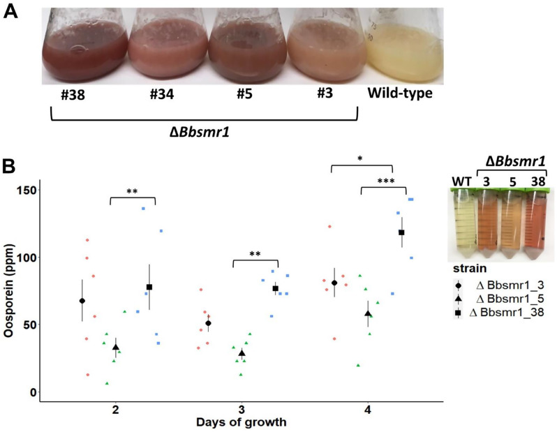
Of the mutants grown in PDB for at least 4 days, 87.8% (36/41) had the red/pink pigmentation phenotype, a strong indication of oosporein production and confirmation that Bbsmr1 represses the biosynthetic pathway of this secondary metabolite in B. bassiana. In contrast, liquid cultures of the parental WT strain were devoid of oosporein even after 10 days of growth and exhibited a yellowish pigmentation instead. Four selected Bbsmr1 mutants (ΔBbsmr1_3, ΔBbsmr1_5, ΔBbsmr1_34, and ΔBbsmr1_38) having a single insertion of the geneticin resistance cassette in the locus were able to produce the red pigment when cultured in PDB medium after 5 days, supporting this transcription factor negatively regulates the secondary metabolite gene cluster responsible for the oosporein biosynthesis (Fig. 2A).
Fig. 2.
Oosporein production during in vitro growth. A) Oosporein production (in ppm) by ΔBbsmr1 strains during four days of growth in 50-mL PDB at 28 °C and 250 rpm. The pink/red color in the culture supernatant of ΔBbsmr1 strains is the evidence of oosporein presence, whilst null oosporein was found in the WT’s liquid cultures. B) Kinetics of oosporein production (ppm) during four days cultivation. Significant differences in oosporein production (mean ± SE, n = 6) between mutants within each time interval are indicated by asterisks (* P < 0.05, ** P < 0.01 or *** P < 0.001) according to Tukey’s test. Different symbols and colors represent the mutants
Three selected mutants were able to secrete oosporein in PDB to varying degrees (F = 17.58, df = 2, 45, P < 0.0001). There was a trend for an increasing oosporein titer over cultivation time for ΔBbsmr1_5 and ΔBbsmr1_38 (F = 8.37, df = 2, 45, P < 0.0001), but not for ΔBbsmr1_3, for which oosporein titers remained virtually unaltered. Notably, the mutant ΔBbsmr1_38 overproduced this compound by yielding 46% and 104% more oosporein than the ΔBbsmr1_3 and ΔBbsmr1_5 strains, respectively, after 4 days of growth (Fig. 2B).
ΔBbsmr1 mutants display increased virulence
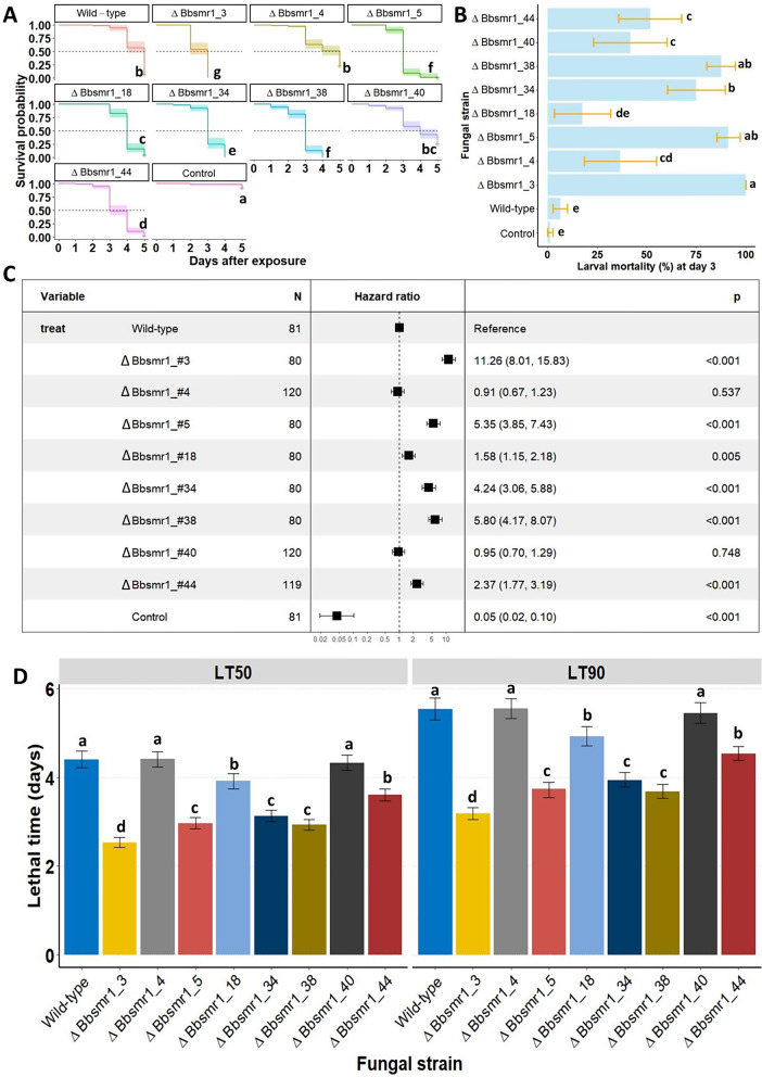
Among eight B. bassiana mutants assayed against the fourth-instar larvae of the greater wax moth (G. mellonella), six out of the eight mutants significantly outperformed the WT strain, indicating a greater virulence attained by these mutants based on their speed of killing the target host (interaction strain-time: χ2 = 32.91, df = 9, P = 0.00013) (Fig. 3A). Mutants ΔBbsmr1_4 and ΔBbsmr1_40 performed similarly to the WT strain in the virulence assay. Independent of the fungal strains, larval mortality significantly increased over time post-exposure (χ2 = 213.45, df = 1, P < 0.0001). When examining survival curves, mutants 3, 5, and 38 induced the fastest decrease in larval survival among all other mutants and WT (χ2 = 662.2, df = 9, P < 0.0001), with a mortality peak found at 3 days post-exposure. Following exposure to fungal treatments at day 3, the larval mortality was over 75% for mutants 3, 5, and 38 and was significantly higher than the other mutants and parental WT (χ2 = 452.13, df = 9, P < 0.0001) (Fig. 3B).
Fig. 3.
Knockout Bbsmr1 mutants of B. bassiana exhibit varied degrees of virulence against the greater wax moth. A) Single-dose survival response of Galleria mellonella larvae following exposure to eight different mutant strains (designed ΔBbsmr1). Inoculum concentration at 1 × 10⁷ blastospores/mL and insects were treated using the direct contact application by exposing larvae for 10 s to 1 mL of blastospore suspension. Natural mortality in controls were attributed to unknown causes with dead larvae appearing all black and putrefied. Survival curves were compared with non-parametric log-rank test and lettering indicates significant differences (P < 0.05). B) Overall mortality confirmed by mycosis of G. mellonella after 3 days post-treatment with blastospores of ΔBbsmr1 and wild-type strains. Means (± SE) not sharing any letter are significantly different by Tukey’s HSD (P < 0.05). C) Vertical bars represent median and 90% lethal times estimated by Weibull parametric model fitted to survival data followed by their respective 95% confidence intervals. Lettering indicates significant differences based on the non-overlapping of confidence intervals. D) Hazard ratio depicting the likelihood of insect death occurring after exposure to B. bassiana mutants in relation to the wild-type. The higher this ratio the greater the risk of death imposed by the mutant strain in comparison to the wild-type. P values are given for all comparisons
According to the hazard ratio test and corroborating the above-mentioned results, ΔBbsmr1 strains 3, 5, 34, and 38 provided the highest ratios (HR > 4) in relation to WT, confirming their virulence in terms of increasing the risk of death to the insect host when exposed to these mutants (Fig. 3C). The less virulent ΔBbsmr1 strains (18 and 44) also exhibited significant hazard ratios (P < 0.05), whereas hazard rations of ΔBbsmr1 strains 4 and 40 were not significantly different in virulence to the insect host (P > 0.05) in relation to WT. Larvae from control groups survived 93% 5 days post-exposure.
Among eight mutants selected, four strains significantly (P < 0.05) reduced the median and 90% lethal times (LT50 and LT90) (Fig. 3D) when compared to WT. Both LT50 and LT90 values were significantly reduced for mutants 3, 5, 34, and 38 in relation to the other mutants and WT. Thus, the lower the lethal times (LT50 and LT90), the more virulent and a faster speed of killing was displayed for the mutant strains. ΔBbsmr1 strains 3, 5, 34, and 38 resulted in 50% insect death within 2.53, 2.97, 3.13, and 2.93 days, while 90% of death occurred within 3.18, 3.73, 3.94, and 3.68 days, corresponding to 43%, 33%, 29%, and 34% faster speed of kill than WT (LC50 = 4.40 days and LC90 = 5.54 days), respectively.
Interestingly, after larval death, cadavers from mutant treatments had a pinkish coloration due to oosporein production by the fungus, and this trend was more pronounced and happened sooner when insects were exposed to the three most highly virulent mutants. Conversely, cadavers from infected larvae with the wild-type strain showed a delayed death coupled with longer time to exhibit the pink color after death (~ 2 days post-death). The few dead larvae found in controls appeared blackened and putrefied, and the cause of death was not related to fungal infection.
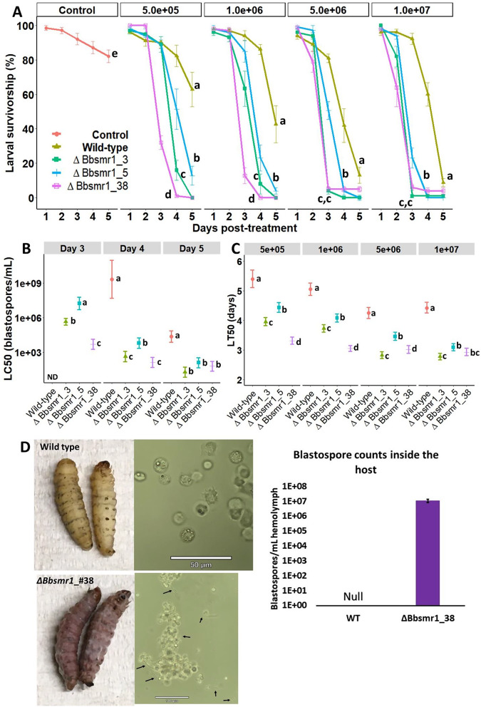
With respect to a multiple-dose-time mortality bioassay, three selected virulent B. bassiana mutants (ΔBbsmr1_3, ΔBbsmr1_5, and ΔBbsmr1_38) outperformed the WT counterpart, indicating a significantly greater virulence attained by these mutants based on their time of killing and inoculum concentration (interaction strain-time-dose: χ2 = 49.6, df = 12, P < 0.0001) (Fig. 4A). Aside from the specific bioactivity of each fungal strain, larval mortality significantly increased with exposure time and inoculum concentration, establishing a time-dose dependent relationship with insect death (interaction time-dose: χ2 = 91.7, df = 4, P < 0.0001) (Fig. S8). All fungal strains (mutants and WT) at all test concentrations differed statistically from the uninoculated control group, and all B. bassiana mutants significantly decreased survival rates by 180-fold of the host insect in comparison to the WT (χ2 = 1004, df = 4, P < 0.0001).
Fig. 4.
B. bassiana mutants display greater and faster killing activity against the greater wax moth compared with the wild-type strain. A) Survival probability of G. mellonella following exposure to crescent concentrations (5 × 10⁵ to 1 × 10⁷ blastospores/mL) of different knockout mutants (ΔBbsmr1) compared with the wild-type strain. Lettering indicates statistical differences based on the log-rank test (P < 0.05) for comparison between survival curves within each fungal concentration. Survival curves with means (± SE) represent data from five replicates (n = 100–140 insects/concentration/strain) derived from two independent bioassays. Lettering represents statistical differences (P < 0.05) based on a log-rank test comparing the Kaplan-Meier survival curves. Natural mortality in controls were attributed to unknown causes with dead larvae appearing all black and putrefied. B) LT50 (± 95% CI) values across different fungal concentrations. ND = not determined due to mortality level did not reach 50%. C) LC50 (± 95% CI) values across different time intervals following exposure to fungal treatments. The LC50 dose for untreated G. mellonella was fixed at zero and reported for all blastospore concentrations for comparison. Lettering of LT50s and LC50s indicates statistical differences based on the non-overlapping of their 95% CIs (P < 0.05). D) Microscopic observation of fungal development and insect immune responses to the WT and mutant strains at different times after cuticle-contact infection. No evidence of aggregation of hemocytes in the hemolymph from wild-type-infected larvae, whereas at within 3 days after exposure to fungal treatments of B. bassiana mutants, blastospores (indicated by arrows) appeared in the hemolymph and induced cellular defense mechanism observed by pronounced hemocyte aggregations to these cells. e) Quantification of free-floating fungal blastospores in insect hemolymph 72 h after cuticle-contact infection
Notably, mutants improved the dose required to kill insects, as they rendered more than a 180 fold and up to 1.5 × 10⁷ fold decrease in LC50 compared to WT across different time intervals (Fig. 4B). Surprisingly, when mortality was assessed 3 days after exposure to fungal treatments, the WT killed less than 10% of insects (LC50 not possible to be determined), whereas the three mutants induced more than 80% mortality, with the lowest LC50 being achieved with ΔBbsmr1_38 requiring as few as 4900 blastospores/mL. The strongest reduction in LC50s by these mutants was attained after 4 days of exposure, which corresponded to only 135–6355 blastospores/mL for mutants in contrast to 2.1 × 10⁹ blastospores/mL estimated for WT, representing a remarkable difference > 330-fold in lethal concentration. The lowest LC50s were observed at 5 days post-exposure to the mutants, as they required a dose of 183 to 1213-fold lower than the parental WT to kill 50% of insects. Overall, mutants ΔBbsmr1_3 and ΔBbsmr1_38 had outstanding performance in yielding both the lowest LT50s and LC50s. Interestingly, blastospores were present in the hemolymph of the insect as soon as 3 days post-infection with B. bassiana mutants, whereas in WT-infected larvae blastospores were not observed at this time point.
Based on speed of kill, all three mutants had an improved LT50 with a reduction in the range of 18–39% relative to WT, after exposing insects to blastospore loads from 5 × 10⁵ to 1 × 10⁷ blastospores/mL (Fig. 4C). The lowest LT50s were attained by the highest inoculum concentration tested, resulting in 2.8 to 3.1 days for mutants to kill 50% of the insect population in contrast to 4.4 days required for WT. Remarkably, all three mutants induced a faster mortality than WT with the lowest blastospore concentration (5 × 10⁵ blastospores/mL), in which the strongest reduction in LT50 was achieved with mutant ΔBbsmr1_38 inciting 50% mortality in only 3.3 days.
Together with the lethal time data, it is evident that blastospores from these mutants displayed a faster infection rate by breaching the insect cuticle and forming blastospores in the hemolymph earlier than WT. The density of free-living blastospores in the hemolymph of the host recorded at 3 days post-inoculation via cuticle infection reached 1.13 × 10⁷ blastospores/mL (SE ± 0.29, n = 6 larvae) of hemolymph (retrieved from infected larvae with ΔBbsmr1_38), whereas blastospores were absent in the hemolymph from WT-infected larvae within this same time interval. This fast infection rate was followed by sooner host death depicting a conspicuous pinkish color in cadavers due to earlier oosporein production compared to the yellowish aspect of recently dead larvae by WT (Fig. 4D).
According to photomicrographs taken with an epifluorescent microscope, it was possible to visualize the germination of blastospores on the G. mellonella larva cuticle at 6 h post-application by a natural cuticle infection route (Fig. S9). Blastospores from the ΔBbsmr1_38 mutant displayed faster germination and longer hyphal extensions on the insect cuticle surface than blastospores from the parental WT isolate, indicating that the higher virulence attained by this mutant could also be related to its accelerated germination speed on the insect host.
Relative expression of key genes in oosporein biosynthetis
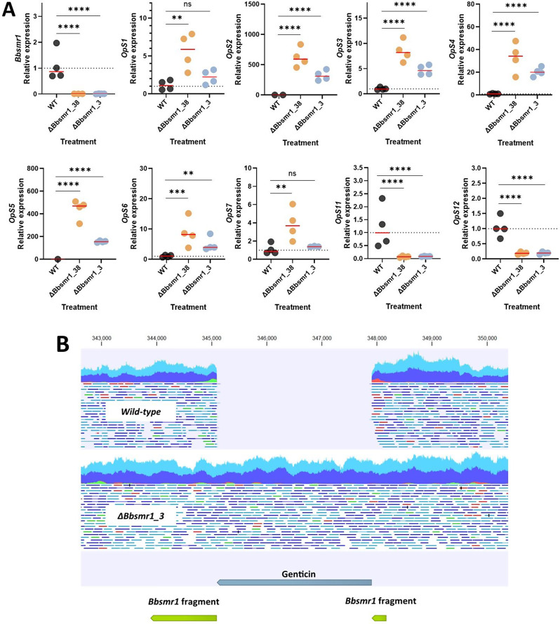
To confirm the knockdown of Bbsmr1 after CRISPR-Cas9 gene-editing and its effects on the oosporein biosynthetic gene cluster, the gene expression analysis was carried out for Bbsmr1 and 9 OpS genes (OpS1-OpS7, OpS11, OpS12) of this cluster from in vitro cultures of WT and two mutant strains grown in PDB with initial pH set to 8.0. Our RT-qPCR data indicated that Bbsmr1 was transcriptionally silent (null expression) in defective Bbsmr1 mutants compared with the parental counterpart (one-way ANOVA: F = 167.1, df = 2, 7, P < 0.0001; Fig. 5A). The disruption of the Bbsmr1 transcription factor led to derepression of ORFs corresponding to OpS1-OpS7 and significant decreased expression of two ORFs located on the 5`-flanking side of OpS1, designated OpS11 and OpS12 (F = 38.72, df = 2, 9, P < 0.0001 and F = 53.36, df = 2, 9, P < 0.0001, respectively; Fig. 5A). Of these, OpS2 and OpS5 exhibited the strongest upregulation with > 100 fold-change (F = 469.7, df = 2, 7, P < 0.0001 and F = 587.9, df = 2, 6, P < 0.0001, respectively), whilst little or no change in expression was seen in the parental WT. This data confirms successful Bbsmr1 gene disruption by the CRISPR-Cas9-RNP system while oosporein production correlated with OpS1-OpS7 expression in the mutants.
Fig. 5.
Oosporein biosynthetic gene cluster regulation in B. bassiana mutants with disrupted Bbsmr1 and off-target detection by whole genome sequencing analysis. A) Fold change in gene expression of Bbsmr1, OpS1-7, OpS11 and OpS12 genes in the ΔBbsmr1 mutants normalized to wild-type (WT) expression levels. Asterisks indicate significant differences between the WT and the mutants (P < 0.05). B) Read mapping of WT and in the Bbsmr1 mutant sequencing reads to the genome of ΔBbsmr1_3, confirming the disruption and location of the insertions
Evaluating potential off-targets effects and the selectivity of the insertion
Whole genome sequencing was used to evaluate the precise integration of the donor DNA and potential off-target effects. Two mutants (ΔBbsmr1_3 and ΔBbsmr1_38) and the parental WT underwent whole genome sequencing revealing the insertion occurred at the expected cleavage site of the guide RNA for both mutants (Fig. 5B). Off-target effects were evaluated in two manners, a guide RNA sequence-dependent approach and a guide RNA sequence-independent approach. For the guide RNA sequence-dependent approach, sites in the genome with a sequence identity of greater than 65% to the guide RNA were manually inspected and found no changes were detected. For the guide RNA sequence-independent approach, the mutant reads were mapped to the WT genome and variants called, and no significant off-targets effects were identified using this approach. Each mutant had less than 10 variants (single or multiple nucleotide polymorphisms, insertions or deletions) and they either resulted in synonymous mutations or were in non-coding regions.
In vitro blastospore production by B. bassiana mutants
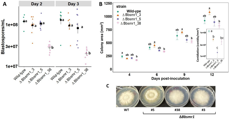
In vitro blastospore production in liquid culture did not increase over time (F = 1.21, df = 1, 51, P = 0.295), indicating that fermentation reached its production peak by day 2 of growth. Nonetheless, blastospore production varied among the WT and mutant strains, of which ΔBbsmr1_38 yielded the lowest blastospore concentration across all time points (F = 36.47, df = 3, 51, P < 0.0001). In comparison to the WT, mutant ΔBbsmr1_38 produced 4.3- and 6.5-fold less blastospores after 2 and 3 days of fermentation, respectively (Fig. 6A). Conversely, mutants ΔBbsmr1_3 and ΔBbsmr1_5 attained similar blastospore yields to the WT.
Fig. 6.
Phenotypic characterization of the mutant strains. A) Blastospore production by knockout B. bassiana mutants (ΔBbsmr1) compared with the wild-type (WT) strain during liquid culture growth at 250 rpm and 28 °C for three days. Lettering of means (± SE) indicates statistical differences based on the Tukey’s test (P < 0.05). Symbols represent observational data in each treatment (n = 8). B. bassiana knockout mutants (ΔBbsmr1) displaying varied growth behavior when grown on potato-dextrose-agar (PDA) without the selectable marker. B) Colony growth kinetics expressed in area (cm²) for three mutants (ΔBbsmr1) and WT, and colony conidiation recorded after 12 days post-inoculation. Lettering of means (± SE, n = 6) within each time interval indicates statistical differences based on the Tukey’s test (P < 0.05). C) Morphology of fungal colonies grown on PDA after 12 days post-inoculation
Colony growth and sporulation by B. bassiana mutants
In general, there was a significant reduction in colony growth between the WT and mutant ΔBbsmr1_38 (F = 10.77, df = 3, 80, P < 0.0001), but the other mutants had similar growth in colony area to the WT. All three mutants produced fewer asexual conidia (spore yield) compared to the WT (F = 25.4, df = 3, 20, P < 0.0001), with a pronounced reduction for mutant ΔBbsmr1_5 (Fig. 6B, C).
Tolerance of B. bassiana mutants to cell chemical stressors
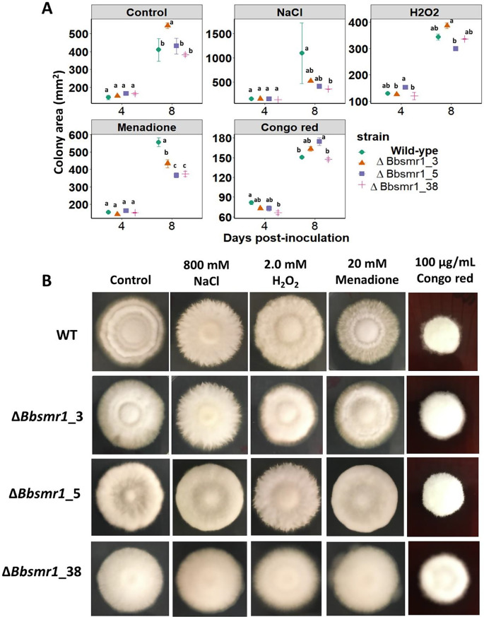
Fungal mutants and WT responded differently when grown in the presence of various cell chemical stressors at different time points (Fig. 7A, B). Greater differences were seen when colonies were measured after 8 days of growth in all cases. Oxidative stress mediated by menadione (F = 17.32, df = 3, 36, P < 0.0001) significantly reduced colony growth of all mutants across all evaluation intervals, while osmotic stress by NaCl (F = 7.28, df = 3, 36, P < 0.0001) only affected the mutant ΔBbsmr1_38 relative to the WT at 8 days post-inoculation. Differences in growth area were attained only 8 days post-inoculation with H2O2 (F = 142.1, df = 1, 36, P < 0.0001) and Congo red (F = 1148.66, df = 1, 36, P < 0.0001). At 4-day post-inoculation, there was reduced colony growth for mutant ΔBbsmr1_38 in relation to the WT when challenged with the cell wall inhibitor Congo red. All mutants exhibited similar growth area to the WT when challenged with oxidative stress caused by H2O2 across all time intervals (F = 1.64, df = 3, 36, P = 0.09). Interestingly, the ΔBbsmr1_3 mutant grew better than the other fungal mutants and WT in PDA without any chemical stressor (control plates) at 8 days post-inoculation (F = 3.87, df = 3, 37, P = 0.017).
Fig. 7.
Multi-stress tolerance displayed by B. bassiana mutants and wild-type (WT) are variable when grown in the presence of various chemical stressors. A) Comparative multi-stress response among B. bassiana mutants and WT strain challenged with osmotic (0.8 M NaCl), oxidative (2.0 M H2O2 and 0.02 M Menadione), and cell wall (100 µg/mL Congo red) stressor agents compared to control cultures grown only in PDA. Inoculum of 5 µL from a fungal suspensions containing 5 × 10⁷ blastospores/mL was added to the center of the agar media and evaluation was carried out across time measuring the area of colonies. Lettering of means (± SE, n = 6) indicates statistical differences between strains, within each time interval, based on the Tukey’s test (P < 0.05). B) Morphological aspects of the mutant and WT colonies when grown in the presence of various cell chemical stressors, after 12 days of incubation
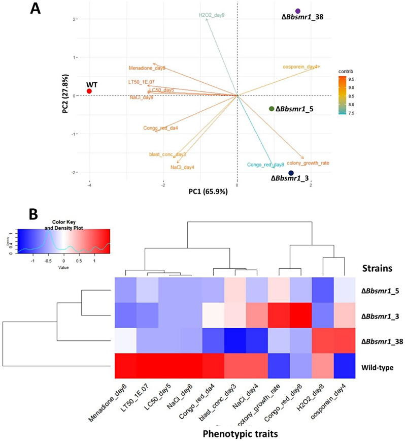
To summarize the results considering the most significant phenotypic variables (pleiotropic effects and virulence parameters), the PCA was highly informative and useful to explain the differences between WT and knockout mutants of B. bassiana (Fig. 8A). Compared to the WT, this multivariate analysis clearly indicated a high correlation of mutants (ΔBbsmr1_3, ΔBbsmr1_5 and ΔBbsmr1_38) with in vitro oosporein overproduction, rapid colony growth rate, high tolerance to cell wall disturbance by Congo red (day 8), and low LT50 and LC50 values attributed to their hypervirulence. However, unlike the WT, these three mutants exhibited less tolerance to certain chemical cell stressors (menadione, NaCl, Congo red day 4) along with lower blastospore production by day 3 of fermentation. According to the heatmap plot (Fig. 8B), the dendrogram assembled for strains indicated that mutants ΔBbsmr1_3 and ΔBbsmr1_5 are more similar to each other than to ΔBbsmr1_38, but all these mutants are highly divergent from the WT based on 11 pleiotropic and virulence variables, previously selected by the PCA.
Fig. 8.
Principal component analysis (PCA) revealing the relationship of pleotropic impacts of growth with virulence traits on B. bassiana mutants and wild-type (WT) strain. A) Biplot consisting of two principal components corresponding to 93.7% of total data variance from 11 meaningful phenotypic variables (arrows with their respective contribution levels) that clearly separate the three B. bassiana mutants (ΔBbsmr1 #3, #5 and #38) from the WT strain (dots colored by strain). B) Heatmap based on Manhatttan distance and Wald method for clustering the variables and strains using dendrograms
Discussion
A precise, efficient, and affordable synthetic CRISPR-Cas9-RNP system was successfully implemented in the broad-insect pathogen B. bassiana and resulted in 93.2% transformation efficiency by disrupting the endogenous transcription factor Bbsmr1, a gene encoding a negative regulator for oosporein biosynthesis. Notably, the resultant mutants displayed phenotypes with overproduction of the red-pigmented metabolite oosporein, increased germination rate on the insect cuticle, and greater cuticle infectivity of blastospores when applied to insects, resulting in enhanced virulence. Interestingly, the hypervirulence of these mutants was also positively correlated with an accelerated colony growth rate and tolerance to cell wall disturbance by Congo red. Whole genome sequencing analysis revealed that this CRISPR-Cas9-RNP transformation system yielded accurate and site-specific gene disruption with no significant off-target mutations identified within the fungal genome. Hence, our CRISPR-Cas9 system based on RNP delivery reduces the risk of off-target mutations and minimizes the risk of altering genes adjacent to the target region. Corroborating the findings from Fan et al. [13], the disruption of the Bbsmr1 transcription factor in the B. bassiana genome was further confirmed through gene expression profiles, including 9 OpS genes. It was shown a strong upregulation of OpS3, which in turn upregulated the rest of the OpS gene cluster, except OpS11 and OpS12. A similar gene disruption transformation protocol via CRISPR-Cas9 with preassembled RNP was previously implemented for the cotton fungal pathogen Fusarium oxysporum with high transformation efficiency, demonstrating that gene disruption of the FoBIK1 encoding a polyketide synthase resulted in the absence of the pigment bikaverin [45]. This study underscores the first evidence of an entomopathogenic fungus modified by CRISPR-Cas9-RNP to develop hypervirulent strains with overproduction of oosporein. These enhanced strains display increased insecticidal activity outperforming the standard parental WT strain, thus offering promising potential for pest biocontrol programs.
After successful gene editing in B. bassiana with CRISPR-Cas9-RNP, it is of utmost importance to investigate potential pleiotropic effects on the fungal biology and development. To this end, we conducted a comprehensive phenotypic characterization of these mutants and compared with the parental WT strain. Mutation of Bbsmr1 resulted in some differences from the WT depending on the mutant strain and evaluation time for colony growth, in vitro blastospore fermentation, conidiation, and abiotic cell stress responses. A prominent reduction in conidiation and increased sensitivity to cellular oxidative stress mediated by menadione were observed in all knockout Bbsmr1, while alteration due to oxidative stress from H2O2 was not observed in these mutants. Blastospore production, osmotic stress due to NaCl, cell wall damage mediated by Congo red were reduced only in a single mutant strain out of the three tested. This is the first report showing a side-effect due to Bbsmr1 gene disruption on reduced blastospore production in vitro, although this feature was not observed in all mutants of B. bassiana. Aligned with our results, defective Bbsmr1 mutants of B. bassiana exhibited reduced mycelial biomass production and conidiation compared with WT, whilst these mutants were more tolerant to oxidative stress induced by H2O2 and cell wall inhibitors like Congo red and fluorescent whitening agent [13, 36]. Another pleiotropic effect was noted for ΔBbsmr1 mutants of B. bassiana, as they exhibited reduced growth in terms of mycelial biomass compared to WT [13]. The asexual conidial production suffered the most detrimental fitness impact due to deletion of Bbsmr1, as this transcription regulator is also involved in the regulation of asexual conidiation and stress response in B. bassiana via the BrlA-AbaA-WetA pathway [36]. This detrimental effect on conidiation could be an obstacle for the mass production of these mutants, although as we showed here it is possible to screen and select mutants with similar conidiation capability to the WT strain.
Making use of the CRISPR-Cas9-RNP transformation strategy, we demonstrate that B. bassiana blastospores are significantly more infective, and kill G. mellonella larvae faster than aerial conidia, corroborating previous data obtained with other insect hosts [24–28]. This cell type formed by entomopathogenic fungi under submerged cultivation or inside the infected host holds a great promise to replace aerial conidia as the active ingredient for the industrial development of commercial fungal biopesticides used in arthropod pest control. Hence, all the virulence bioassays with the ΔBbsmr1 mutants were performed using blastospores as the main inoculum source. Six of the eight ΔBbsmr1 mutants displayed superior virulence in relation to the parental WT, indicating virulence bioassays are important to identify the hypervirulent fungal mutants.
Another important approach is to eliminate the heterokaryon phenotypes by selecting single-spored mutants in order to obtain homokaryons with having a single nucleus carrying the mutation, thus rendering consistent phenotypic results. Among eight mutants screened against G. mellonella larvae, three highly virulent engineered homokaryon strains (e.g., ΔBbsmr1_3, ΔBbsmr1_5, and ΔBbsmr1_38) exhibited strong virulence to G. mellonella larvae, killing these insects more quickly (up to a 43% reduction in LT50,90) and more efficiently lethal, thereby requiring lower inoculum loads (> 2 log-fold reduction in LC50) than the parental counterpart. These mutant strains were capable of killing the entire insect cohort in the experiments in less than 4 days post-inoculation, indicating remarkable virulence. In contrast to our findings, Fan et al. [13] only described the virulence of a single mutant strain of ΔBbsmr1 based on the LT50, and found that this mutant exhibited less pronounced virulence after topically applying 1 × 10⁷ conidia/mL that led to 100% mortality of G. mellonella larvae after 5 days with only ∼20% reduction in LT50 in relation to the WT parent. Although oosporein has been claimed not to be directly involved in virulence of B. bassiana [13, 20], our defective Bbsmr1 mutants depicted hypervirulence with overproduction of oosporein, which could be related to other virulence mechanisms unlocked by this gene mutation. There are several possible explanations for these data. First, we observed that these mutants germinated much faster on the insect cuticle than the WT parent, thus suggesting the involvement of enhanced activity of cuticle-degrading enzymes during the cuticle penetration. Besides that, another assumption arises with the expression of other secondary metabolites and enzymes during the infection stage in the hemolymph where the mutant also formed earlier free-floating in vivo blastospores along with the overproduction of oosporein in infected insects, thus efficiently evading the insect immune system and accelerating the insect mortality. Once inside the host, another mechanism concerns the overproduction of oosporein triggered by the disruption of Bbsmr1, as this metabolite could play a synergistic role with the rapid formation of blastospores in accelerating the host immune evasion, and consequently increasing mortality levels in a faster fashion. In agreement with this observation, the role of oosporein in promoting infection rather than directly killing insects is probably mediated by reducing the number of insect hemocytes, with the consequent alteration of the humoral immune system [37]. Furthermore, a reverse-genetic-based study demonstrated that the null mutant of BbOpS1 (ΔBbOpS1) delayed hemocyte encapsulation by 12 h, thus confirming the link of oosporein with the evasion of insect host immunity [20]. Further transcriptomeic analysis could shed light into the underlying molecular mechanisms of these knockout Bbsmr1 mutants and identify the main metabolic pathways unlocked by this gene disruption.
Interestingly, 2–3 days post-inoculation of G. mellonella larvae during the host colonization stage, ΔBbsmr1 mutants formed numerous blastospores in the host hemolymph, whereas these propagules were absent in the hemolymph from larvae infected with the parental WT. This indicates that B. bassiana mutants were able to propagate as yeast-like budding blastospores in the host hemocoel at an earlier stage of infection than the WT strain, an important trait associated with enhanced virulence [22]. In addition, we performed epifluorescence microscopy that revealed blastospores from deficient Bbsmr1 strains germinating faster on the larva cuticle than blastospores of the parental WT, which concurs with an earlier observation of accelerated in vitro spore germination by ΔBbsmr1 strains [36]. Another phenotypic feature displayed by these mutants regards their ability to turn cadavers into pinkish or reddish sooner after death than cadavers infected with the WT. This observation suggests that earlier host death could be associated with overproduction of oosporein in infected insects with hypervirulent ΔBbsmr1 mutants.
Surprisingly, the application of CRISPR-Cas9 technology in fungal biocontrol agents of agricultural importance is still in its infancy and awaits full development in the near future as new knowledge is gathered about their genomes and gene functions coupled to affordable gene editing technologies. For instance, Chen et al. [30] used uridine auxotrophy and URA5 as a selectable marker with a blastospore-based transformation system to establish a highly efficient, low false-positive, and cost-effective CRISPR-Cas9-mediated gene editing system in B. bassiana. In our study, we made attempts to transform blastospores instead of protoplasts of B. bassiana but were not successful, but instead protoplasts of B. bassiana were successfully transformed through our CRISPR-Cas9-RNP system. Other filamentous entomopathogenic fungi exploited for biocontrol strategies in pest control may be edited using this flexible and effective CRISPR-Cas9-RNP system, and may contribute to generating novel genetically improved strains for the biological control market.
In our study, virulence of the ΔBbsmr1 mutants outperformed the cpf-overexpression strains [15] not only in terms of lower LT50 but also by having a lower lethal dose required to kill the same host insect. Another important attribute regarding fungal virulence is the ability of ΔBbsmr1 mutants to speed up the formation and proliferation of blastospores in the insect hemocoel compared with the parental WT. This facilitated formation and proliferation of hyphal bodies or blastospores in host hemocoel was also noticed by Mou et al. [15], and this trait is strongly linked with the ability of the fungus to colonize the host and induce host mummification and death [22]. Although not investigated here, we suspect that the high virulence attribute to the ΔBbsmr1 mutants may be linked to increased activity of secreted proteases, chitinases, and antioxidant enzymes crucial for the collapse of the insect immune defense, acceleration of hemocoel localization, in vivo proliferation, and probable overexpression of other secondary metabolites during fungal colonization in host hemocoel, leading to faster killing and reduced lethal inoculum loads of the mycopathogen. Our future goals will focus on implementing a marker-free CRISPR system to develop a genetically improved, non-transgenic B. bassiana ΔBbsmr1 mutant, enabling field studies and potential commercialization of enhanced mycoinsecticides in Brazil.
Oosporein is a high value bioactive molecule with important applications in cell therapy to combat proliferation of cancer cells as well as harbouring potential antifungal and antibacterial activities against plant pathogens [18]. Hence, having mutant strains with the capacity to overproduce this biomolecule using inexpensive culture medium after only a few days of fermentation represents a step toward its economically large-scale production. The overproduction of oosporein by B. bassiana mutants has the potential to be used as a natural eco-friendly biofungicide against economically important plant fungal pathogens, such as Fusarium oxysporum f. sp. cubense [58] and Giberella moniliformis [59]. Notably, we identified three deficient Bbsmr1 mutants capable of overproducing oosporein, in the range of 33 to 68 ppm, in only 2 days of fermentation, whereas the WT cultures resulted in no detectable oosporein during in vitro growth. Comparatively, our B. bassiana ΔBbsmr1_38 mutant was considered highly productive for this metabolite reaching up to 118 ppm after 4 days of cultivation, whereas WT strain PQ2 of B. bassiana from another study only reached its production peak after 7 days of cultivation yielding 183 ppm [50]. Genetic improvement for overexpression of fungal secondary metabolites represents an important strategy toward the expansion and diversification of these bioactive compounds for use as agricultural biopesticides. In this sense, CRISPR-Cas9 can contribute to unlocking novel secondary metabolites in entomopathogenic fungi by broadening their biochemical repertoire for exploitation in biological control of agricultural pests [21]. This can be done by precisely editing endogenous genes rather than integrating in the fungus genome exogenous DNA for heterologous expression, which may facilitate the regulatory process of registering non-transgenic improved fungal strains for use in biological control programs tackling plant pests and diseases.
The technology for cost-effective mass production of B. bassiana blastospores via submerged liquid fermentation using large-scale bioreactors has been established and is available for industrial exploitation, offering advantages over traditional solid-substrate fermentation methods for the production of aerial conidia [27, 28, 60, 61]. Therefore, combining the powerful tool based on CRISPR-Cas9 gene editing technology targeting endogenous genes involved in fungal secondary metabolism pathways with the use of fast-acting blastospores as the primary active ingredient entails a promising approach to enhance the efficiency of entomopathogenic fungi as next generation biocontrol agents in agriculture. Additionally, this genetic engineering approach will pave the way for the development of novel commercial fungal strains with desired phenotypic traits meeting key industrial needs in a rapid and cost-effective manner.
Electronic supplementary material
Below is the link to the electronic supplementary material.
Author contributions
GMM and JJC conceptualized the study. GMM and JJC designed the sgRNA to the target gene. GMM developed and optimized transformation methods, established the endogenous promoters, and vectors for transformation, produced and purified the Cas9 through heterologous expression, synthetized in vitro the sgRNA, and engineered the final strains. GMM, MVCBC and SS obtained the homokaryon strains and conducted genotypic and phenotypic characterization experiments. JLR and CAD conducted qPCR experiments and analysis. CAD conducted genome sequencing and bioinformatics analysis, including genome annotation and assembly. All authors revised and approved the final manuscript.
Funding
The first author thanks São Paulo Research Foundation for the scholarship (FAPESP grant 2022/00242-3). This project was funded by EMBRAPA (grant SEG 30.21.90.026.00.02.005 to GMM), and the Alabama Cotton Commission and Cotton Inc. (23-552AL to JJC). This work was supported in part by the U.S. Department of Agriculture, Agricultural Research Service (Project Number: 5010-22410-023-00-D to CAD and JLR). Any opinions, findings, conclusions, or recommendations expressed in this publication are those of the author(s) and do not necessarily reflect the view of the U.S. Department of Agriculture. The mention of firm names or trade products does not imply they are endorsed or recommended by the USDA over other firms or similar products not mentioned. USDA is an equal opportunity provider and employer.
Data availability
Sequencing data that support the findings of this study are available under GenBank Bioproject PRJNA1093450.
Declarations
Competing interests
Holding, or currently applying for, patents relating to the content of the manuscript: G.M.M. and J.J.C. are listed as inventors on a provisional patent that relates to the methods and composition described in the engineering of oosporein metabolism in an entomopathogenic fungus for improved virulence.
Footnotes
Publisher’s note
Springer Nature remains neutral with regard to jurisdictional claims in published maps and institutional affiliations.
References
- 1.Gullino ML, Albales R, Al-Jboory I, Angelotti F, Chakraborty S, Garrett KA, Hurley BP, Juroszek P, Makkouk K, Stephenson T. Scientific review of the impact of climate change on plant pests: a global challenge to prevent and mitigate plant pest risks in agriculture, forestry and ecosystems. Rome: FAO: IPCC,; 2021. p. 72. 10.4060/cb4769en. [Google Scholar]
- 2.Pathak VM, Verma VK, Rawat BS, Kaur B, Babu N, Sharma A, Dewali S, Yadav M, Kumari R, Singh S, Mohapatra A, Pandey V, Rana N, Cunill JM. Current status of pesticide effects on environment, human health and it’s eco-friendly management as bioremediation: a comprehensive review. Front Microbiol. 2022;13:962619. 10.3389/fmicb.2022.962619. [DOI] [PMC free article] [PubMed] [Google Scholar]
- 3.Sabarwal A, Kumar K, Singh RP. Hazardous effects of chemical pesticides on human health-Cancer and other associated disorders. Environ Toxicol Pharmacol. 2018;63:103–14. 10.1016/j.etap.2018.08.018. [DOI] [PubMed] [Google Scholar]
- 4.Glare T, Caradus J, Gelernter W, Jackson T, Keyhani N, Köhl J, Marrone P, Morin L, Stewart A. Have biopesticides come of age? Trends Biotechnol. 2012;30(5):250–8. 10.1016/j.tibtech.2012.01.003. [DOI] [PubMed] [Google Scholar]
- 5.Pimentel D, Perkins JH. Pest Control: Cultural and Environmental aspects. CRC; 2019.
- 6.Whalon ME, Mota-Sanchez D, Hollingworth RM. In: Whalon ME, Mota-Sanchez D, Hollingworth RM, editors. Global Pesticide Resistance in Arthropods. CABI; 2008. pp. 5–31.
- 7.Root Analysis. Agricultural Biologicals Market. 2024. https://www.rootsanalysis.com/reports/agricultural-biologicals-market.html
- 8.Mascarin GM, Lopes RB, Delalibera Junior I, Fernandes EKK, Luz C, Faria M. Current status and perspectives of fungal entomopathogens used for microbial control of arthropod pests in Brazil. J Invertebr Pathol. 2019;165:46–53. 10.1016/j.jip.2018.01.001. [DOI] [PubMed] [Google Scholar]
- 9.Lovett B, St. Leger RJ. Genetically engineering better fungal biopesticides. Pest Manag Sci. 2017;74:781–9. 10.1002/ps.4734. [DOI] [PubMed] [Google Scholar]
- 10.Gibson DM, Donzelli BGG, Krasnoff SB, Keyhani NO. Discovering the secondary metabolite potential encoded within entomopathogenic fungi. Nat Prod Rep. 2014;31:1287–305. 10.1039/C4NP00054D. [DOI] [PubMed] [Google Scholar]
- 11.Pedrini N. The entomopathogenic fungus Beauveria bassiana shows its toxic side within insects: expression of genes encoding secondary metabolites during pathogenesis. J Fungi. 2022;8:488. 10.3390/jof8050488. [DOI] [PMC free article] [PubMed] [Google Scholar]
- 12.Zhang L, Fasoyin OE, Molnár I, Xu Y. Secondary metabolites from hypocrealean entomopathogenic fungi: novel bioactive compounds. Nat Prod Rep. 2020;37:1181–206. 10.1039/C9NP00065H. [DOI] [PMC free article] [PubMed] [Google Scholar]
- 13.Fan Y, Liu X, Keyhani NO, Tang G, Pei Y, Zhang W, et al. Regulatory cascade and biological activity of Beauveria bassiana oosporein that limits bacterial growth after host death. Proc Natl Acad Sci USA. 2017;114(9):E1586. 10.1073/pnas.1616543114. [DOI] [PMC free article] [PubMed] [Google Scholar]
- 14.St. Leger RJ, Wang C. Genetic engineering of fungal biocontrol agents to achieve greater efficacy against insect pests. Appl Microbiol Biotechnol. 2010;85:901–7. 10.1007/s00253-009-2306-z. [DOI] [PubMed] [Google Scholar]
- 15.Mou YN, Ren K, Tong SM, Ying SH, Feng MG. Fungal insecticidal activity elevated by non-risky markerless overexpression of an endogenous cysteine-free protein gene in Beauveria bassiana. Pest Manag Sci. 2022;78(7):3164–72. 10.1002/ps.6946. [DOI] [PubMed] [Google Scholar]
- 16.Ma M, Luo J, Li C, Eleftherianos I, Zhang W, Xu L. A life-and-death struggle: interaction of insects with entomopathogenic fungi across various infection stages. Front Immunol. 2024;14:1329843. 10.3389/fimmu.2023.1329843. [DOI] [PMC free article] [PubMed] [Google Scholar]
- 17.Boucias DG, Zhou Y, Huang S, Keyhani NO. Microbiota in insect fungal pathology. Appl Microbiol Biotechnol. 2018;102(14):5873–88. 10.1007/s00253-018-9089-z. [DOI] [PubMed] [Google Scholar]
- 18.Wang H, Peng H, Li W, Cheng P, Gong M. The toxins of Beauveria bassiana and the strategies to improve their virulence to insects. Front Microbiol. 2021;12:705343. 10.3389/fmicb.2021.705343. [DOI] [PMC free article] [PubMed] [Google Scholar]
- 19.Butt TM, Coates CJ, Dubovskiy IM, Ratcliffe NA. Entomopathogenic fungi: new insights into host-pathogen interactions. Adv Genet. 2016;94:307–64. 10.1016/bs.adgen.2016.01.006. [DOI] [PubMed] [Google Scholar]
- 20.Feng P, Shang Y, Kai C, Wang C. Fungal biosynthesis of the bibenzoquinone oosporein to evade insect immunity. Proc Natl Acad Sci USA. 2015;112:11365–70. 10.1073/pnas.1503200112. [DOI] [PMC free article] [PubMed] [Google Scholar]
- 21.Oliveira LR, Gonçalves AR, Quintela ED, Colognese L, Cortes MVCB, Filippi MCC. Genetic engineering of filamentous fungi: prospects for obtaining fourth-generation biological products. Appl Microbiol. 2024;4:794–810. 10.3390/applmicrobiol4020055. [Google Scholar]
- 22.Zhang AX, Mouhoumed AZ, Tong SM, Ying SH, Feng MG. BrlA and AbaA govern virulence-required dimorphic switch, conidiation, and pathogenicity in a fungal insect pathogen. mSystems. 2019;4(4). 10.1128/mSystems.00140-19. [DOI] [PMC free article] [PubMed]
- 23.Alkhaibari AM, Carolino AT, Bull JC, Samuels RI, Butt TM. Differential pathogenicity of Metarhizium blastospores and conidia against larvae of three mosquito species. J Med Entomol. 2017;54:696–704. 10.1093/jme/tjw223. [DOI] [PubMed] [Google Scholar]
- 24.Bernardo CC, Barreto LP, Silva CSR, Luz C, Arruda W, Fernandes EKK. Conidia and blastospores of Metarhizium spp. and Beauveria bassiana s.l.: their development during the infection process and virulence against the tick Rhipicephalus microplus. Ticks Tick Borne Dis. 2018; 9:1334–1342. 10.1016/j.ttbdis.2018.06.001 [DOI] [PubMed]
- 25.Corrêa B, da Silveira-Duarte V, Silva DM, Mascarin GM, Delalibera I Jr. Comparative analysis of blastospore production and virulence of Beauveria bassiana and Cordyceps fumosorosea against soybean pests. Biocontrol. 2020;65:323–37. 10.1007/s10526-020-09999-6. [Google Scholar]
- 26.Lopes RB, Souza TAD, Mascarin GM, Souza DA, Bettiol W, Souza HR, et al. Akanthomyces diversity in Brazil and their pathogenicity to plant-sucking insects. J Invertebr Pathol. 2023;200:107955. 10.1016/j.jip.2023.107955. [DOI] [PubMed] [Google Scholar]
- 27.Mascarin GM, Jackson MA, Kobori NN, Behle RW, Delalibera Junior I. Liquid culture fermentation for rapid production of desiccation tolerant blastospores of Beauveria bassiana and Isaria fumosorosea strains. J Invertebr Pathol. 2015a;127:11–20. 10.1016/j.jip.2014.12.001. [DOI] [PubMed] [Google Scholar]
- 28.Mascarin GM, Jackson MA, Kobori NN, Behle RW, Dunlap CA, Delalibera Junior I. Glucose concentration alters dissolved oxygen levels in liquid cultures of Beauveria bassiana and affects formation and bioefficacy of blastospores. Appl Microbiol Biotechnol. 2015b;99:6653–65. 10.1007/s00253-015-6620-3. [DOI] [PubMed] [Google Scholar]
- 29.Vega FE, Jackson MA, McGuire MR. Germination of conidia and blastospores of Paecilomyces fumosoroseus on the cuticle of the silverleaf whitefly, Bemisia argentifolii. Mycopathologia. 1999;47:33–5. 10.1023/A:1007011801491. [DOI] [PubMed] [Google Scholar]
- 30.Chen J, Lai Y, Wang L, Zhai S, Zou G, Zhou Z, et al. CRISPR/Cas9-mediated efficient genome editing via blastospore-based transformation in entomopathogenic fungus Beauveria Bassiana. Sci Rep. 2017;8:45763. 10.1038/srep45763. [DOI] [PMC free article] [PubMed] [Google Scholar]
- 31.Davis KA, Sampson JK, Panaccione DG. Genetic reprogramming of the ergot alkaloid pathway of Metarhizium brunneum. Appl Environ Microbiol. 2020;86(19). 10.1128/AEM.01251-20. [DOI] [PMC free article] [PubMed]
- 32.Oliveira TC. Metodologias para a geração de mutantes funcionais em Metarhizium anisopliae: CRISPR/Cas9 e RNAi. Masters Thesis (in Portuguese). UFRGS; 2016. https://lume.ufrgs.br/handle/10183/150695
- 33.Bilgo E, Lovett B, Fang W, Bende N, King GF, Diabate A, et al. Improved efficacy of an arthropod toxin expressing fungus against insecticide-resistant malaria-vector mosquitoes. Sci Rep. 2017;7:3433. 10.1038/s41598-017-03399-0. [DOI] [PMC free article] [PubMed] [Google Scholar]
- 34.Zeng S, Lin Z, Yu X, Zhang J, Zou Z. Expressing parasitoid venom protein VRF1 in an entomopathogen Beauveria bassiana enhances virulence toward cotton bollworm Helicoverpa armigera. Appl Environ Microbiol. 2023;89. 10.1128/aem.00705-23. [DOI] [PMC free article] [PubMed]
- 35.Cortes MVCB, Barbosa ET, Oliveira MIS, Maciel LHR, Lobo-Junior M, Contesini FJ, et al. Trichoderma harzianum marker-free strain construction based on efficient CRISPR/Cas9 recyclable system: A helpful tool for the study of biological control agents. Biol Control. 2023;184:105281. 10.1016/j.biocontrol.2023.105281.
- 36.Chen JF, Liu Y, Tang GR, Jin D, Chen X, Pei Y, et al. The secondary metabolite regulator, BbSmr1, is a central regulator of conidiation via the BrlA-AbaA-WetA pathway in Beauveria bassiana. Environ Microbiol. 2021;23(2):810–25. 10.1111/1462-2920.15155. [DOI] [PubMed] [Google Scholar]
- 37.Mc Namara L, Dolan SK, Walsh JMD, Stephens JC, Glare TR, Kavanagh K, et al. Oosporein, an abundant metabolite in Beauveria caledonica, with a feedback induction mechanism and a role in insect virulence. Fungal Biol. 2019;123:601–10. 10.1016/j.funbio.2019.01.004. [DOI] [PubMed] [Google Scholar]
- 38.Qin Y, Ying SH, Chen Y, Shen ZC, Feng MG. Integration of insecticidal protein Vip3Aa1 into Beauveria bassiana enhances fungal virulence to Spodoptera litura larvae by cuticle and per os infection. Appl Environ Microbiol. 2010;76(14):4611–8. 10.1128/AEM.00302-10. [DOI] [PMC free article] [PubMed] [Google Scholar]
- 39.Wang C, St. Leger RJ. A scorpion neurotoxin increases the potency of a fungal insecticide. Nat Biotechnol. 2007;25:1455–6. 10.1038/nbt1357. [DOI] [PubMed] [Google Scholar]
- 40.Xiao G, Ying SH, Zheng P, Wang ZL, Zhang S, Xie XQ, et al. Genomic perspectives on the evolution of fungal entomopathogenicity in Beauveria bassiana. Sci Rep. 2012;2:483. 10.1038/srep00483. [DOI] [PMC free article] [PubMed] [Google Scholar]
- 41.Grigoriev IV, Roman N, Sajeet H, Kuo A, Ohm R, Otillar R, Riley R, Salamov A, Zhao X, Korzeniewski F, Smirnova T, Nordberg H, Dubchak I, Shabalov I. MycoCosm Portal: gearing up for 1000 fungal genomes. Nucleic Acids Res. 2014;42–704. 10.1093/nar/gkt1183. [DOI] [PMC free article] [PubMed]
- 42.Stemmer M, Thumberger T, del Sol Keyer M, Wittbrodt J, Mateo JL. CCTop: an intuitive, flexible and reliable CRISPR/Cas9 target prediction tool. PLoS ONE. 2015;10(4). 10.1371/journal.pone.0124633. [DOI] [PMC free article] [PubMed]
- 43.Sievers F, Wilm A, Dineen D, Gibson TJ, Karplus K, Li W, Lopez R, McWilliam H, Remmert M, Söding J, Thompson JD, Higgins DG. Fast, scalable generation of high-quality protein multiple sequence alignments using Clustal Omega. Mol Syst Biol. 2011;7:539. 10.1038/msb.2011.75. [DOI] [PMC free article] [PubMed] [Google Scholar]
- 44.Sambrook J, Russell DW. Molecular cloning: a Laboratory Manual. Cold Spring Harbor, NY: Cold Spring Harbor Laboratory Press; 2001. [Google Scholar]
- 45.Wang Q, Cobine PA, Coleman JJ. Efficient genome editing in Fusarium oxysporum based on CRISPR/Cas9 ribonucleoprotein complexes. Fungal Genet Biol. 2018;117:21–9. 10.1016/j.fgb.2018.05.003. [DOI] [PMC free article] [PubMed] [Google Scholar]
- 46.Pokhrel A, Seo S, Wang Q, Coleman JJ. Targeted gene disruption via CRISPR/Cas9 ribonucleoprotein complexes in Fusarium oxysporum. In: Coleman JJ, editor. Fusarium Wilt: Methods and Protocols. Methods in Molecular Biology, vol. 2391. 2021. 10.1007/978-1-0716-1795-3_7 [DOI] [PubMed]
- 47.Cortés-Maldonado L, Marcial-Quino J, Gómez-Manzo S, Fierro F, Tomasini A. A method for the extraction of high quality fungal RNA suitable for RNA-seq. J Microbiol Methods. 2020;170:105855. 10.1016/j.mimet.2020.105855. [DOI] [PubMed] [Google Scholar]
- 48.Livak KJ, Schmittgen TD. Analysis of relative gene expression data using real-time quantitative PCR and the 2(-Delta Delta C(T)) method. Methods. 2001;25:402–8. 10.1006/meth.2001.1262. [DOI] [PubMed] [Google Scholar]
- 49.Watanabe M, Lee K, Goto K, Kumagai S, Sugita-Konishi Y, Hara-Kudo Y. Rapid and effective DNA extraction method with bead grinding for a large amount of fungal DNA. J Food Prot. 2010;73(6):1077–84. 10.4315/0362-028X-73.6.1077. [DOI] [PubMed] [Google Scholar]
- 50.Lara-Juache HR, Ávila-Hernández JG, Rodríguez-Durán LV, Michel MR, Wong-Paz JE, Muñiz-Márquez DB, Veana F, Aguilar-Zárate M, Ascacio-Valdés JA, Aguilar-Zárate P. Characterization of a biofilm bioreactor designed for the single-step production of aerial conidia and oosporein by Beauveria Bassiana PQ2. J Fungi. 2021;7(8):582. 10.3390/jof7080582. [DOI] [PMC free article] [PubMed] [Google Scholar]
- 51.Guo CT, Luo XC, Tong SM, Zhou Y, Ying SH, Feng MG. FluG and FluG-like FlrA coregulate manifold gene sets vital for fungal insect-pathogenic lifestyle but not involved in asexual development. mSystems. 2022;7(4). 10.1128/msystems.00318-22. [DOI] [PMC free article] [PubMed]
- 52.Ritz C, Baty F, Streibig JC. Gerhard. Dose-response analysis using R. PLoS ONE. 2015;10:e0146021. 10.1371/journal.pone.0146021. [DOI] [PMC free article] [PubMed] [Google Scholar]
- 53.Kassambara A, Kosinski M, Biecek P, Fabian S. Package ‘survminer’. Drawing survival curves using ‘ggplot2’ (R package version 0.4.9); 2024. 10.32614/CRAN.package.survminer. https://rpkgs.datanovia.com/survminer/index.html
- 54.Jackson C. Flexsurv: a platform for Parametric Survival modeling in R. J Stat Softw. 2016;70(8):1–33. 10.18637/jss.v070.i08. [DOI] [PMC free article] [PubMed] [Google Scholar]
- 55.Lenth RV, Emmeans. Estimated marginal means, aka least-squares means [Computer software manual], 2020. Available online: http://cran.project.org/package=emmeans (R package version 1.4.1).
- 56.Kassambara A, Mundt F, Factoextra. Extract and Visualize the Results of Multivariate Data Analyses. R Package Version 1.0.7; 2020. https://CRAN.R-project.org/package=factoextra
- 57.Warnes GR, Bolker B, Bonebakker L, Gentleman R, Huber W, Liaw A, et al. Gplots: various R programming tools for plotting data. R Package Version 2. 2009;4:1. [Google Scholar]
- 58.Mascarin GM, Marinho-Prado JS, Assalin MR, Martins LG, Braga ES, Tasic L, et al. Natural occurrence of Beauveria caledonica, pathogenicity to Cosmopolites sordidus, and antifungal activity against Fusarium oxysporum f. sp. cubense. Pest Manag Sci. 2022;78(11):4458–70. 10.1002/ps.7063. [DOI] [PubMed] [Google Scholar]
- 59.Ávila-Hernández JG, Aguilar-Zárate P, Carrillo-Inungaray ML, Michel MR, Wong-Paz JE, Muñiz-Márquez DB, et al. The secondary metabolites from Beauveria bassiana PQ2 inhibit the growth and spore germination of Gibberella moniliformis LIA. Braz J Microbiol. 2022;53(1):143–52. 10.1007/s42770-021-00668-z. [DOI] [PMC free article] [PubMed] [Google Scholar]
- 60.Mascarin GM, Jackson MA. Stable fungal blastospores and methods for their production, stabilization, and use. WO2016044091A1; 2016.
- 61.Mascarin GM, Jackson MA, Behle RW, Kobori NN, Delalibera Junior I. Improved shelf life of dried Beauveria bassiana blastospores using convective drying and active packaging processes. Appl Microbiol Biotechnol. 2016;100(19):8359–70. 10.1007/s00253-016-7597-2. [DOI] [PubMed] [Google Scholar]
Associated Data
This section collects any data citations, data availability statements, or supplementary materials included in this article.
Supplementary Materials
Data Availability Statement
Sequencing data that support the findings of this study are available under GenBank Bioproject PRJNA1093450.


