

Abstract
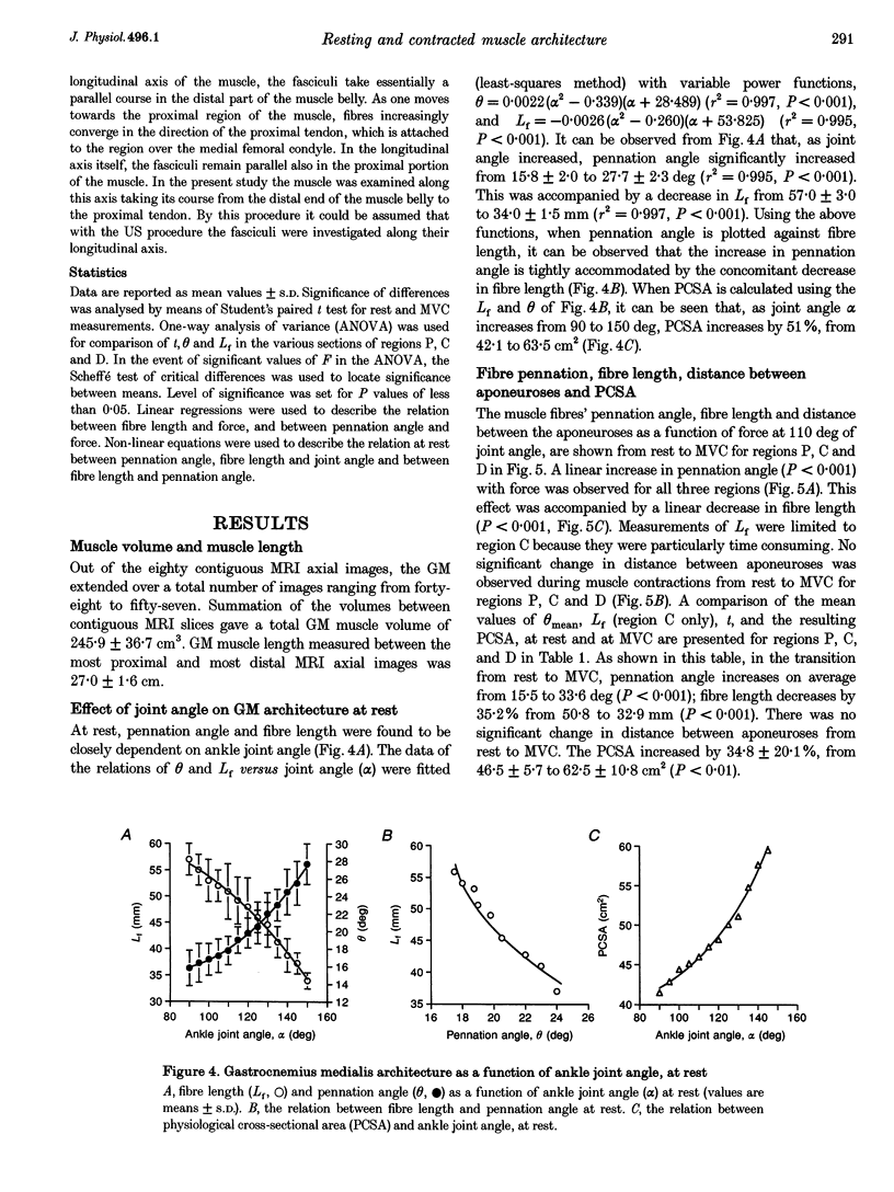
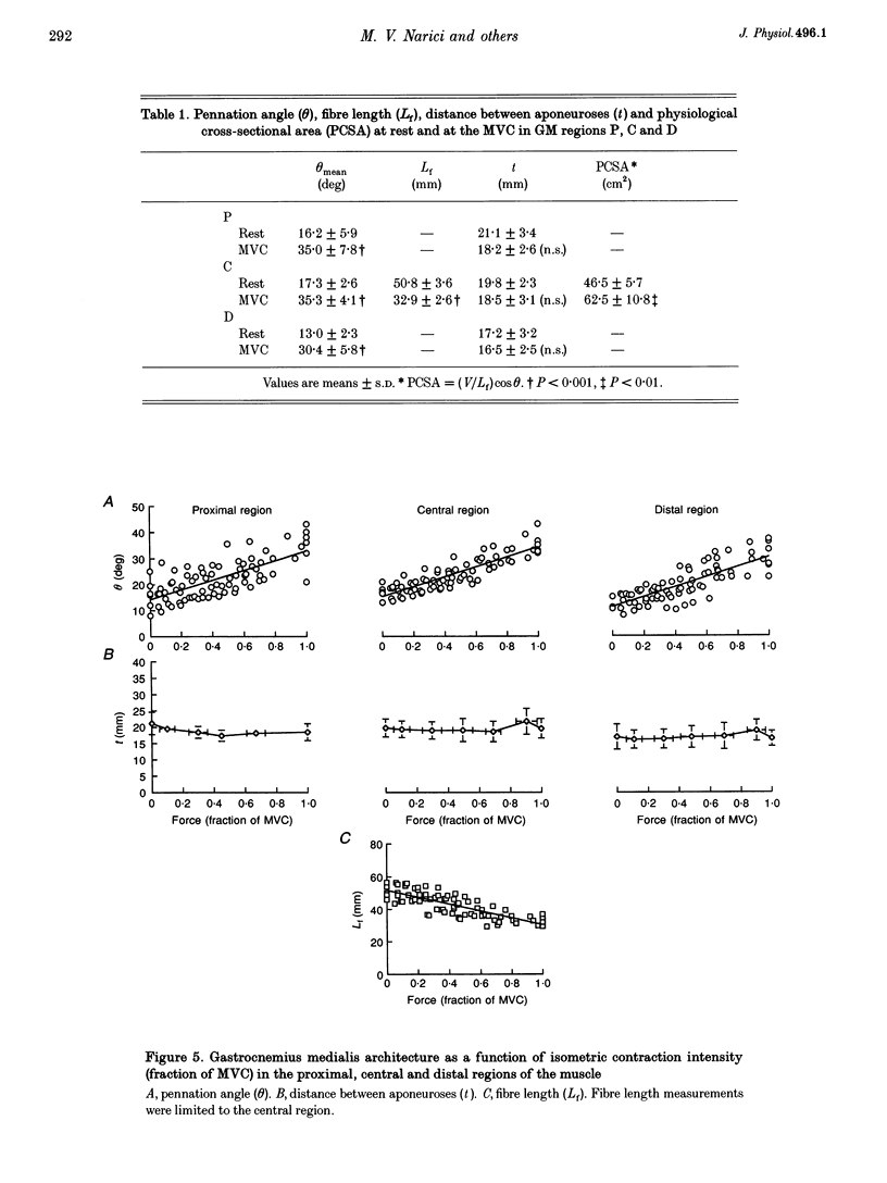
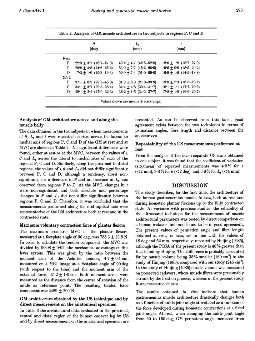
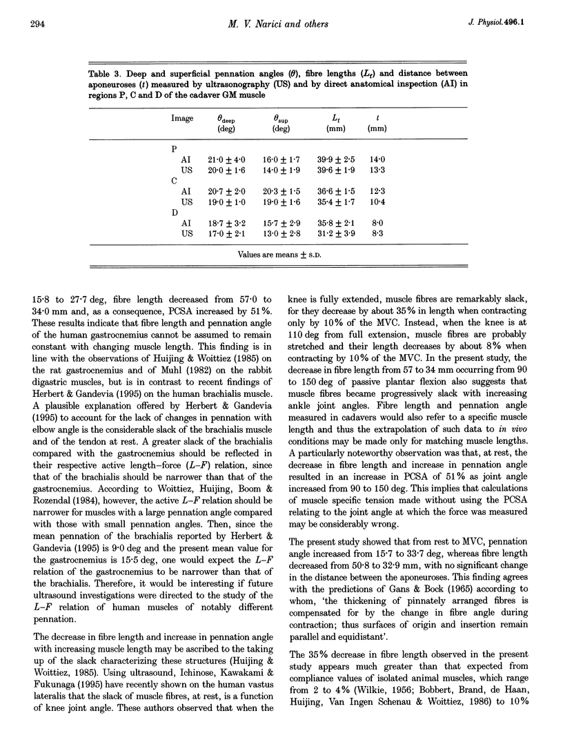
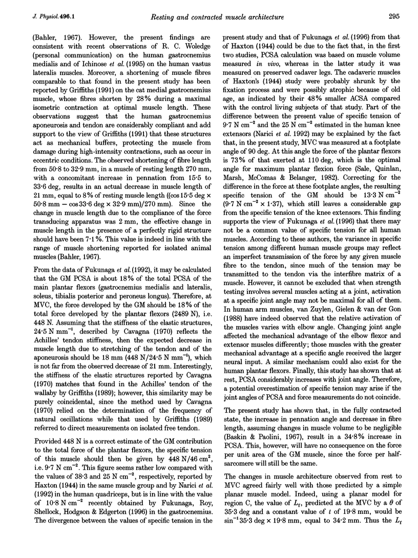
1. Human gastrocnemius medialis architecture was analysed in vivo, by ultrasonography, as a function of joint angle at rest and during voluntary isometric contractions up to the maximum force (MCV). maximum force (MVC). 2. At rest, as ankle joint angle increased from 90 to 150 deg, pennation increased from 15.8 to 27.7 deg, fibre length decreased from 57.0 to 34.0 mm and the physiological cross-sectional area (PCSA) increased from 42.1 to 63.5 cm2. 3. From rest to MVC, at a fixed ankle joint angle of 110 deg, pennation angle increased from 15.5 to 33.6 deg and fibre length decreased from 50.8 to 32.9 mm, with no significant change in the distance between the aponeuroses. As a result of these changes the PCSA increased by 34.8%. 4. Measurements of pennation angle, fibre length and distance between the aponeuroses of the gastrocnemius medialis were also performed by ultrasound on a cadaver leg and found to be in good agreement with direct anatomical measurements. 5. It is concluded that human gastrocnemius medialis architecture is significantly affected both by changes of joint angle at rest and by isometric contraction intensity. The remarkable shortening observed during isometric contraction suggests that, at rest, the gastrocnemius muscle and tendon are considerably slack. The extrapolation of muscle architectural data obtained from cadavers to in vivo conditions should be made only for matching muscle lengths.
Full text
PDF
Images in this article
Selected References
These references are in PubMed. This may not be the complete list of references from this article.
- Bahler A. S. Series elastic component of mammalian skeletal muscle. Am J Physiol. 1967 Dec;213(6):1560–1564. doi: 10.1152/ajplegacy.1967.213.6.1560. [DOI] [PubMed] [Google Scholar]
- Baskin R. J., Paolini P. J. Volume change and pressure development in muscle during contraction. Am J Physiol. 1967 Oct;213(4):1025–1030. doi: 10.1152/ajplegacy.1967.213.4.1025. [DOI] [PubMed] [Google Scholar]
- Cavagna G. A. Elastic bounce of the body. J Appl Physiol. 1970 Sep;29(3):279–282. doi: 10.1152/jappl.1970.29.3.279. [DOI] [PubMed] [Google Scholar]
- Fukashiro S., Itoh M., Ichinose Y., Kawakami Y., Fukunaga T. Ultrasonography gives directly but noninvasively elastic characteristic of human tendon in vivo. Eur J Appl Physiol Occup Physiol. 1995;71(6):555–557. doi: 10.1007/BF00238560. [DOI] [PubMed] [Google Scholar]
- Fukunaga T., Roy R. R., Shellock F. G., Hodgson J. A., Day M. K., Lee P. L., Kwong-Fu H., Edgerton V. R. Physiological cross-sectional area of human leg muscles based on magnetic resonance imaging. J Orthop Res. 1992 Nov;10(6):928–934. doi: 10.1002/jor.1100100623. [DOI] [PubMed] [Google Scholar]
- Fukunaga T., Roy R. R., Shellock F. G., Hodgson J. A., Edgerton V. R. Specific tension of human plantar flexors and dorsiflexors. J Appl Physiol (1985) 1996 Jan;80(1):158–165. doi: 10.1152/jappl.1996.80.1.158. [DOI] [PubMed] [Google Scholar]
- Gans C., Bock W. J. The functional significance of muscle architecture--a theoretical analysis. Ergeb Anat Entwicklungsgesch. 1965;38:115–142. [PubMed] [Google Scholar]
- Griffiths R. I. Shortening of muscle fibres during stretch of the active cat medial gastrocnemius muscle: the role of tendon compliance. J Physiol. 1991 May;436:219–236. doi: 10.1113/jphysiol.1991.sp018547. [DOI] [PMC free article] [PubMed] [Google Scholar]
- Haxton H. A. Absolute muscle force in the ankle flexors of man. J Physiol. 1944 Dec 15;103(3):267–273. doi: 10.1113/jphysiol.1944.sp004075. [DOI] [PMC free article] [PubMed] [Google Scholar]
- Henriksson-Larsén K., Wretling M. L., Lorentzon R., Oberg L. Do muscle fibre size and fibre angulation correlate in pennated human muscles? Eur J Appl Physiol Occup Physiol. 1992;64(1):68–72. doi: 10.1007/BF00376443. [DOI] [PubMed] [Google Scholar]
- Herbert R. D., Gandevia S. C. Changes in pennation with joint angle and muscle torque: in vivo measurements in human brachialis muscle. J Physiol. 1995 Apr 15;484(Pt 2):523–532. doi: 10.1113/jphysiol.1995.sp020683. [DOI] [PMC free article] [PubMed] [Google Scholar]
- Huijing P. A. Architecture of the human gastrocnemius muscle and some functional consequences. Acta Anat (Basel) 1985;123(2):101–107. doi: 10.1159/000146047. [DOI] [PubMed] [Google Scholar]
- Kuno S., Fukunaga T. Measurement of muscle fibre displacement during contraction by real-time ultrasonography in humans. Eur J Appl Physiol Occup Physiol. 1995;70(1):45–48. doi: 10.1007/BF00601807. [DOI] [PubMed] [Google Scholar]
- Muhl Z. F. Active length-tension relation and the effect of muscle pinnation on fiber lengthening. J Morphol. 1982 Sep;173(3):285–292. doi: 10.1002/jmor.1051730305. [DOI] [PubMed] [Google Scholar]
- Narici M. V., Landoni L., Minetti A. E. Assessment of human knee extensor muscles stress from in vivo physiological cross-sectional area and strength measurements. Eur J Appl Physiol Occup Physiol. 1992;65(5):438–444. doi: 10.1007/BF00243511. [DOI] [PubMed] [Google Scholar]
- Otten E. Concepts and models of functional architecture in skeletal muscle. Exerc Sport Sci Rev. 1988;16:89–137. [PubMed] [Google Scholar]
- Rack P. M., Westbury D. R. The effects of length and stimulus rate on tension in the isometric cat soleus muscle. J Physiol. 1969 Oct;204(2):443–460. doi: 10.1113/jphysiol.1969.sp008923. [DOI] [PMC free article] [PubMed] [Google Scholar]
- Rutherford O. M., Jones D. A. Measurement of fibre pennation using ultrasound in the human quadriceps in vivo. Eur J Appl Physiol Occup Physiol. 1992;65(5):433–437. doi: 10.1007/BF00243510. [DOI] [PubMed] [Google Scholar]
- Sale D., Quinlan J., Marsh E., McComas A. J., Belanger A. Y. Influence of joint position on ankle plantarflexion in humans. J Appl Physiol Respir Environ Exerc Physiol. 1982 Jun;52(6):1636–1642. doi: 10.1152/jappl.1982.52.6.1636. [DOI] [PubMed] [Google Scholar]
- Van Leeuwen J. L., Spoor C. W. Modelling mechanically stable muscle architectures. Philos Trans R Soc Lond B Biol Sci. 1992 May 29;336(1277):275–292. doi: 10.1098/rstb.1992.0061. [DOI] [PubMed] [Google Scholar]
- WILKIE D. R. Measurement of the series elastic component at various times during a single muscle twitch. J Physiol. 1956 Dec 28;134(3):527–530. doi: 10.1113/jphysiol.1956.sp005662. [DOI] [PMC free article] [PubMed] [Google Scholar]
- Woittiez R. D., Huijing P. A., Boom H. B., Rozendal R. H. A three-dimensional muscle model: a quantified relation between form and function of skeletal muscles. J Morphol. 1984 Oct;182(1):95–113. doi: 10.1002/jmor.1051820107. [DOI] [PubMed] [Google Scholar]
- van Zuylen E. J., Gielen C. C., Denier van der Gon J. J. Coordination and inhomogeneous activation of human arm muscles during isometric torques. J Neurophysiol. 1988 Nov;60(5):1523–1548. doi: 10.1152/jn.1988.60.5.1523. [DOI] [PubMed] [Google Scholar]