



Abstract
Empathy is a critical component of social interaction that enables individuals to understand and share the emotions of others. We report a preregistered experiment in which 240 participants, including adolescents, young adults, and older adults, viewed images depicting hands and feet in physically or socially painful situations (versus nonpainful). Empathy was measured using imagined pain ratings and EEG mu suppression. Imagined pain was greater for physical versus social pain, with young adults showing particular sensitivity to social pain events compared to adolescents and older adults. Mu desynchronization was greater to pain versus no-pain situations, but the physical/social context did not modulate pain responses. Brain responses to painful situations increased linearly from adolescence to young and older adulthood. These findings highlight shared activity across the core empathy network for both physical and social pain contexts, and an empathic response that develops over the lifespan with accumulating social experience.
Keywords: empathy, aging, physical and social pain, EEG, sensorimotor mirror system
Empathy refers to the ability to share and understand others’ emotional states, thoughts, and feelings (Davis 1980), and therefore plays a crucial role in human social behaviour (Ferguson and Wimmer 2023). Empathy can be divided into affective and cognitive branches. Affective empathy refers to the ability to share another’s emotional state, including feeling compassion for others and experiencing personal distress (i.e. self-focused emotional responding; Beadle and de la Vega 2019, Bailey et al. 2020), while cognitive empathy involves an evaluation and understanding of another’s experience [thus implicating perspective-taking and Theory of Mind (ToM) processes]. Both affective and cognitive empathy enable accurate responding to social situations (Frith and Frith 2005, Stietz et al. 2019). While a great deal of research has been conducted to examine empathic responses among healthy young adults, how they develop in infancy, or how they are disrupted in clinical disorders, the extent to which empathy responses and neural signatures change over the healthy lifespan remains relatively unexplored. In this paper, we examine how the brain’s responses to others experiencing pain (i.e. affective empathy) develops from adolescence through to young and older adulthood.
Empathizing with others in pain
Neuroscientific research has identified a neural network in the sensorimotor cortex, commonly known as the mirror neuron system (Di Pellegrino et al. 1992, Gallese et al. 1996), that is especially active when humans are understanding or imitating others’ actions or empathizing with others in pain (Jackson et al. 2005, Schulte-Rüther et al. 2007, Cattaneo and Rizzolatti 2009, Ferrari and Rizzolatti 2014, Lamm and Majdandžić 2015, Woodruff 2018, Arnett et al. 2019). It has been suggested that the mirror neuron system underlies empathy because seeing or imagining another person in pain activates our own experience of a similar situation, and this generates a shared physiological response (Preston and De Waal 2002). Indeed, brain imaging research has revealed overlapping brain activation patterns over the anterior insula (AI) and anterior cingulate cortex (ACC) when people observe others experiencing physical pain and when they experience physical pain themselves (Singer et al. 2004, Adolphs 2009, Craig 2009, Lamm et al. 2010).
Electroencephalography (EEG) provides a reliable means of measuring neural changes underlying empathy (Pineda 2005, Puzzo et al. 2011, Fox et al. 2016, Woodruff 2018). The mu rhythm—which has been proposed to reflect activity of the mirror neuron system (Pineda 2005)—is elicited by the sensorimotor and premotor areas of the brain around the central sulcus within the alpha frequency range of 8–13 Hz, and also within the beta frequency range of 13–35 Hz. When the sensorimotor areas become activated, the mu rhythm is suppressed, known as event-related desynchronization (ERD; Pfurtscheller and Neuper 1997). Observing others’ actions or empathizing with them, particularly empathy for pain or unpleasant events, creates motor resonance in the sensorimotor and premotor areas of the brain, which causes suppression of the mu rhythm (Jackson et al. 2005, Lepage and Théoret 2006, Yang et al. 2009, Lamm et al. 2011, Woodruff et al. 2011, Chen et al. 2014, Fan et al. 2014, Fabi and Leuthold 2017).
Studies that have examined empathy for others’ pain typically present participants with static images or short video clips depicting hands and feet in physically painful (e.g. a needle piercing skin) and nonpainful (e.g. a cotton bud pressing on skin) situations, and have revealed consistent evidence that mu desynchronization is greater in response to painful versus nonpainful stimuli (Perry et al. 2010, Cheng et al. 2017, Fan et al. 2014, Arnett et al. 2019). Interestingly, the brain’s empathic response to others’ pain is known to be modulated by a range of factors including in-group effects (Hein et al. 2010, Fox et al. 2013, Lübke et al. 2020), the intensity of pain (Lamm et al. 2010), observer gender (Yang et al. 2009), and bodily self-attribution (Riečanský et al. 2020; i.e. how much the actor’s hand is perceived to be the observer’s own).
While the majority of research on empathy has focused on responses to physical pain, a number of studies have shown that similar neural circuits are activated when people experience social pain (Eisenberger 2012a; e.g. rejection, exclusion, embarrassment, death of a loved one). For example, Eisenberger and Lieberman (2004; see also Bolling et al., 2011, Eisenberger et al., 2003) conducted a study in which participants were excluded from a virtual ball-toss game (cyberball) and Krach et al. (2015; see also Kross et al., 2007) recorded fMRI while participants were shown static images of situations depicting social pain. Across these studies, social exclusion and viewing others in social pain led to activation in the dorsal ACC and AI, similar to that seen in response to physical pain (Eisenberger et al. 2003). Moreover, participants who self-reported greater feelings of social pain or who were predisposed to be more sensitive to social pain also showed greater brain activity in these areas. A processing overlap between physical and social pain is further demonstrated by research which has found that behavioural and neural responses to social exclusion are lowered when physical pain thresholds have been pharmaceutically suppressed (DeWall et al. 2010).
Processing of both physical and social pain requires multimodal cognitive processes, including detecting a threat and reacting appropriately in the given context, and both rely on a circuit of brain regions in engaging this processing (e.g. the ‘social brain network’ and the ‘pain network’; Dalgleish et al. 2017; Iannetti et al. 2013). Despite the strong evidence for a functional overlap between empathy for physical and social pain, to our knowledge, no studies to date have directly compared the two in the same participants using a matched design and stimuli (but see Flasbeck et al. 2023 for a comparison of ERP responses to viewing physical and psychological interactions). In the current study, we addressed these questions using EEG as a measure of the neural changes underlying empathy since it has been shown to be sensitive to both physical and social pain. For example, Fraser et al. (Fraser et al. 2020) found that mu suppression was increased when children viewed films depicting social injustice/victimization (a form of social pain) relative to neutral film segments. It has also been suggested that the somatosensory cortex is preferentially activated in response to physical pain (Akitsuki and Decety 2009), and that pain ratings are greater when observing others in physical than social pain (Flasbeck et al. 2023). These findings suggest that, while brain regions responding to both social and physical pain overlap, neural and behavioural responses might be greater when empathizing with people in physical than social pain, reflecting adaptation to potential danger present in different contexts, such as perceiving physical pain as posing a more immediate threat than social pain (e.g. Akitsuki and Decety 2009; Kross et al. 2011).
Developing empathy across the lifespan
Developmental studies on empathy have largely relied on behavioural measures and have revealed that the affective component of empathy first emerges in early childhood (from ∼3 years old; Decety 2010, Decety and Michalska 2010, Chen et al. 2014), continues to develop throughout adolescence (from ∼10 years old; Burnett et al. 2009, Levy and Feldman 2017, Kim et al. 2020), then remains stable or increases though adulthood and older age (Sze et al. 2012, Ze et al. 2014, Sun et al. 2018, Beadle and de la Vega 2019, Bailey et al. 2020). However, the majority of these studies have examined developmental changes in a single age group (e.g. adolescence) or compared across just two age groups. To our knowledge, no experimental studies have systematically tested the development of affective empathy across the lifespan (De Lillo and Ferguson 2023, Dorris et al. 2022; e.g. childhood to old age; though such studies have been conducted on cognitive empathy), making it difficult to reliably infer the trajectory of affective empathy as it changes with age or whether the empathic responses to physical and social pain develop along the same lifespan trajectory.
Adolescence is a period of significant social and emotional development, and learning to empathize with others is a crucial aspect of this process. Research has shown that adolescents experience difficulties relative to adults in understanding the emotions of others (Steinberg and Morris 2001, Blakemore 2008, Dumontheil et al. 2010), and show a decline in prosocial behaviour during adolescence (i.e. between 13 and 17 years old) before increasing again into young adulthood and beyond (Carlo et al. 2007, Matsumoto et al. 2016). Changes in empathic responses during adolescence have been attributed in part to ongoing development of the prefrontal cortex (Blakemore and Choudhury 2006), which impacts their understanding of complex emotions, such as those involving moral reasoning or social norms. Adolescents report experiencing more intense and unstable emotions than adults and can be more emotionally reactive (Bailen et al. 2018). Together, these findings suggest that affective empathy follows an extended period of development through adolescence and young adulthood (i.e. that empathy responses increase from adolescence to adulthood), and that adolescents may be especially variable in their empathy responses.
Adulthood is characterized by greater emotional stability and cognitive development, which leads to increased empathic ability. Adults tend to be more skilled at understanding others’ emotions and are better able to regulate their own emotions in response to others (Eisenberg and Fabes 1990), in perhaps due to increasing life experience and a peak socio-cognitive functioning in young adulthood (De Lillo et al. 2021; Bradford et al. 2023, De Lillo and Ferguson 2023). Evidence is more mixed regarding affective empathy development in later life. Some studies suggest that affective empathy remains fairly stable with increasing age (Beadle and de la Vega 2019, Stietz et al. 2019). Older adults report higher state emotional empathy (Richter and Kunzmann 2011) and enhanced facial mimicry (Bailey et al. 2020) compared to younger adults when viewing empathy-eliciting film clips. In contrast, neuroimaging studies have shown that older adults consistently elicit a reduced or even absent neural response to others in pain compared with young adults (Chen et al. 2014, Guay et al. 2018, Riva et al. 2018), which suggests that the neural mechanisms that underlie affective empathy may decline in older age in parallel with declines in cognitive empathy (ToM; Bailey and Henry 2008, Bernstein et al. 2011, Henry et al. 2013, De Lillo et al. 2021, Bradford et al. 2023, De Lillo and Ferguson 2023). It remains unclear how these age-related reductions in empathic neural activity are associated with the relatively intact behavioural responses and enhanced prosocial behaviours seen in older adults (Charles and Carstensen 2010, Lockwood et al. 2021).
The current study
In this paper, we aimed to examine how empathic responses to other people in physical and social pain changes across a wide age range that spanned adolescence (10–19 years), young adulthood (20–40 years), and older adulthood (≥60 years). We adapted a paradigm that has been commonly used to investigate empathy for pain, in which participants viewed photographs of hands and feet in physically or socially painful (e.g. a needle piercing skin or a hand resting on a coffin) and nonpainful (e.g. a cotton bud pressing on skin or a hand resting on a table) situations. Empathy was measured using behavioural ratings of imagined pain and EEG measures of mu suppression (alpha and low beta ranges), and the effect of age was modelled as a continuous variable.
Replicating the basic effects seen in previous studies (Jackson et al. 2005, Cheng et al. 2008, Perry et al. 2010), we predicted that behavioural pain ratings and mu desynchronization would reflect the stimuli’s affective content, with higher ratings and greater mu desynchronization for pictures that show painful situations compared to no-pain situations. Moreover, based on recent research that has examined neural responses to vicarious social pain (Krach et al. 2011, 2015, Fraser et al. 2020, Flasbeck et al. 2023), we predicted that this pain versus no-pain difference would be elicited by both physical and social content, but expected to see greater pain ratings for physical than social pain, and distinct neural responses between the two (i.e. a greater pain difference and increased sensorimotor desynchronization for physical versus social content, and possibly more widespread neural responses for social pain reflecting a broader circuit of brain networks for these complex emotions). Finally, in line with evidence for a peak in social cognitive and empathizing ability in young adulthood (Decety and Michalska 2010, Chen et al. 2014, De Lillo and Ferguson 2023), we predicted that responses to pain (i.e. greater differences in pain versus no-pain ratings and greater mu desynchronization in response to painful stimuli) would be greatest during young adulthood compared to adolescence and older adulthood.
Methods
All methodological procedures were preregistered on the Open Science Framework (OSF) web pages (https://osf.io/guf6k).
Participants
A total of 273 participants, aged between 10 and 19 years (adolescents), 20 and 40 years (younger adults), and 60 and 80 years old (older adults) were recruited for this study. Middle-aged adults (aged 41–59 years) were not collected in this study due to time-restrictions for the project, and the focus of our research questions on developmental changes between adolescence and young adults, and young to older adulthood (informed by prior research indicating key time periods for change, e.g. Bradford et al. 2023; De Lillo and Ferguson 2023). Participants were eligible for inclusion if they were in the relevant age range, had normal or corrected to normal vision, were native English speakers, had no learning disabilities, no current mental health diagnoses, no diagnoses of autism, epilepsy or dementia, and had no history of stroke. Of the total sample, 33 participants were excluded according to preregistered criteria: nine were excluded for having MoCA scores below 23 (Carson et al. 2018), 22 were excluded due to excessive noise on the EEG recordings or too few segments for the EEG analysis (>25% data loss), and two were excluded due to computer failure. This resulted in a final sample of 240 participants. Table 1 presents participant characteristics in three age groups, though data was analysed with age as a continuous variable: 74 adolescents, 83 younger adults, and 83 older adults. We note that our preregistration planned to include N = 80 in each age range, however, we were unable to meet the target in the adolescent group due to lab closures during the COVID-19 pandemic. The total planned sample size was therefore achieved by including an additional three participants in each of the young and older adult age groups. Participants completed the empathy task as part of a larger task battery and were paid £50 for their time. Participants were recruited from a community sample in the local area of Kent, UK, using a variety of recruitment strategies (e.g. newspaper adverts, local groups, word-of-mouth, Kent Child Development Unit). Of the participants who took part, 217 reported their ethnicity: 88% were white, 4% were Asian, 2% were black, 5% were mixed/multiple ethnic groups, and 1% stated ‘other’ (details not provided). Sample size was preregistered based on previous research, and time constraints to complete a PhD. The Ethical Committee of the School of Psychology, University of Kent, UK, approved the study.
Table 1.
Participant characteristics by age group (mean values, with standard deviations in parenthesis).
| Adolescents | Young Adults | Older Adults | |
|---|---|---|---|
| N | 74 | 83 | 83 |
| Age (years) | 15.1 (2.8) | 27.3 (5.5) | 67.7 (5.1) |
| Gender (F:M) | 44:30 | 55:28 | 55:28 |
| SES index | 9.8 (3.9) | 10.6 (2.8) | 11.1 (2.6) |
| Full scale IQ | 102.8 (10.5) | 101.3 (13.1) | 109.9 (11.2) |
| Verbal IQ | 100.7 (8.9) | 99.2 (9.4) | 107.3 (12.7) |
| Perceptual reasoning IQ | 105.7 (11.8) | 103.4 (11.8) | 110.2 (13.4) |
| MoCA | 28.1 (1.8) | 27.9 (1.7) | 27.2 (1.8) |
SES = Socio-economic status; IQ (assessed using the Wechsler Abbreviated Scale of Intelligence; Wechsler 1999) = Intelligence Quotient; MoCA = Montreal Cognitive Assessment (Nasreddine et al. 2005).
Physical and social pain stimuli
The main task was based on the basic design used in Jackson et al. (2005); participants viewed images depicting others in painful and nonpainful situations, while brain activity was measured using EEG. Specifically, we compared the brain’s response to images of hands and feet in physical and social pain, as an indicator of empathy for others. Stimuli were taken from stock images online and photographed events with real actors. Physical pain images depicted pain caused by pressure, thermal, sharp objects, etc., and social pain images depicted situations of embarrassment, grief, misery, etc. (see Table 2). Each pain image was paired with a no-pain image that depicted an equivalent scene (e.g. a hand in a candle flame versus a hand lighting a candle). All images were selected through a pretest (see Supplementary Materials) and edited to the same size (320 × 240 pixels).
Table 2.
Example stimuli used to depict physical and social pain and their corresponding no-pain images.
| Content | |||||
|---|---|---|---|---|---|
| Physical | Social | ||||
| Type | Pain |

|

|

|

|

|

|

|

|
||
| No Pain |

|

|

|

|
|

|

|

|

|
||
In sum, the task employed a mixed design, crossing the between-subjects factor Age Group (adolescents versus young adults versus older adults) with the within-subjects factors Type (Painful versus Nonpainful) and Content (Physical versus Social). Three dependent variables were analysed: explicit pain ratings, mu/alpha (8–13 Hz), and mu/beta (13–35 Hz) suppression.
Procedure
Participants were informed about the EEG procedure and experimental tasks. The empathy for pain task consisted of 160 trials, 40 in each of the four conditions, and EEG activity was recorded throughout. Each image was shown four times over the experiment. As shown in Fig. 1, trials began with a central fixation cross for 500 ms, followed by an image for 3000 ms. On 25% of trials (i.e. once per image), a subsequent screen prompted participants to rate the level of pain that the person in the picture was feeling on a visual analogue scale from 0 (no pain) to 100 (worst possible pain); responses were made using the mouse. A blank screen was presented between trials using a variable inter-stimulus interval between 500 and 1500 ms to prevent expectancy effects on oscillatory rhythm.
Figure 1.
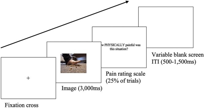
Schematic trial sequence used to present stimuli in the pain rating task. Note that participants were only prompted to rate pain on 25% of trials.
Trials were presented in a randomized order, over four blocks (two showing physical pain/no pain images and two showing social pain/no pain images); each image appeared twice in each of the relevant blocks. Social and physical pain images were presented in separate blocks (the rating question asked either ‘how PHYSICALLY painful was this situation?’ or how SOCIALLY painful was this situation?’, for the relevant the block) in a counterbalanced order. This task lasted 40 min on average, including EEG setup.
EEG recording and analysis
Electroencephalographic (EEG) activity was recorded during the empathy for physical and social pain task from 30 active electrodes using a Brain Vision Quickamp amplifier system, and subsequently processed using Brain Vision Analyzer 2.1. EEG activity containing blinks was corrected using a semi-automatic ocular ICA correction approach (see Supplementary Materials for full details of EEG preprocessing steps).
EEG data were time-locked to the onset of each stimulus image, and data were segmented into a 500 ms baseline period (−500 to 0 ms from stimulus onset) and a 2s pain observation period (500–2,500 ms from stimulus onset), as shown in Fig. 1. Semi-automatic artefact detection software was run, to identify and discard segments with non-ocular artefacts (drifts, channel blockings, EEG activity exceeding ± 50 µV). A fast-fourier transformation, with 10% Hanning window, was then applied to each segment, and the signal was averaged for each condition and electrode.
The average mu/alpha (8–13 Hz) and mu/beta (13–35 Hz) power for each condition was calculated for the electrodes of interest over the central (C3, Cz, C4) and occipital electrodes (O1, Oz, O2). This allowed us to test whether changes in alpha and beta desynchronization were specific to empathy-related influences on sensorimotor processing (i.e. over central sites) and distinct from alpha and beta desynchronization over occipital sites (Perry et al. 2010, Whitmarsh et al. 2011, Hobson and Bishop 2017a, 2017b). A measure of the percentage change in power was calculated for each experimental condition (physical pain, physical no-pain, social pain trials, and social no-pain) relative to the baseline period in that same condition for each electrode of interest in both alpha and beta bands, using the formula: ((baseline-experimental)/baseline)*100. Data from electrodes C3, Cz, and C4 was averaged for the central electrode site, and data from electrodes O1, Oz, and O2 was averaged for the occipital electrode site. Positive values indicate mu/alpha and mu/beta desynchronization, and negative values indicate mu/alpha and mu/beta synchronization.
Results
Analysis procedures were preregistered, (Note that our preregistered analysis plan proposed to use ANOVAs, with age group as a between subjects variable, with three levels (adolescents, young adults, and older adults). However, in line with more recent statistical norms in the field (Baayen et al. 2008), we adapted this plan to use linear mixed models since this allowed us to include random effects for both participants and image, and to apply a maximal random effects structure. We also adapted the model to include age as a continuous predictor since discretizing continuous variables reduces statistical power (cf. Rucker et al. 2015). The preregistered ANOVA analysis is reported in Supplementary Materials for transparency, with statistics for age-group analyses in Tables A and B.) and the full datasets and analysis scripts are available on the OSF web pages (https://osf.io/f9c2r/). Statistical analyses were conducted in R version 4.2.2.
Pain ratings
Pain ratings were analysed using a general linear mixed effects model (since the rating data were found to be non-normally distributed, Shapiro–Wilk test W = 0.86, P < .001), using the glmer function in the lme4 package in R (Bates et al. 2015). The model included the within-subjects variables Type (pain versus no-pain) and Content (physical versus social) as fixed effects (using deviation contrast coding), random effects for participant and image, and random slopes for Type and Content on the participant random effect. Age was entered into the model as a continuous predictor variable, including both linear and quadratic terms to examine the nature of developmental changes (i.e. consistent a linear increase/decrease with age, or quadratic increase/decrease to a vertex in young adulthood). Model comparison showed that including the quadratic age term significantly improved model fit, X2 = 20.92, P < .001). The pain ratings data are plotted in Fig. 2 and full statistical effects are reported in Table 3.
Figure 2.
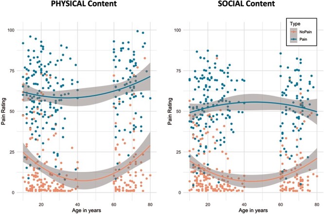
Pain ratings for each condition and across the age range. The plots show raw data points (averaged across trials for each participant for visualization), a quadratic line of best fit for age (orange line = no pain, blue line = pain), and the standard error around this line of best fit (grey shading).
Table 3.
Statistical effects for pain ratings.
| β | SE | t | P | |
|---|---|---|---|---|
| Age (linear) | 80.33 | 56.14 | 1.43 | .152 |
| Age (quadratic) | 183.67 | 55.61 | 3.30 | <.001*** |
| Type | 42.08 | 1.43 | 29.48 | <.001*** |
| Content | −5.64 | 1.24 | −4.55 | <.001*** |
| Age (linear) × Type | 224.68 | 95.20 | 2.36 | .018* |
| Age (quadratic) × Type | −253.99 | 94.16 | −2.70 | .007** |
| Age (linear) × Content | −119.00 | 66.01 | −1.80 | .071 |
| Age (quadratic) × Content | −224.15 | 65.05 | −3.45 | <0.001*** |
| Type × Content | −6.99 | 2.26 | −3.10 | 0.002** |
| Age (linear) × Type × Content | −136.84 | 86.84 | −1.57 | 0.116 |
| Age (quadratic) × Type × Content | −261.32 | 84.62 | −3.09 | 0.002** |
Asterisks show significance of effects, where *P < .05;
P < .01;
P < .001.
Results revealed significant effects for the Type and Content of pain. The effect of Type revealed that participants judged images depicting pain as more painful (M = 57.4) than no-pain images (M = 14.7), and the effect of Content showed that participants judged physical stimuli as more painful (M = 38.9) than social stimuli (M = 33.1). In addition, the effect of age was significant on the quadratic model, showing that younger adults gave higher overall ratings of pain than adolescents and older adults. As predicted, the Type × Content interaction was significant. Follow-up analyses showed that the Type effect (pain minus no-pain) was larger when participants rated physical stimuli (MDiff = 45.4) compared to social stimuli (MDiff = 39.5), V = 9586, P < .001.
The 3-way interaction between Age (quadratic), Type, and Content was significant, and Age (quadratic) modulated the effects of Type and Content separately. Post-hoc analysis of this three-way interaction revealed that the Type effect (pain minus no-pain) showed a significant quadratic fit with age for social content (β = −72.46, SE = 20.09, t = −3.61, P < .001) but this quadratic effect of age did not reach significance for physical content (β = −43.70, SE = 23.39, t = −1.87, P = .063). That is, young adults showed a larger Type effect (i.e. larger difference in pain ratings between pain and no pain images) to social images compared to adolescents and older adults, but the Type effect was stable across the age range for physical images.
Mu desynchronization
Alpha (8–13 Hz) and beta (13–35 Hz) desynchronization was analysed using separate general linear mixed effects models (since the data were non-normally distributed, Shapiro–Wilk test W = 0.98, P < .001 and W = 0.99, P < .001, for alpha and beta bands respectively), using the glmer function in the lme4 package in R. (Reliability of the alpha and beta desynchronization data were verified by running a split-half analysis. This reliability check is summarized in the Supplementary Materials; condition effects are consistent across the full sample and the two split-half samples.) The model included the within-subjects variables Type (pain versus no-pain) and Content (physical versus social) as fixed effects (using deviation contrast coding), random effects for participant, and random slopes for Type, Content and Electrode on the participant random effect. Image was not included as a random effect in the analysis of EEG data because percentage change in power needed to be calculated over trials in each experimental condition; there were not enough instances of each image to reliably calculate power change from baseline (maximum segments per image/participant = 4). We report analyses with Age as a continuous linear predictor variable since model comparisons showed that including the quadratic age term did not significantly improve model fit (alpha: X2 = 13.3, P = .103; beta: X2 = 9.15, P = .330). Data are plotted for alpha and beta in Figs 3 and 4, respectively, and full statistical effects are reported in Table 4.
Figure 3.
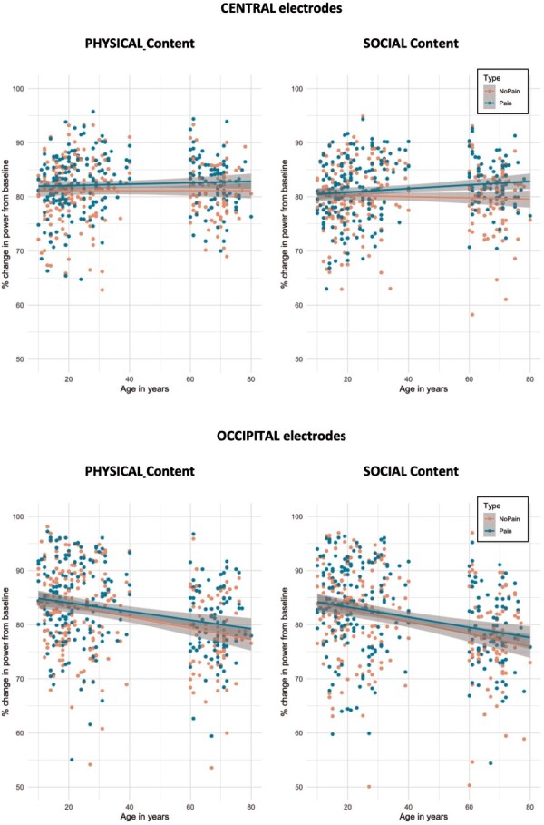
Alpha desynchronization for each electrode site and condition across the age range. The plots show raw data points (averaged across trials for each participant for visualization), a linear line of best fit for age (orange line = no pain, blue line = pain), and the standard error around this line of best fit (grey shading).
Figure 4.
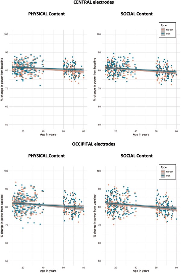
Beta desynchronization for each electrode site and condition across the age range. The plots show raw data points (averaged across trials for each participant for visualization), a linear line of best fit for age (orange line = no pain, blue line = pain), and the standard error around this line of best fit (grey shading).
Table 4.
Statistical effects for alpha and beta wavebands.
| Β | SE | t | P | ||
|---|---|---|---|---|---|
| Alpha desynchronization | Age (linear) | −1.02 | 0.22 | −4.63 | <.001*** |
| Type | 1.17 | 0.23 | 5.09 | <.001*** | |
| Content | −1.07 | 0.26 | −4.19 | <.001** | |
| Electrode | 0.37 | 0.33 | 1.12 | .264 | |
| Age (linear) × Type | 0.62 | 0.23 | 2.68 | .007** | |
| Age (linear) × Content | −1.11 | 0.26 | −0.41 | .679 | |
| Age (linear) × Electrode | −2.48 | 0.33 | −7.54 | <.001*** | |
| Type × Content | 0.22 | 0.29 | 0.78 | .435 | |
| Type × Electrode | −0.23 | 0.29 | −0.80 | .424 | |
| Content × Electrode | −0.16 | 0.29 | −0.57 | .568 | |
| Age (linear) × Type × Content | 0.36 | 0.28 | 1.28 | .200 | |
| Age (linear) × Type × Electrode | −0.13 | 0.28 | −0.47 | .641 | |
| Age (linear) × Content × Electrode | −0.40 | 0.28 | −1.42 | .155 | |
| Type × Content × Electrode | −0.06 | 0.57 | −0.10 | .919 | |
| Age (linear) × Type × Content × Electrode | −0.83 | 0.57 | −1.47 | .142 | |
| Beta desynchronisation | Age (linear) | −0.91 | 0.12 | −7.67 | <.001*** |
| Type | 0.33 | 0.12 | 2.83 | .005** | |
| Content | −0.44 | 0.13 | −3.46 | <.001*** | |
| Electrode | 0.24 | 0.17 | 1.37 | .170 | |
| Age (linear) × Type | 0.29 | 0.12 | 2.48 | <.013* | |
| Age (linear) × Content | −0.12 | 0.13 | −0.95 | .341 | |
| Age (linear) × Electrode | −0.20 | 0.17 | −1.19 | .236 | |
| Type × Content | 0.15 | 0.15 | 1.02 | .307 | |
| Type × Electrode | −0.12 | 0.15 | −0.79 | .433 | |
| Content × Electrode | 0.28 | 0.15 | 1.87 | .061 | |
| Age (linear) × Type × Content | −0.29 | 0.15 | −1.94 | .052 | |
| Age (linear) × Type × Electrode | −0.06 | 0.15 | −0.39 | .700 | |
| Age (linear) × Content × Electrode | 0.06 | 0.15 | 0.40 | .690 | |
| Type × Content × Electrode | 0.30 | 0.30 | 1.00 | .318 | |
| Age (linear) × Type × Content × Electrode | −0.17 | 0.30 | −0.56 | .573 |
Asterisks show significance of effects, where *P < .05;
P < .01;
P < .001.
Alpha Analysis of alpha oscillations revealed a significant effect of Type, reflecting greater mu/alpha desynchronization for images that depicted pain (M = 82.0%) compared to no-pain (M = 80.9%). A significant effect of Content showed that alpha desynchronization was greater for physical stimuli (M = 82.0%) than social stimuli (M = 80.9%). A significant effect of Age revealed that overall, alpha desynchronization decreased linearly with advancing age.
Crucially, Age significantly modulated alpha desynchronization in response to Type of pain. Follow-up analyses revealed that the size of the Type effect (pain minus no-pain) increased linearly with advancing age (β = 0.59, SE = 0.18, t = 3.34, P < .001); older adults showed a larger difference in alpha desynchronization between pain and no pain images than adolescents. The Age × Electrode interaction was also significant: overall alpha desynchronization decreased linearly with advancing age over the occipital electrodes (β = −2.26, SE = 0.48, t = −4.76, P < .001), but did not change with age over the central electrodes (β = 0.20, SE = 0.30, t = 0.67, P = .50).
Beta Analysis of beta oscillations revealed a significant effect of Type, reflecting greater beta desynchronization for pictures that depicted pain (M = 81.0%) than no-pain (M = 80.7%). The significant effect of Content showed that beta desynchronization was greater for physical (M = 81.1%) than social stimuli (M = 80.6%). The significant effect of Age revealed that overall, beta desynchronization decreased linearly with advancing age.
Once again, Age significantly modulated beta desynchronization in response to Type of pain. Follow-up analyses revealed that the size of the Type effect (pain minus no-pain) increased linearly with advancing age (β = 0.29, SE = 0.09, t = 3.22, P = .001); older adults showed a larger difference in beta desynchronization between pain and no pain images than adolescents. In addition, the three-way interaction between Age, Type, and Content just missed significance (P = .052); since this was a key predicted effect we ran exploratory analyses to examine the underlying patterns. These post-hoc analyses revealed that the Type effect (pain minus no-pain) showed a significant linear fit with age for physical content (β = 0.44, SE = 0.13, t = 3.49, P < .001) but not for social content (β = 0.14, SE = 0.12, t = 1.20, P = .228). That is, older adults showed a larger Type effect (i.e. larger difference in beta desynchronization between pain and no pain images) to physical images compared to adolescents, but the Type effect was stable across the age range for social images.
Correlations between pain ratings and mu desynchronization
To test whether individuals’ subjective ratings of pain in physical and social contexts were related to their neural responses to pain, we computed correlations between the Type effects (pain minus no-pain) on each measure. Given the number of variables included in the correlation, the alpha level for significance was set to 0.01. Significant associations are shown in Fig. 5. Analyses revealed a strong correlation between behavioural ratings for physical and social pain; however, these behavioural ratings of pain did not correlate with pain responses on any of the neural measures. (Note that when correlations were run separately for each age group, there was a significant positive correlation between behavioural ratings of social pain and alpha desynchronization for social pain among adolescents [r(74) = 0.268, P = .021], such that the greater adolescents rated the social pain in the image, the more alpha desynchronization they exhibited. No other correlations were found between behavioural ratings and mu desynchronization among adolescents, young or older adults.) Mu desynchronization correlated moderately between the alpha and beta bands for both physical and social pain, and a weak correlation was found between physical and social pain in the alpha band.
Figure 5.
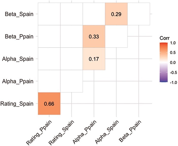
Correlation matrix between behavioural pain ratings and mu desynchronization (alpha and beta bands), separately for physical (P) and social (S) content. Coloured cells indicate a significant correlation (P < .01), and values show the correlation coefficient (r).
General discussion
Previous research has shown that seeing other people in pain activates brain areas associated with empathy (Eisenberger 2011, 2012b, Lamm et al. 2010, Singer et al. 2004; i.e. AI, ACC, and somatosensory cortex), and that these neural circuits are sensitive to both physical and social pain. In this paper, we employed a pain ratings EEG task to directly compare empathic responses to other people in physical and social pain, and examined whether these responses change from adolescence (10–19 years) to young (20–40 years) and older (≥60years) adulthood. We predicted that empathy responses would reflect the stimuli’s affective content, with higher ratings and greater mu desynchronization for pictures that show painful situations compared to no-pain situations. In addition, we predicted that this pain versus no-pain difference would be elicited by both physical and social content, but expected to see distinct neural responses between the two (i.e. a greater pain difference and increased sensorimotor desynchronization for physical versus social content). Finally, we predicted that young adults would show greater responses to pain (i.e. greater differences in pain versus no-pain ratings and greater mu desynchronization in response to painful stimuli) compared to both adolescents and older adults.
Overall, our results replicated the basic findings from previous studies that have tested empathic responses to others in pain: participants clearly distinguished between pain and no-pain stimuli (Yang et al. 2009, Perry et al. 2010, Chen et al. 2014). Behavioural ratings revealed higher scores for images that depicted pain versus no-pain, and EEG mu rhythms revealed greater alpha and beta desynchronization for images that depicted pain versus no-pain. Unlike some studies that have reported neural responses to physical pain localized over the sensorimotor cortex (Perry et al. 2010, Whitmarsh et al. 2011), we found that effects of Type were evident over both central and occipital sites. It is likely that this reflects methodological differences between studies (i.e. Perry et al. and Whitmash et al. recorded oscillations using magnetoencephalography and analysed the data using time-frequency and source localization procedures). It is also possible that by including both physical and social pain images together in the current study, a wider and more complex empathy-social cognitive network was activated. Association analyses showed that mu activity for the pain effect was correlated across alpha and beta bands (for both physical and social pain), which suggests that neural activity in both wavebands reflects similar socio-cognitive responses, and therefore both provide reliable and complementary EEG measures of sensorimotor activity (and mirror neuron system sensitivity) in response to others in pain.
It is notable, however, that overall mu activity did not correlate with behavioural responses, which suggests that individuals’ subjective ratings of others in pain did not influence the strength of their real-time neural responses while observing others in pain. This contrasts with previous studies that have reported significant associations between behavioural and neural empathy responses (Jackson et al. 2006, Saarela et al. 2007, Wu and Han 2021). It is likely that differences in the rating prompt between studies explain these differences. In our study, participants were asked to rate ‘how (socially/physically) painful the situation was’, whereas most previous studies that have found an association asked participants to imagine and rate the intensity of pain/distress/emotion that a pictured person is experiencing. The latter clearly prompts participants to infer another person’s affective mental state to answer the question, while the rating question used in the current study could be answered without mentalizing. As such, it seems likely that reported correlations between subjective ratings of others’ pain and neural responses to observing others’ pain rely upon a shared activation of the mentalizing system (Frith and Frith 2006, Brass et al. 2007, Van Overwalle and Baetens 2009), and that this system needs to be explicitly activated by task constraints.
Importantly, our data revealed both shared and distinct patterns of empathy responses to seeing others in physical and social pain. Iannetti et al. (2013) highlight the importance of examining the neural overlap and distinctions between processing of physical and social pain, arguing that these are likely to be at least partially sub-served by different neural activity. In the current study, neural responses recorded while participants were directly viewing the images clearly distinguished pain and no-pain events in both physical and social contexts, and this effect of pain on real-time mu desynchronization was not modulated by the physical or social context {though global differences emerged between physical and social stimuli [This global difference between physical and social content likely reflects lower-level visual differences between stimuli, as well as a generally higher response to physical images where the potential for pain was more salient across both image types (i.e,. a foot next to upturned wall tacks or a hand close to the hot iron).]}. Behavioural ratings following the images also clearly distinguished pain and no-pain events in both physical and social contexts; however, in line with our predictions, pain ratings were significantly higher for physical pain than for social pain. This pattern suggests that perceiving others in pain initially activates comparable neural responses in brain areas underlying sensorimotor resonance (i.e. the sensorimotor cortex), but when a broader brain network is engaged over a longer-lasting period of reflection about how painful the situation was, behavioural responses distinguished different intensities for physical and social pain. Our experiment employed a carefully controlled design that directly compared empathy for others in physical and social pain in the same participants using matched stimuli (the only other study to date that has tested responses to both physical and social pain used vastly different stimuli in each case, and could not directly compare the two, Krach et al. 2015). Thus, we can infer that real-time sensorimotor resonance reflects a common neural response to others in pain across the core empathy network (Fan et al. 2011; i.e. the AI and ACC), including brain regions that underlie affective sharing between the self and other (Singer et al. 2004, Jackson et al. 2005, Lamm et al. 2011), and that this common empathy network is comparably activated by both physical and social pain contexts. When additional processing time is given for participants to reflect on how painful the depicted situation was, this activates functional connections between the sensorimotor and affective sharing brain areas with key hubs (that are at least partially distinct for physical and social contexts) to explicitly interpret pain intensity within the physical and social context. It is likely that these key hubs include differential activation of the mentalizing system [the medial prefrontal cortex (mPFC) and temporoparietal junction (TPJ), (Gallagher and Frith 2003, Samson et al. 2004, Saxe and Kanwisher 2013)] and regions associated with learning and memory retrieval (the hippocampus, Squire et al. 2004, Krach et al. 2015). Further research using spatially sensitive neuroimaging methods and statistical approaches that may be more sensitive to detecting small differences between physical and social pain responses (i.e. multivariate pattern analysis, Iannetti et al. 2013) is needed to identify these neural mechanisms that distinguish ongoing empathy processing for physical and social pain.
Our data also provide novel evidence about the developmental trajectory of empathy beyond early childhood. Behavioural ratings revealed distinct developmental patterns for physical and social pain. That is, while responses to others in physical pain were stable across the age range, responses to others in social pain (i.e. ratings of pain versus no-pain images) peaked in young adulthood, suggesting that young adults were especially sensitive to social pain events and inferred more intense social pain for the other person than adolescents or older adults did. This heightened sensitivity to social pain in young adulthood likely reflects the extended development of prosocial empathy behaviours—prosociality is reported to decline during adolescence (i.e. between 13 and 17 years old) before increasing again into young adulthood and beyond (Carlo et al. 2007, Matsumoto et al. 2016)—and this period is thought to evoke increasing sensitivity to one’s social environment (Peper and Dahl 2013). In contrast, the effect of pain on mu (alpha and low beta) desynchronization increased linearly from adolescence through to young and older adults; in fact, the data plotted in Figs 3 and 4 suggest that adolescents (and to some extent, young adults) did not distinguish pain and no-pain conditions at all in neural oscillations. These findings demonstrate that the oscillatory mu rhythm is sensitive to age-related changes in empathy (Ruffman et al. 2008, Isaacowitz and Stanley 2011), and are consistent with the proposal that empathy brain networks continue to develop through childhood (Decety 2010) and adolescence (Decety and Michalska 2010, Levy and Feldman 2017), then remain stable or increase though adulthood and older age (Beadle and de la Vega 2019). Importantly, in contrast to the age-related decline observed in other areas of social cognition (Henry et al. 2013, Moran 2013), data in this affective area converge to suggest that older adults are not impaired at recognizing and responding to others’ pain.
The finding that older adults showed clear, or even enhanced compared to the younger age groups, affective empathy responses across behavioural and neural measures is in line with research showing relatively spared affective ToM in older age (Pardini and Nichelli 2009, Castelli et al. 2010, Henry et al. 2013, Mahy et al. 2014, Bottiroli et al. 2016). There are a number of potential explanations for these results. For instance, empathy for others might increase in older age due to accumulating experience in social situations and exposure to pain-related scenarios over the lifespan, which strengthens activity in the empathy network and facilitates older adults’ ability to share affective experiences with others (Hess et al. 2005, Leclerc and Hess 2007, De Lillo and Ferguson 2023). Indeed, people who are predisposed to be more sensitive to pain exhibit stronger reactions to social pain and greater brain activity in empathy areas (Eisenberger et al. 2003, 2006). Increasing experience with the recipient of pain has also been shown to influence individuals’ empathy responses (Young et al. 2018). The lack of decline in older age is consistent with the predictions from Apperly and Butterfill’s model (2009), that some sub-components of social cognition are relatively automatic and cognitively efficient so are less susceptible to age decline. Numerous studies have reported that empathy neural networks are activated even when participants have not been prompted to engage these responses. For example, Singer et al. (2004) found overlapping brain activation in the empathy/pain brain network when participants experienced a painful stimulus themselves and when they observed an arbitrary cue that indicated their loved one was receiving the same painful stimulus (i.e. ruling out the possibility that the effect was driven by a general response to an emotional cue). In addition, the enhanced sensorimotor alpha/beta rhythm in older age parallels that seen in previous research that has found over-activation of motor areas during action execution in older adults (Vallesi and Stuss 2010, Rossiter et al. 2014, Schmiedt-Fehr et al. 2016, Heinrichs-Graham et al. 2018, Brunsdon et al. 2019). This change could either reflect the activity of an enhanced specialization of the empathy network in older age (due to accumulating experience) or increasing compensatory neural activity to maintain task performance when cognitive capacities are declining (Ward 2006).
Finally, while we interpret the finding that empathy responses to pain increase from adolescence to adulthood as reflecting an immature/inexperienced empathy network that is less effective at sharing and understanding others’ emotional states, thoughts, and feelings (Burnett et al. 2009, Kim et al. 2020), it is important to consider that some features of the stimuli themselves might have contributed to this effect. Specifically, all images included adult actors, which may have enhanced social closeness for our adult participants (Gutsell and Inzlicht 2010) but elicited out-group effects for our adolescent participants (Simpson and Todd 2017). Previous research has shown that an own-age bias can enhance performance in a range of social perception tasks (Melinder et al. 2010, Slessor et al. 2014, Ferguson et al. 2018), and that other cues about out-group status (e.g. race, football team or University affiliations) can alter individuals’ propensity to help and sensorimotor sensitivity to others’ pain (Avenanti et al. 2010, Hein et al. 2010, Hackel et al. 2017, Cao et al. 2019). It is therefore possible that behavioural judgements about others’ pain were facilitated among young adults because the actor was from participants’ in-group but reduced among adolescents because the actor was from participants’ out-group. Since our images depicted only actors’ hands and feet (not faces or full bodies), we expect that age-biases were unlikely to have had a significant influence on our results (since the age of the actors was not clear or salient); however, further research is needed to systematically manipulate this variable. Additionally, while efforts were made to create images that captured the range of physical and social experiences encountered across the age range, our pretest of the images was completed by predominantly young adult participants, and thus these experiences might not be equally representative across the wide age range tested here.
The current study’s use of EEG and behavioural ratings to test empathy responses while viewing real-life photos depicting pain/no-pain improves on some previous studies that elicited empathy for others’ pain using context-free facial emotions or cartoon drawings of social situations (Eisenberger et al. 2003, Eisenberger and Lieberman 2004, Krach et al. 2015). However, this task remains limited in ecological validity due to the lack of a genuine social interaction between co-present social partners/stimuli or availability of wider environmental cues to contextualise painful events. This is particularly important for social pain, which is typically embedded within a richer context. Findings in these controlled lab-based contexts may therefore not represent the same processes that are activated in naturalistic settings, since interactivity is known to alter sensitivity to others’ perspectives and influence communication success in other domains of social cognition (Surtees et al. 2016, Kuhlen and Rahman 2022). This is an important consideration for future empathy research: how to create more ecologically valid situations in which to assess empathy responses in more ‘real-world’ scenarios, allowing results to be more generalizable to the types of empathy processes involved in interactions throughout our daily lives (Ochsner 2004, Schilbach et al. 2006, 2013, Schilbach 2015).
One emerging area of research that offers exciting possibilities to enhance our understanding of empathy is ‘second-person’ neuroscience (Dumas 2011, Konvalinka and Roepstorff 2012, Froese et al. 2014), which involves the examination of coordinated behaviour and brain activity of two (or more) individuals in a real-time interaction, rather than a single individual engaging in an observation task. This second-person neuroscience approach has begun to reveal promising results in other domains, showing, for example, that neural activity in key social brain areas synchronizes between cooperating partners (and not between others) when they are working towards a shared goal (Jiang et al. 2015, Astolfi et al. 2020) or interacting through speech (Pérez et al. 2017). A recent review of the second-person neuroscience approach to social interaction revealed differences in the neural mechanisms that support real-time reciprocal social interaction and those involved in social observation, highlighting a key role for the mentalizing network in this distinction (Redcay and Schilbach 2019). Therefore, future empathy studies should take advantage of designs that include engaged participants and simultaneous recordings of synchronized behaviour and brain activity to further elucidate the mechanisms of empathy.
In conclusion, this study provides valuable insights into the complex nature of empathic responses to others in pain, highlighting activity in the core affective empathy network that is shared across physical and social pain contexts. The study’s careful design adds to our understanding of the neural mechanisms underlying empathy, allowing us to directly compare empathic processing of physical and social pain, and to examine the developmental trajectory of empathy from adolescence to young and older adulthood. Our results revealed that while observers across the 10–80 years age range showed distinct responses to others in pain versus no-pain, young adults are more sensitive in their ratings of social pain compared to adolescents and older adults, and real-time neural responses to physical and social pain increase linearly across the lifespan (i.e. older adults exhibit heightened neural affective empathy responses compared to younger counterparts). These patterns show the extended period of affective empathy development from adolescence to adulthood and challenge the notion of universal age-related social cognitive decline in older age. This enhancement in empathic engagement among older adults aligns with theories of accumulated social experience fostering affective resonance and reflects the intricate interplay between cognitive and emotional processes across the lifespan. Overall, this research contributes to a deeper understanding of how affective empathy operates within different contexts and age groups, unravelling the intricate interplay between neural activation, emotional processing, and socio-cognitive development. As we continue to uncover the subtleties of empathic responses, this study provides a stepping-stone for future investigations that could help refine our comprehension of human interactions and pave the way for interventions targeting empathy-related deficits or enhancements.
Supplementary Material
Acknowledgements
The preregistration, datasets, and R code supporting this article are available on the Open Science Framework (https://osf.io/f9c2r/). All work for this project was carried out at the University of Kent.
Contributor Information
Heather J Ferguson, School of Psychology, University of Kent, Canterbury, Kent CT2 7NP, United Kingdom.
Martina De Lillo, School of Psychology, University of Kent, Canterbury, Kent CT2 7NP, United Kingdom.
Camilla Woodrow-Hill, School of Psychology, University of Kent, Canterbury, Kent CT2 7NP, United Kingdom.
Rebecca Foley, School of Psychology, University of Kent, Canterbury, Kent CT2 7NP, United Kingdom.
Elisabeth E F Bradford, School of Psychology and Neuroscience, University of St Andrews, St Andrews KY16 9JP, United Kingdom.
Supplementary data
Supplementary data is available at SCAN online.
Conflict of interest
None declared.
Funding
This work was carried out with the support of a European Research Council grant to H.F. (Ref: CogSoCoAGE; 636458).
References
- Adolphs R. The social brain: neural basis of social knowledge. Annu Rev Psychol 2009;60:693–716. doi: 10.1146/annurev.psych.60.110707.163514 [DOI] [PMC free article] [PubMed] [Google Scholar]
- Akitsuki Y, Decety J. Social context and perceived agency affects empathy for pain: an event-related fMRI investigation. Neuroimage 2009;47:722–34. doi: 10.1016/j.neuroimage.2009.04.091 [DOI] [PubMed] [Google Scholar]
- Apperly IA, Butterfill SA. Do humans have two systems to track beliefs and belief-like states? Psychol Rev 2009;116:953–70. doi: 10.1037/a0016923 [DOI] [PubMed] [Google Scholar]
- Arnett K, Roach A, Elzy M. et al. Childhood emotional invalidation and right hemispheric mu suppression during a pain empathy task: an EEG study. Soc Neurosci 2019;14:236–50. doi: 10.1080/17470919.2018.1441905 [DOI] [PubMed] [Google Scholar]
- Astolfi L, Toppi J, Ciaramidaro A. et al. Raising the bar: can dual scanning improve our understanding of joint action? NeuroImage 2020;216:116813. doi: 10.1016/j.neuroimage.2020.116813 [DOI] [PubMed] [Google Scholar]
- Avenanti A, Sirigu A, Aglioti SM. Racial bias reduces empathic sensorimotor resonance with other-race pain. Curr Biol 2010;20:1018–22. doi: 10.1016/j.cub.2010.03.071 [DOI] [PubMed] [Google Scholar]
- Baayen RH, Davidson DJ, Bates DM. Mixed-effects modeling with crossed random effects for subjects and items. J Memory Lang 2008;59:390–412. doi: 10.1016/j.jml.2007.12.005 [DOI] [Google Scholar]
- Bailen NH, Green LM, Thompson RJ. Understanding emotion in adolescents: a review of emotional frequency, intensity, instability, and clarity. Emotion Rev 2018;11:63–73. doi: 10.1177/1754073918768878 [DOI] [Google Scholar]
- Bailey PE, Brady B, Ebner NC. et al. Effects of age on emotion regulation, emotional empathy, and prosocial behavior. J Gerontol B 2020;75:802–10. [DOI] [PubMed] [Google Scholar]
- Bailey PE, Henry JD. Growing less empathic with age: disinhibition of the self-perspective. J Gerontol B Psychol Sci Soc Sci 2008;63:219–P226. doi: 10.1093/geronb/63.4.P219 [DOI] [PubMed] [Google Scholar]
- Bates D, Mächler M, Bolker B. et al. Fitting linear mixed-effects models using lme4. J Stat Soft 2015;67:1–48. doi: 10.18637/jss.v067.i01 [DOI] [Google Scholar]
- Beadle JN, de la Vega CE. Impact of aging on empathy: review of psychological and neural mechanisms. Front Psychiatry 2019;10:331. doi: 10.3389/fpsyt.2019.00331 [DOI] [PMC free article] [PubMed] [Google Scholar]
- Bernstein DM, Thornton WL, Sommerville JA. Theory of mind through the ages: older and middle-aged adults exhibit more errors than do younger adults on a continuous false belief task. Exp Aging Res 2011;37:481–502. doi: 10.1080/0361073X.2011.619466 [DOI] [PubMed] [Google Scholar]
- Blakemore SJ. The social brain in adolescence. Nat Rev Neurosci 2008;9:267–77. doi: 10.1038/nrn2353 [DOI] [PubMed] [Google Scholar]
- Blakemore SJ, Choudhury S. Development of the adolescent brain: implications for executive function and social cognition. J Child Psychol Psychiatry 2006;47:296–312. doi: 10.1111/j.1469-7610.2006.01611.x [DOI] [PubMed] [Google Scholar]
- Bolling DZ, Pitskel NB, Deen B. et al. Development of neural systems for processing social exclusion from childhood to adolescence. Dev Sci 2011;14:1431–44. doi: 10.1111/j.1467-7687.2011.01087.x [DOI] [PMC free article] [PubMed] [Google Scholar]
- Bottiroli S, Cavallini E, Ceccato I. et al. Theory of mind in aging: comparing cognitive and affective components in the faux pas test. Arch Gerontol Geriatrics 2016;62:152–62. doi: 10.1016/j.archger.2015.09.009 [DOI] [PubMed] [Google Scholar]
- Bradford EEF, Brunsdon VEA, Ferguson HJ. Cognitive mechanisms of perspective-taking across adulthood: an eye-tracking study using the director task. J Exp Psychol Learn Mem Cogn 2023;49:959–73. doi: 10.1037/xlm0001190 [DOI] [PubMed] [Google Scholar]
- Brass M, Schmitt RM, Spengler S. et al. Investigating action understanding: inferential processes versus action simulation. Curr Biol 2007;17:2117–21. doi: 10.1016/j.cub.2007.11.057 [DOI] [PubMed] [Google Scholar]
- Brunsdon VE, Bradford EEF, Ferguson HJ. Sensorimotor mu rhythm during action observation changes across the lifespan independently from social cognitive processes. Dev Cogn Neurosci 2019;38:100659. doi: 10.1016/j.dcn.2019.100659 [DOI] [PMC free article] [PubMed] [Google Scholar]
- Burnett S, Bird G, Moll J. et al. Development during adolescence of the neural processing of social emotion. J Cogn Neurosci 2009;21:1736–50. doi: 10.1162/jocn.2009.21121 [DOI] [PMC free article] [PubMed] [Google Scholar]
- Cao Y, Yusri NM, Powell T. et al. Neural and behavioral markers of observed pain of older adults. Neuropsychologia 2019;131:84–90. doi: 10.1016/j.neuropsychologia.2019.04.012 [DOI] [PubMed] [Google Scholar]
- Carlo G, Crockett LJ, Randall BA. et al. A latent growth curve analysis of prosocial behavior among rural adolescents. J Res Adolescence 2007;17:301–24. doi: 10.1111/j.1532-7795.2007.00524.x [DOI] [Google Scholar]
- Carson N, Leach L, Murphy KJ. A re‐examination of Montreal Cognitive Assessment (MoCA) cutoff scores. Int J Geriatr Psychiatry 2018;33:379–88. doi: 10.1002/gps.4756 [DOI] [PubMed] [Google Scholar]
- Castelli I, Baglio F, Blasi V. et al. Effects of aging on mindreading ability through the eyes: an fMRI study. Neuropsychologia 2010;48:2586–94.doi: 10.1016/j.neuropsychologia.2010.05.005 [DOI] [PubMed] [Google Scholar]
- Cattaneo L, Rizzolatti G. The mirror neuron system. Arch Neurol 2009;66:557–60. doi: 10.1001/archneurol.2009.41 [DOI] [PubMed] [Google Scholar]
- Charles ST, Carstensen LL. Social and emotional aging. Annu Rev Psychol 2010;61:383–409. doi: 10.1146/annurev.psych.093008.100448 [DOI] [PMC free article] [PubMed] [Google Scholar]
- Chen YC, Chen CC, Decety J. et al. Aging is associated with changes in the neural circuits underlying empathy. Neurobiol Aging 2014;35:827–36. doi: 10.1016/j.neurobiolaging.2013.10.080 [DOI] [PubMed] [Google Scholar]
- Cheng Y, Chen C, Decety J. How situational context impacts empathic responses and brain activation patterns. Front Behav Neurosci 2017;11:165. doi: 10.3389/fnbeh.2017.00165 [DOI] [PMC free article] [PubMed] [Google Scholar]
- Cheng Y, Yang CY, Lin CP. et al. The perception of pain in others suppresses somatosensory oscillations: a magnetoencephalography study. Neuroimage 2008;40:1833–40. doi: 10.1016/j.neuroimage.2008.01.064 [DOI] [PubMed] [Google Scholar]
- Craig AD. How do you feel—now? The anterior insula and human awareness. Nat Rev Neurosci 2009;10:59–70. doi: 10.1038/nrn2555 [DOI] [PubMed] [Google Scholar]
- Dalgleish T, Walsh N, Mobbs D. et al. Social pain and social gain in the adolescent brain: A common neural circuitry underlying both positive and negative social evaluation. Sci Rep 2017;7:42010. doi: 10.1038/srep42010 [DOI] [PMC free article] [PubMed] [Google Scholar]
- Davis MH. A multidimensional approach to individual differences in empathy. JSAS Catalog Selected Documents Psychol 1980;10:85. [Google Scholar]
- Decety J. The neurodevelopment of empathy in humans. Dev Neurosci 2010;32:257–67. doi: 10.1159/000317771 [DOI] [PMC free article] [PubMed] [Google Scholar]
- Decety J, Michalska KJ. Neurodevelopmental changes in the circuits underlying empathy and sympathy from childhood to adulthood. Dev Sci 2010;13:886–99. doi: 10.1111/j.1467-7687.2009.00940.x [DOI] [PubMed] [Google Scholar]
- De Lillo M, Ferguson HJ. Perspective-taking and social inferences in adolescents, young adults and older adults. J Exp Psychol Gen 2023;152:1420–38. doi: 10.1037/xge0001337 [DOI] [PubMed] [Google Scholar]
- De Lillo M, Foley R, Fysh MC. et al. Tracking developmental differences in real-world social attention across adolescence, young adulthood and older adulthood. Nat Human Behav 2021;5:1381–90.doi: 10.1038/s41562-021-01113-9 [DOI] [PMC free article] [PubMed] [Google Scholar]
- DeWall CN, MacDonald G, Webster GD. et al. Acetaminophen reduces social pain: behavioral and neural evidence. Psychol Sci 2010;21:931–37.doi: 10.1177/0956797610374741 [DOI] [PubMed] [Google Scholar]
- Di Pellegrino G, Fadiga L, Fogassi L. et al. Understanding motor events: a neurophysiological study. Exp Brain Res 1992;91:176–80. doi: 10.1007/BF00230027 [DOI] [PubMed] [Google Scholar]
- Dorris L, Young D, Barlow J. et al. Cognitive empathy across the lifespan. Dev Med Child Neurol 2022;64:1524–31. doi: 10.1111/dmcn.15263 [DOI] [PMC free article] [PubMed] [Google Scholar]
- Dumas G. Towards a two-body neuroscience. Commun Integr Biol 2011;4:349–52. doi: 10.4161/cib.4.3.15110 [DOI] [PMC free article] [PubMed] [Google Scholar]
- Dumontheil I, Apperly IA, Blakemore SJ. Online usage of theory of mind continues to develop in late adolescence. Dev Sci 2010;13:331–38. doi: 10.1111/j.1467-7687.2009.00888.x [DOI] [PubMed] [Google Scholar]
- Eisenberg N, Fabes RA. Empathy: conceptualization, measurement, and relation to prosocial behavior. Motiv Emot 1990;14:131–49. doi: 10.1007/BF00991640 [DOI] [Google Scholar]
- Eisenberger NI. The neural basis of social pain: findings and implications. In: MacDonald G, Jensen-Campbell LA (eds), Social Pain: Neuropsychological and Health Implications of Loss and Exclusion. Washington: American Psychological Association, 2011, 53–78. [Google Scholar]
- Eisenberger NI. Broken hearts and broken bones: a neural perspective on the similarities between social and physical pain. Curr Dir Psychol Sci 2012a;21:42–47. doi: 10.1177/0963721411429455 [DOI] [Google Scholar]
- Eisenberger NI. The neural bases of social pain: evidence for shared representations with physical pain. Psychosomatic Med 2012b;74:126. doi: 10.1097/PSY.0b013e3182464dd1 [DOI] [PMC free article] [PubMed] [Google Scholar]
- Eisenberger NI, Jarcho JM, Lieberman MD. et al. An experimental study of shared sensitivity to physical pain and social rejection. Pain 2006;126:132–38. doi: 10.1016/j.pain.2006.06.024 [DOI] [PubMed] [Google Scholar]
- Eisenberger NI, Lieberman MD. Why rejection hurts: a common neural alarm system for physical and social pain. Trends Cogn Sci 2004;8:294–300. doi: 10.1016/j.tics.2004.05.010 [DOI] [PubMed] [Google Scholar]
- Eisenberger NI, Lieberman MD, Williams KD. Does rejection hurt? An fMRI study of social exclusion. Science 2003;302:290–92. doi: 10.1126/science.1089134 [DOI] [PubMed] [Google Scholar]
- Fabi S, Leuthold H. Empathy for pain influences perceptual and motor processing: evidence from response force, ERPs, and EEG oscillations. Soc Neurosci 2017;12:701–16. doi: 10.1080/17470919.2016.1238009 [DOI] [PubMed] [Google Scholar]
- Fan Y, Duncan NW, De Greck M. et al. Is there a core neural network in empathy? An fMRI based quantitative meta-analysis. Neurosci Biobehav Rev 2011;35:903–11. doi: 10.1016/j.neubiorev.2010.10.009 [DOI] [PubMed] [Google Scholar]
- Fan YT, Chen C, Chen SC. et al. Empathic arousal and social understanding in individuals with autism: evidence from fMRI and ERP measurements. Soc Cognit Affective Neurosci 2014;9:1203–13. doi: 10.1093/scan/nst101 [DOI] [PMC free article] [PubMed] [Google Scholar]
- Ferguson HJ, Brunsdon VE, Bradford EE. Age of avatar modulates the altercentric bias in a visual perspective-taking task: ERP and behavioral evidence. Cognit Affective Behav Neurosci 2018;18:1298–319. doi: 10.3758/s13415-018-0641-1 [DOI] [PMC free article] [PubMed] [Google Scholar]
- Ferguson HJ, Wimmer L. A psychological exploration of empathy. In: Mezzenzana F, Peluso D (eds), Conversations on Empathy: Interdisciplinary Perspectives on Imagination and Radical Othering. London: Routledge, 2023. [Google Scholar]
- Ferrari PF, Rizzolatti G. Mirror neuron research: the past and the future. Philos Trans R Soc B 2014;369:20130169. doi: 10.1098/rstb.2013.0169 [DOI] [PMC free article] [PubMed] [Google Scholar]
- Flasbeck V, Matthiessen A, Alabowitz A. et al. Is your pain my pain? A study exploring the relation between pain sensitivity, pain thresholds and empathy for somatic and psychological pain. Br J Clin Psychol 2023;62:748–61. [DOI] [PubMed] [Google Scholar]
- Fox GR, Sobhani M, Aziz-Zadeh L. Witnessing hateful people in pain modulates brain activity in regions associated with physical pain and reward. Front Psychol 2013;4:772. doi: 10.3389/fpsyg.2013.00772 [DOI] [PMC free article] [PubMed] [Google Scholar]
- Fox NA, Bakermans-Kranenburg MJ, Yoo KH. et al. Assessing human mirror activity with EEG mu rhythm: a meta-analysis. Psychol Bull 2016;142:291–313.doi: 10.1037/bul0000031 [DOI] [PMC free article] [PubMed] [Google Scholar]
- Fraser AM, Hampton RS, Spinrad TL. et al. Children’s mu suppression is sensitive to witnessing others’ social victimization. Soc Neurosci 2020;15:348–54.doi: 10.1080/17470919.2020.1722220 [DOI] [PubMed] [Google Scholar]
- Frith CD, Frith U. The neural basis of mentalizing. Neuron 2006;50:531–34. doi: 10.1016/j.neuron.2006.05.001 [DOI] [PubMed] [Google Scholar]
- Frith C, Frith U. Theory of mind. Curr Biol 2005;15:R644–R645. doi: 10.1016/j.cub.2005.08.041 [DOI] [PubMed] [Google Scholar]
- Froese T, Iizuka H, Ikegami T. Embodied social interaction constitutes social cognition in pairs of humans: a minimalist virtual reality experiment. Sci Rep 2014;4:3672. doi: 10.1038/srep03672 [DOI] [PMC free article] [PubMed] [Google Scholar]
- Gallagher HL, Frith CD. Functional imaging of ‘theory of mind’. Trends Cogn Sci 2003;7:77–83. doi: 10.1016/S1364-6613(02)00025-6 [DOI] [PubMed] [Google Scholar]
- Gallese V, Fadiga L, Fogassi L. et al. Action recognition in the premotor cortex. Brain 1996;119:593–609. doi: 10.1093/brain/119.2.593 [DOI] [PubMed] [Google Scholar]
- Guay S, Héroux J, Joubert S. et al. Effects of age and sex on neurophysiological substrates of empathy: a cross-sectional EEG study. 2018. doi: 10.31234/osf.io/cpd7f [DOI]
- Gutsell JN, Inzlicht M. Empathy constrained: prejudice predicts reduced mental simulation of actions during observation of outgroups. J Exp Soc Psychol 2010;46:841–45. doi: 10.1016/j.jesp.2010.03.011 [DOI] [Google Scholar]
- Hackel LM, Zaki J, Van Bavel JJ. Social identity shapes social valuation: evidence from prosocial behavior and vicarious reward. Soc Cognit Affective Neurosci 2017;12:1219–28. doi: 10.1093/scan/nsx045 [DOI] [PMC free article] [PubMed] [Google Scholar]
- Hein G, Silani G, Preuschoff K. et al. Neural responses to ingroup and outgroup members’ suffering predict individual differences in costly helping. Neuron 2010;68:149–60. doi: 10.1016/j.neuron.2010.09.003 [DOI] [PubMed] [Google Scholar]
- Heinrichs-Graham E, McDermott TJ, Mills MS. et al. The lifespan trajectory of neural oscillatory activity in the motor system. Dev Cogn Neurosci 2018;30:159–68.doi: 10.1016/j.dcn.2018.02.013 [DOI] [PMC free article] [PubMed] [Google Scholar]
- Henry JD, Phillips LH, Ruffman T. et al. A meta-analytic review of age differences in theory of mind. Psychol Aging 2013;28:826. doi: 10.1037/a0030677 [DOI] [PubMed] [Google Scholar]
- Hess TM, Osowski NL, Leclerc CM. Age and experience influences on the complexity of social inferences. Psychol Aging 2005;20:447. doi: 10.1037/0882-7974.20.3.447 [DOI] [PubMed] [Google Scholar]
- Hobson HM, Bishop DVM. The interpretation of mu suppression as an index of mirror neuron activity: past, present and future. Royal Soc Open Sci 2017a;4:160662. doi: 10.1098/rsos.160662 [DOI] [PMC free article] [PubMed] [Google Scholar]
- Hobson HM, Bishop DVM. Reply to Bowman et al.: building the foundations for moving mu suppression research forward. Cortex 2017b;96:126–28. doi: 10.1016/j.cortex.2017.06.012 [DOI] [PubMed] [Google Scholar]
- Iannetti GD, Salomons TV, Moayedi M. et al. Beyond metaphor: contrasting mechanisms of social and physical pain. TRENDS Cog Sci 2013;17:371–78. doi: 10.1016/j.tics.2013.06.002 [DOI] [PubMed] [Google Scholar]
- Isaacowitz DM, Stanley JT. Bringing an ecological perspective to the study of aging and recognition of emotional facial expressions: past, current, and future methods. J Nonverbal Behav 2011;35:261–78. doi: 10.1007/s10919-011-0113-6 [DOI] [PMC free article] [PubMed] [Google Scholar]
- Jackson PL, Meltzoff AN, Decety J. How do we perceive the pain of others? A window into the neural processes involved in empathy. Neuroimage 2005;24:771–79. doi: 10.1016/j.neuroimage.2004.09.006 [DOI] [PubMed] [Google Scholar]
- Jackson PL, Rainville P, Decety J. To what extent do we share the pain of others? Insight from the neural bases of pain empathy. Pain 2006;125:5–9. doi: 10.1016/j.pain.2006.09.013 [DOI] [PubMed] [Google Scholar]
- Jiang J, Chen C, Dai B. et al. Leader emergence through interpersonal neural synchronization. Proc Natl Acad Sci 2015;112:4274–79.doi: 10.1073/pnas.1422930112 [DOI] [PMC free article] [PubMed] [Google Scholar]
- Kim EJ, Son JW, Park SK. et al. Cognitive and emotional empathy in young adolescents: an fMRI study. J Korean Acad Child Adolesc Psychiatry 2020;31:121.doi: 10.5765/jkacap.200020 [DOI] [PMC free article] [PubMed] [Google Scholar]
- Konvalinka I, Roepstorff A. The two-brain approach: how can mutually interacting brains teach us something about social interaction? Front Human Neurosci 2012;6:215. doi: 10.3389/fnhum.2012.00215 [DOI] [PMC free article] [PubMed] [Google Scholar]
- Krach S, Cohrs JC, de Echeverría Loebell NC. et al. Your flaws are my pain: linking empathy to vicarious embarrassment. PloS One 2011;6:e18675.doi: 10.1371/journal.pone.0018675 [DOI] [PMC free article] [PubMed] [Google Scholar]
- Krach S, Kamp‐Becker I, Einhäuser W. et al. Evidence from pupillometry and fMRI indicates reduced neural response during vicarious social pain but not physical pain in autism. Human Brain Mapp 2015;36:4730–44.doi: 10.1002/hbm.22949 [DOI] [PMC free article] [PubMed] [Google Scholar]
- Kross E, Berman MG, Mischel W. et al. Social rejection shares somatosensory representations with physical pain. Proc Natl Acad Sci U S A. 2011;108:6270–5. doi: 10.1073/pnas.1102693108 [DOI] [PMC free article] [PubMed] [Google Scholar]
- Kross E, Egner T, Ochsner K. et al. Neural dynamics of rejection sensitivity. J Cogn Neurosci 2007;19:945–56. doi: 10.1162/jocn.2007.19.6.945 [DOI] [PubMed] [Google Scholar]
- Kuhlen AK, Rahman RA. Mental chronometry of speaking in dialogue: semantic interference turns into facilitation. Cognition 2022;219:104962. doi: 10.1016/j.cognition.2021.104962 [DOI] [PubMed] [Google Scholar]
- Lamm C, Decety J, Singer T. Meta-analytic evidence for common and distinct neural networks associated with directly experienced pain and empathy for pain. Neuroimage 2011;54:2492–502. doi: 10.1016/j.neuroimage.2010.10.014 [DOI] [PubMed] [Google Scholar]
- Lamm C, Majdandžić J. The role of shared neural activations, mirror neurons, and morality in empathy–a critical comment. Neurosci Res 2015;90:15–24. doi: 10.1016/j.neures.2014.10.008 [DOI] [PubMed] [Google Scholar]
- Lamm C, Meltzoff AN, Decety J. How do we empathize with someone who is not like us? A functional magnetic resonance imaging study. J Cogn Neurosci 2010;22:362–76. doi: 10.1162/jocn.2009.21186 [DOI] [PubMed] [Google Scholar]
- Leclerc CM, Hess TM. Age differences in the bases for social judgments: tests of a social expertise perspective. Exp Aging Res 2007;33:95–120. doi: 10.1080/03610730601006446 [DOI] [PubMed] [Google Scholar]
- Lepage JF, Théoret H. EEG evidence for the presence of an action observation–execution matching system in children. Eur J Neurosci 2006;23:2505–10. doi: 10.1111/j.1460-9568.2006.04769.x [DOI] [PubMed] [Google Scholar]
- Levy J, Feldman R. Can teenagers feel the pain of others? Peeking into the teenage brain to find empathy. Front Young Minds 2017;5:1–8. doi: 10.3389/frym.2017.00059 [DOI] [Google Scholar]
- Lockwood PL, Abdurahman A, Gabay AS. et al. Aging increases prosocial motivation for effort. Psychol Sci 2021;32:668–81.doi: 10.1177/0956797620975781 [DOI] [PMC free article] [PubMed] [Google Scholar]
- Lübke KT, Sachse C, Hoenen M. et al. Mu-suppression as an indicator of empathic processes in lesbian, gay, and heterosexual adults. Arch Sex Behav 2020;49:635–44. doi: 10.1007/s10508-019-01491-2 [DOI] [PMC free article] [PubMed] [Google Scholar]
- Mahy CE, Vetter N, Kühn-Popp N. et al. The influence of inhibitory processes on affective theory of mind in young and old adults. Aging Neuropsychol Cognit 2014;21:129–45. doi: 10.1080/13825585.2013.789096 [DOI] [PubMed] [Google Scholar]
- Matsumoto Y, Yamagishi T, Li Y. et al. Prosocial behavior increases with age across five economic games. PloS One 2016;11:e0158671. doi: 10.1371/journal.pone.0158671 [DOI] [PMC free article] [PubMed] [Google Scholar]
- Melinder A, Gredebäck G, Westerlund A. et al. Brain activation during upright and inverted encoding of own‐and other‐age faces: ERP evidence for an own‐age bias. Dev Sci 2010;13:588–98. doi: 10.1111/j.1467-7687.2009.00910.x [DOI] [PMC free article] [PubMed] [Google Scholar]
- Moran JM. Lifespan development: the effects of typical aging on theory of mind. Behav Brain Res 2013;237:32–40. doi: 10.1016/j.bbr.2012.09.020 [DOI] [PubMed] [Google Scholar]
- Nasreddine ZS, Phillips NA, Bédirian V. et al. The Montreal Cognitive Assessment, MoCA: a brief screening tool for mild cognitive impairment. J Am Geriatr Soc 2005;53:695–99.doi: 10.1111/j.1532-5415.2005.53221.x [DOI] [PubMed] [Google Scholar]
- Ochsner KN. Current directions in social cognitive neuroscience. Curr Opin Neurobiol 2004;14:254–58. doi: 10.1016/j.conb.2004.03.011 [DOI] [PubMed] [Google Scholar]
- Pardini M, Nichelli PF. Age-related decline in mentalizing skills across adult life span. Exp Aging Res 2009;35:98–106. doi: 10.1080/03610730802545259 [DOI] [PubMed] [Google Scholar]
- Peper JS, Dahl RE. The teenage brain. Curr Dir Psychol Sci 2013;22:134–39. doi: 10.1177/0963721412473755 [DOI] [PMC free article] [PubMed] [Google Scholar]
- Pérez A, Carreiras M, Duñabeitia JA. Brain-to-brain entrainment: EEG interbrain synchronization while speaking and listening. Sci Rep 2017;7:4190. doi: 10.1038/s41598-017-04464-4 [DOI] [PMC free article] [PubMed] [Google Scholar]
- Perry A, Bentin S, Bartal IBA. et al. “Feeling” the pain of those who are different from us: modulation of EEG in the mu/alpha range. Cognit Affective Behav Neurosci 2010;10:493–504. doi: 10.3758/CABN.10.4.493 [DOI] [PubMed] [Google Scholar]
- Pfurtscheller G, Neuper C. Motor imagery activates primary sensorimotor area in humans. Neurosci Lett 1997;239:65–68. doi: 10.1016/S0304-3940(97)00889-6 [DOI] [PubMed] [Google Scholar]
- Pineda JA. The functional significance of mu rhythms: translating “seeing” and “hearing” into “doing”. Brain Res Rev 2005;50:57–68. doi: 10.1016/j.brainresrev.2005.04.005 [DOI] [PubMed] [Google Scholar]
- Preston SD, De Waal FB. Empathy: its ultimate and proximate bases. Behav Brain Sci 2002;25:1–20. doi: 10.1017/S0140525X02000018 [DOI] [PubMed] [Google Scholar]
- Puzzo I, Cooper NR, Cantarella S. et al. Measuring the effects of manipulating stimulus presentation time on sensorimotor alpha and low beta reactivity during hand movement observation. NeuroImage 2011;57:1358–63. doi: 10.1016/j.neuroimage.2011.05.071 [DOI] [PubMed] [Google Scholar]
- Redcay E, Schilbach L. Using second-person neuroscience to elucidate the mechanisms of social interaction. Nat Rev Neurosci 2019;20:495–505. doi: 10.1038/s41583-019-0179-4 [DOI] [PMC free article] [PubMed] [Google Scholar]
- Richter D, Kunzmann U. Age differences in three facets of empathy: performance-based evidence. Psychol Aging 2011;26:60. doi: 10.1037/a0021138 [DOI] [PubMed] [Google Scholar]
- Riečanský I, Lengersdorff LL, Pfabigan DM. et al. Increasing self-other bodily overlap increases sensorimotor resonance to others’ pain. Cognit Affective Behav Neurosci 2020;20:19–33. doi: 10.3758/s13415-019-00724-0 [DOI] [PMC free article] [PubMed] [Google Scholar]
- Riva F, Tschernegg M, Chiesa PA. et al. Age-related differences in the neural correlates of empathy for pleasant and unpleasant touch in a female sample. Neurobiol Aging 2018;65:7–17.doi: 10.1016/j.neurobiolaging.2017.12.028 [DOI] [PubMed] [Google Scholar]
- Rossiter HE, Davis EM, Clark EV. et al. Beta oscillations reflect changes in motor cortex inhibition in healthy ageing. Neuroimage 2014;91:360–65. doi: 10.1016/j.neuroimage.2014.01.012 [DOI] [PMC free article] [PubMed] [Google Scholar]
- Rucker DD, McShane BB, Preacher KJ. A researcher’s guide to regression, discretization, and median splits of continuous variables. J Consum Psychol 2015;25:666–78. doi: 10.1016/j.jcps.2015.04.004 [DOI] [Google Scholar]
- Ruffman T, Henry JD, Livingstone V. et al. A meta-analytic review of emotion recognition and aging: implications for neuropsychological models of aging. Neurosci Biobehav Rev 2008;32:863–81. doi: 10.1016/j.neubiorev.2008.01.001 [DOI] [PubMed] [Google Scholar]
- Saarela MV, Hlushchuk Y, Williams ACDC. et al. The compassionate brain: humans detect intensity of pain from another’s face. Cereb Cortex 2007;17:230–37. doi: 10.1093/cercor/bhj141 [DOI] [PubMed] [Google Scholar]
- Samson D, Apperly IA, Chiavarino C. et al. Left temporoparietal junction is necessary for representing someone else’s belief. Nat Neurosci 2004;7:499–500. doi: 10.1038/nn1223 [DOI] [PubMed] [Google Scholar]
- Saxe R, and Kanwisher N. People thinking about thinking people: the role of the temporo-parietal junction in “theory of mind”. In: Social Neuroscience. London: Psychology Press, 2013, 171–82. [DOI] [PubMed] [Google Scholar]
- Schilbach L. Eye to eye, face to face and brain to brain: novel approaches to study the behavioral dynamics and neural mechanisms of social interactions. Curr Opin Behav Sci 2015;3:130–35. doi: 10.1016/j.cobeha.2015.03.006 [DOI] [Google Scholar]
- Schilbach L, Timmermans B, Reddy V. et al. Toward a second-person neuroscience1. Behav Brain Sci 2013;36:393–414.doi: 10.1017/S0140525X12000660 [DOI] [PubMed] [Google Scholar]
- Schilbach L, Wohlschlaeger AM, Kraemer NC. et al. Being with virtual others: neural correlates of social interaction. Neuropsychologia 2006;44:718–30.doi: 10.1016/j.neuropsychologia.2005.07.017 [DOI] [PubMed] [Google Scholar]
- Schmiedt-Fehr C, Mathes B, Kedilaya S. et al. Aging differentially affects alpha and beta sensorimotor rhythms in a go/nogo task. Clin Neurophysiol 2016;127:3234–42. doi: 10.1016/j.clinph.2016.07.008 [DOI] [PubMed] [Google Scholar]
- Schulte-Rüther M, Markowitsch HJ, Fink GR. et al. Mirror neuron and theory of mind mechanisms involved in face-to-face interactions: a functional magnetic resonance imaging approach to empathy. J Cogn Neurosci 2007;19:1354–72. doi: 10.1162/jocn.2007.19.8.1354 [DOI] [PubMed] [Google Scholar]
- Simpson AJ, Todd AR. Intergroup visual perspective-taking: shared group membership impairs self-perspective inhibition but may facilitate perspective calculation. Cognition 2017;166:371–81. doi: 10.1016/j.cognition.2017.06.003 [DOI] [PubMed] [Google Scholar]
- Singer T, Seymour B, O’Doherty J. et al. Empathy for pain involves the affective but not sensory components of pain. Science 2004;303:1157–62. doi: 10.1126/science.1093535 [DOI] [PubMed] [Google Scholar]
- Slessor G, Phillips LH, Ruffman T. et al. Exploring own-age biases in deception detection. Cogn Emot 2014;28:493–506. doi: 10.1080/02699931.2013.839438 [DOI] [PubMed] [Google Scholar]
- Squire LR, Stark CE, Clark RE. The medial temporal lobe. Annu Rev Neurosci 2004;27:279–306. doi: 10.1146/annurev.neuro.27.070203.144130 [DOI] [PubMed] [Google Scholar]
- Steinberg L, Morris AS. Adolescent development. Annu Rev Psychol 2001;52:83–110. doi: 10.1146/annurev.psych.52.1.83 [DOI] [PubMed] [Google Scholar]
- Stietz J, Jauk E, Krach S. et al. Dissociating empathy from perspective-taking: evidence from intra-and inter-individual differences research. Front Psychiatry 2019;10:126. doi: 10.3389/fpsyt.2019.00126 [DOI] [PMC free article] [PubMed] [Google Scholar]
- Sun B, Luo Z, Zhang W. et al. Age-related differences in affective and cognitive empathy: self-report and performance-based evidence. Aging Neuropsychol Cognit 2018;25:655–72. doi: 10.1080/13825585.2017.1360835 [DOI] [PubMed] [Google Scholar]
- Surtees A, Apperly I, Samson D. I’ve got your number: spontaneous perspective-taking in an interactive task. Cognition 2016;150:43–52. doi: 10.1016/j.cognition.2016.01.014 [DOI] [PubMed] [Google Scholar]
- Sze JA, Gyurak A, Goodkind MS. et al. Greater emotional empathy and prosocial behavior in late life. Emotion 2012;12:1129. doi: 10.1037/a0025011 [DOI] [PMC free article] [PubMed] [Google Scholar]
- Vallesi A, Stuss DT. Excessive sub-threshold motor preparation for non-target stimuli in normal aging. Neuroimage 2010;50:1251–57. doi: 10.1016/j.neuroimage.2010.01.022 [DOI] [PubMed] [Google Scholar]
- Van Overwalle F, Baetens K. Understanding others’ actions and goals by mirror and mentalizing systems: a meta-analysis. Neuroimage 2009;48:564–84. doi: 10.1016/j.neuroimage.2009.06.009 [DOI] [PubMed] [Google Scholar]
- Ward NS. Compensatory mechanisms in the aging motor system. Ageing Res Rev 2006;5:239–54. doi: 10.1016/j.arr.2006.04.003 [DOI] [PubMed] [Google Scholar]
- Wechsler D. Wechsler Abbreviated Scale of Intelligence (WASI). San Antonio: Harcourt Assessment, 1999. [Google Scholar]
- Whitmarsh S, Nieuwenhuis IL, Barendregt H. et al. Sensorimotor alpha activity is modulated in response to the observation of pain in others. Front Human Neurosci 2011;5:91. doi: 10.3389/fnhum.2011.00091 [DOI] [PMC free article] [PubMed] [Google Scholar]
- Woodruff CC. Reflections of others and of self: the mirror neuron system’s relationship to empathy. In: The Neuroscience of Empathy, Compassion, and Self-Compassion. Cambridge, MA: Academic Press, 2018, 157–87. [Google Scholar]
- Woodruff CC, Martin T, Bilyk N. Differences in self-and other-induced Mu suppression are correlated with empathic abilities. Brain Res 2011;1405:69–76. doi: 10.1016/j.brainres.2011.05.046 [DOI] [PubMed] [Google Scholar]
- Wu T, Han S. Neural mechanisms of modulations of empathy and altruism by beliefs of others’ pain. Elife 2021;10:e66043. doi: 10.7554/eLife.66043 [DOI] [PMC free article] [PubMed] [Google Scholar]
- Yang CY, Decety J, Lee S. et al. Gender differences in the mu rhythm during empathy for pain: an electroencephalographic study. Brain Res 2009;1251:176–84. doi: 10.1016/j.brainres.2008.11.062 [DOI] [PubMed] [Google Scholar]
- Young A, Khalil KA, Wharton J. Empathy for animals: a review of the existing literature. Curator: Mus J 2018;61:327–43. doi: 10.1111/cura.12257 [DOI] [Google Scholar]
- Ze O, Thoma P, Suchan B. Cognitive and affective empathy in younger and older individuals. Aging Mental Health 2014;18:929–35. doi: 10.1080/13607863.2014.899973 [DOI] [PubMed] [Google Scholar]
Associated Data
This section collects any data citations, data availability statements, or supplementary materials included in this article.