Abstract
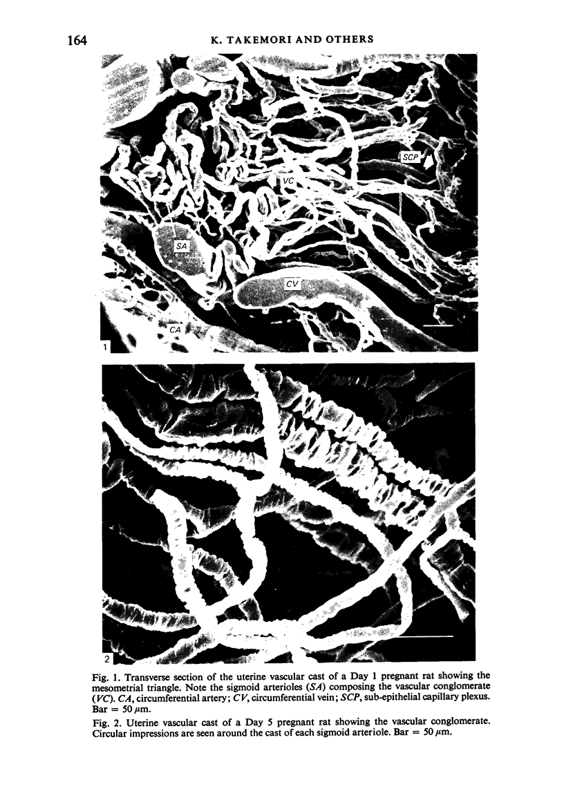
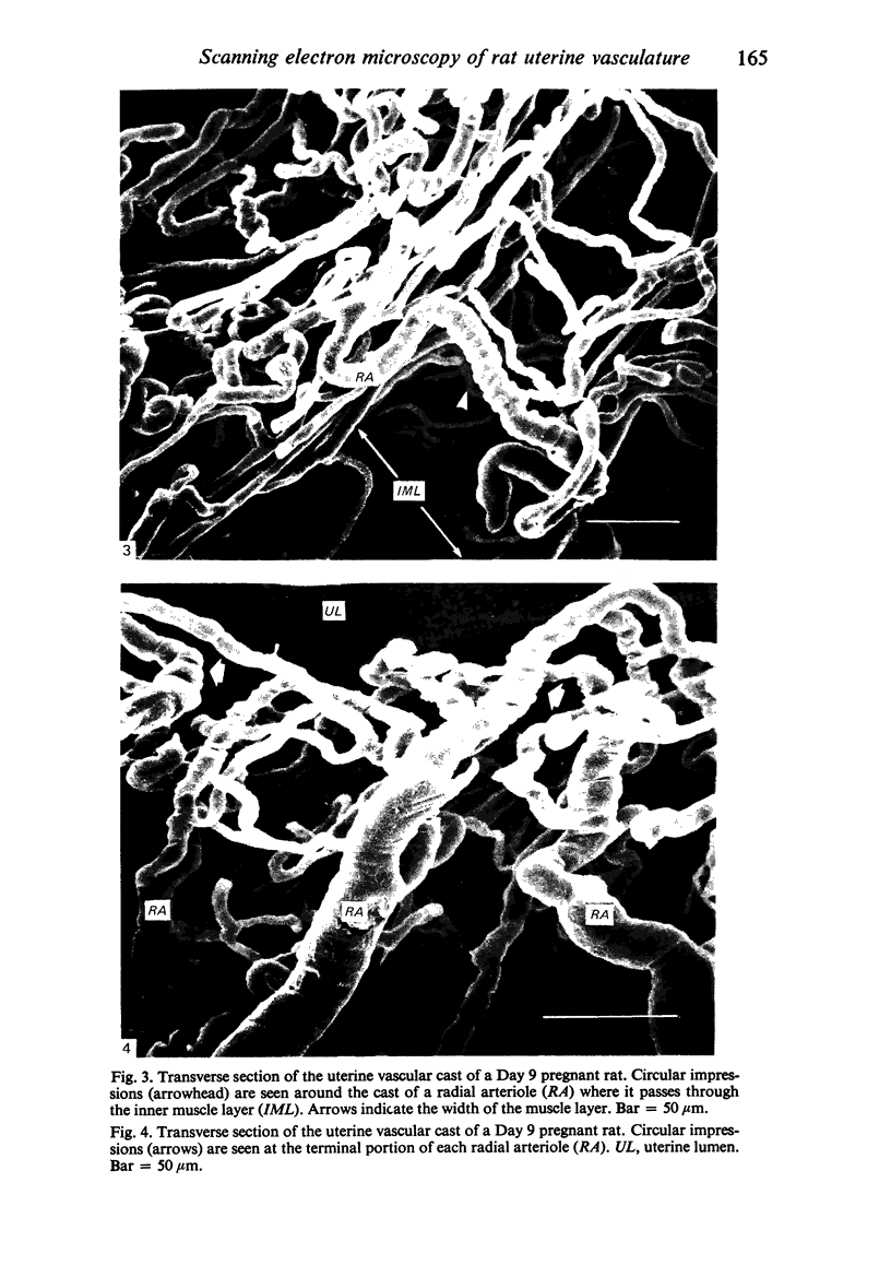
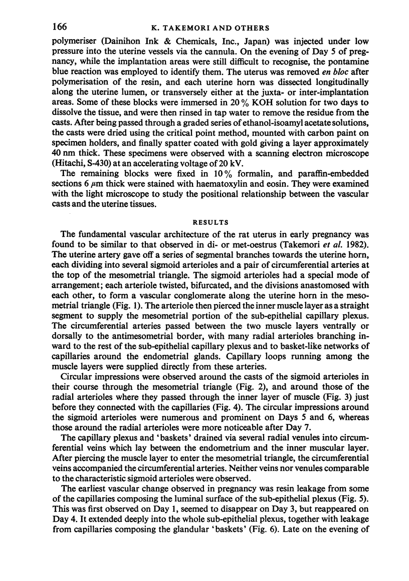
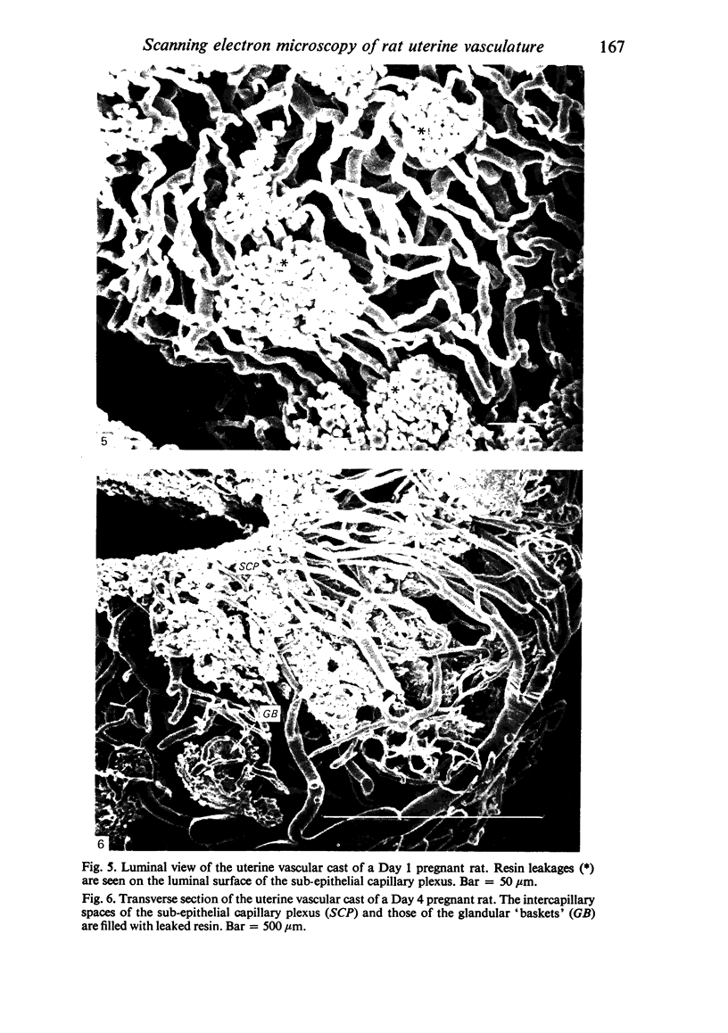
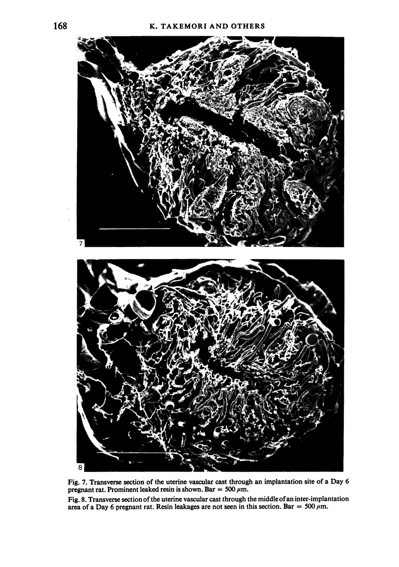
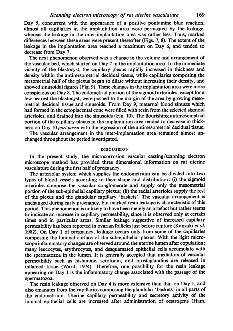
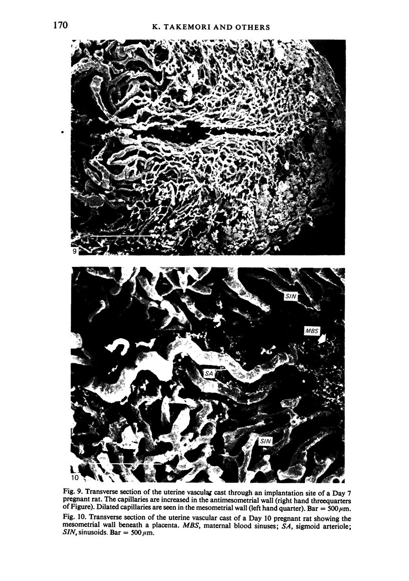
Vascular changes in the rat uterus during the first half of pregnancy were studied by the microcorrosion casting/scanning electron microscope method. Arterioles supplying the endometrium were divided into two groups according to differences in their shape, distribution, and development. Circular impressions indicative of the presence of vascular sphincters were observed around the casts of each group of arterioles. Resin leakage, suggesting an increase in vascular permeability, was observed from capillaries composing the sub-epithelial plexus and the glandular 'baskets'. Although leakage was noted throughout the endometrium before implantation, it tended to be localised around each blastocyst after the onset of implantation. The present results suggest the existence of a special control mechanism of the uterine blood flow, and that the uterine vasculature is affected by the conceptus from the beginning of implantation.
Full text
PDF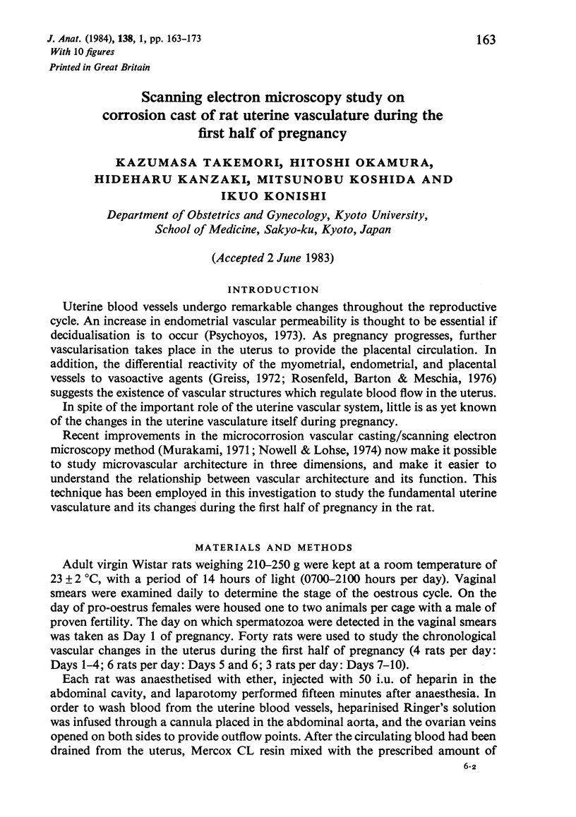
Images in this article
Selected References
These references are in PubMed. This may not be the complete list of references from this article.
- Adham N., Schenk E. A. Autonomic innervation of the rat vagina, cervix, and uterus and its cyclic cariation. Am J Obstet Gynecol. 1969 Jun 15;104(4):508–516. doi: 10.1016/s0002-9378(16)34239-9. [DOI] [PubMed] [Google Scholar]
- Anderson B. G., Anderson W. D. Scanning electron microscopy of microcorrosion casts; intracranial and abdominal microvasculature in domestic animals. Am J Anat. 1978 Dec;153(4):523–536. doi: 10.1002/aja.1001530404. [DOI] [PubMed] [Google Scholar]
- Czekanowski R. Investigations into the spontaneous contractile activity of isolated human uterine arteries in vitro. Am J Obstet Gynecol. 1975 Mar 1;121(5):718–722. doi: 10.1016/0002-9378(75)90479-2. [DOI] [PubMed] [Google Scholar]
- Dickmann Z., Gupta J. S., Dey S. K. Does "blastocyst estrogen" initiate implantation? Science. 1977 Feb 18;195(4279):687–688. doi: 10.1126/science.841306. [DOI] [PubMed] [Google Scholar]
- Finn C. A., Martin L. Hormonal control of the secretion of the endometrial glands in the mouse. J Endocrinol. 1976 Nov;71(2):273–274. doi: 10.1677/joe.0.0710273. [DOI] [PubMed] [Google Scholar]
- Greiss F. C., Jr Differential reactivity of the myoendometrial and placental vasculatures: adrenergic responses. Am J Obstet Gynecol. 1972 Jan 1;112(1):20–30. doi: 10.1016/0002-9378(72)90524-8. [DOI] [PubMed] [Google Scholar]
- Ham K. N., Hurley J. V., Lopata A., Ryan G. B. A combined isotopic and electron microscopic study of the response of the rat uterus to exogenous oestradiol. J Endocrinol. 1970 Jan;46(1):71–81. doi: 10.1677/joe.0.0460071. [DOI] [PubMed] [Google Scholar]
- Hashimoto I., Henricks D. M., Anderson L. L., Melampy R. M. Progesterone and pregn-4-en-20 alpha-ol-3-one in ovarian venous blood during various reproductive states in the rat. Endocrinology. 1968 Feb;82(2):333–341. doi: 10.1210/endo-82-2-333. [DOI] [PubMed] [Google Scholar]
- Kanzaki H., Okamura H., Okuda Y., Takenaka A., Morimoto K., Nishimura T. Scanning electron microscopic study of rabbit ovarian follicle microvasculature using resin injection-corrosion casts. J Anat. 1982 Jun;134(Pt 4):697–704. [PMC free article] [PubMed] [Google Scholar]
- Kennedy T. G. Evidence for a role for prosaglandins in the initiation of blastocyst implantation in the rat. Biol Reprod. 1977 Apr;16(3):286–291. doi: 10.1095/biolreprod16.3.286. [DOI] [PubMed] [Google Scholar]
- Murakami T. Application of the scanning electron microscope to the study of the fine distribution of the blood vessels. Arch Histol Jpn. 1971 Feb;32(5):445–454. doi: 10.1679/aohc1950.32.445. [DOI] [PubMed] [Google Scholar]
- Rosenfeld C. R., Barton M. D., Meschia G. Effects of epinephrine on distribution of blood flow in the pregnant ewe. Am J Obstet Gynecol. 1976 Jan 15;124(2):156–163. doi: 10.1016/s0002-9378(16)33292-6. [DOI] [PubMed] [Google Scholar]
- Surani M. A. Uterine luminal proteins at the time of implantation in rats. J Reprod Fertil. 1976 Sep;48(1):141–145. doi: 10.1530/jrf.0.0480141. [DOI] [PubMed] [Google Scholar]
- Takemori K., Okamura H., Kanzaki H., Okuda Y., Fujii S. [Scanning electron microscopic observations of microcorrosion casts for non pregnant rat uterine vascular architecture (author's transl)]. Nihon Sanka Fujinka Gakkai Zasshi. 1982 Jun;34(6):729–737. [PubMed] [Google Scholar]
- Ward P. A. The inflammatory mediators. Ann N Y Acad Sci. 1974;221:290–298. doi: 10.1111/j.1749-6632.1974.tb28228.x. [DOI] [PubMed] [Google Scholar]
- Yoshinaga K., Hawkins R. A., Stocker J. F. Estrogen secretion by the rat ovary in vivo during the estrous cycle and pregnancy. Endocrinology. 1969 Jul;85(1):103–112. doi: 10.1210/endo-85-1-103. [DOI] [PubMed] [Google Scholar]


