Abstract
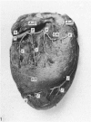
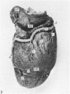
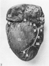
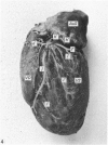
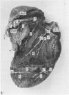
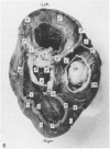
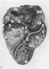
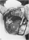
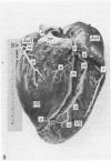
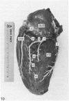
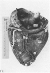
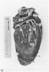
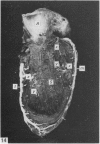
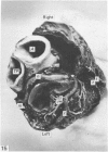
The distribution of the coronary arteries of the ostrich is described and compared with existing accounts of other species of birds. The blood supply to the ventricular walls, part of the interventricular septum and atria comes from the superficial branches of the left and right coronary arteries. The deep branches are small, supplying most of the interventricular septum and part of the right atrioventricular valve. The left and right coronary arteries are of equal size, forming a balanced circulation. Numerous homocoronary and intercoronary anastomoses are present. The venous drainage of the ostrich heart corresponds in the main to that of the fowl. Four major systems of veins are seen with multiple anastomoses between them. The major trunks are located underneath the epicardium and apart from some of the ventral cardiac veins, are concomitant veins of the arteries. The intra-atrial openings of the left cardiac, left cardiac circumflex and dorsal cardiac veins lie near to but separate from each other in a sinus below the intra-atrial opening of the left cranial vena cava. The dorsal cardiac vein consists of two branches. In some hearts the two branches do not unite, in which case the right branch opens separately into the right atrium, dorsal to the sinus, while the left branch opens into the sinus. Many luminal cardiac veins are seen, draining the interventricular septum, right atrioventricular valve and to a lesser extent the right atrium. The right atrioventricular valve is drained mainly by a subendocardial vein, opening directly into the right atrium or into a ventral cardiac vein.
Full text
PDF
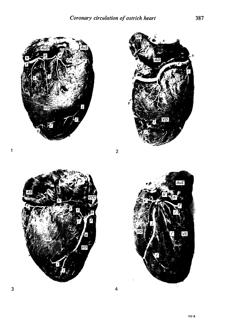
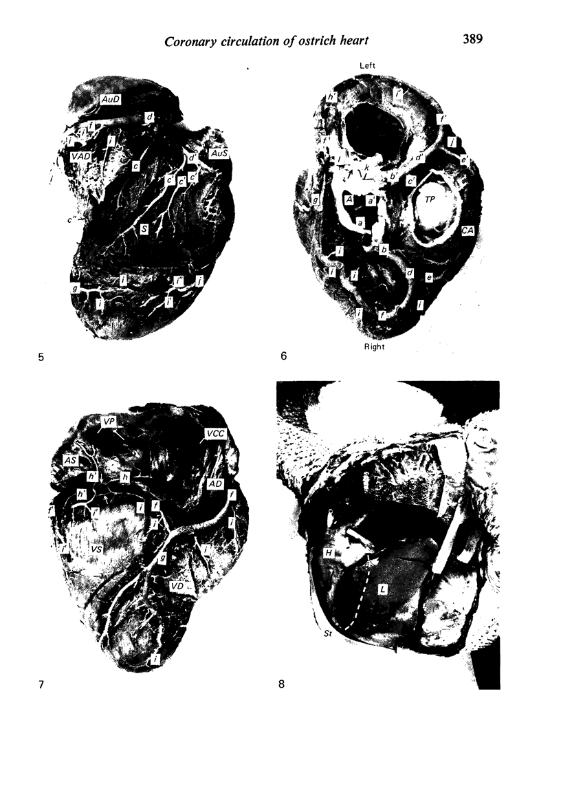
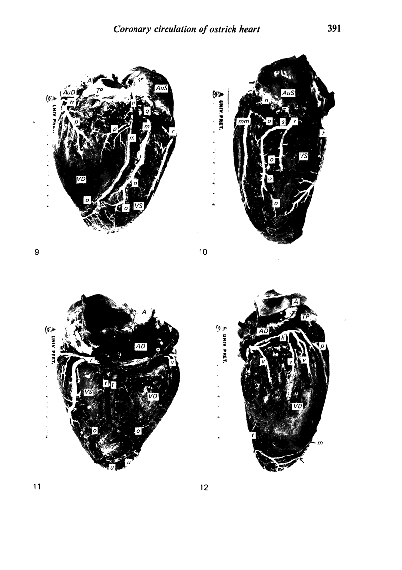
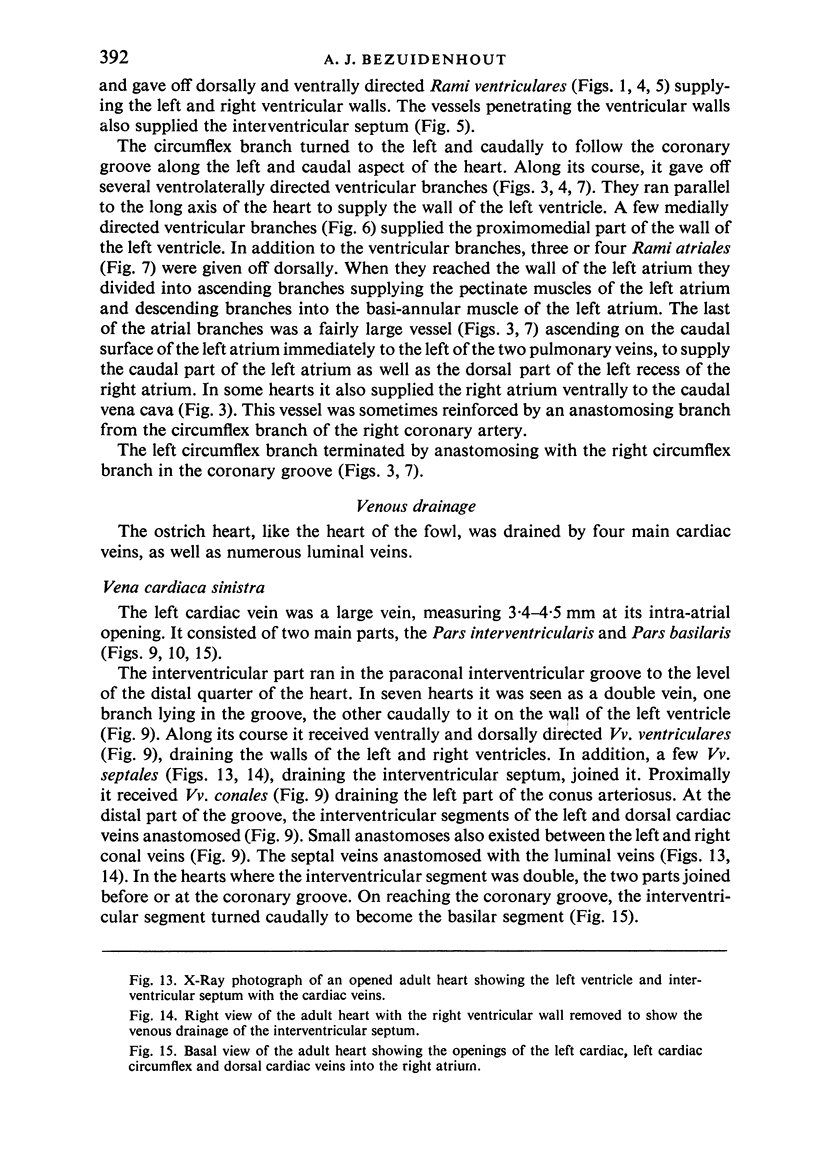
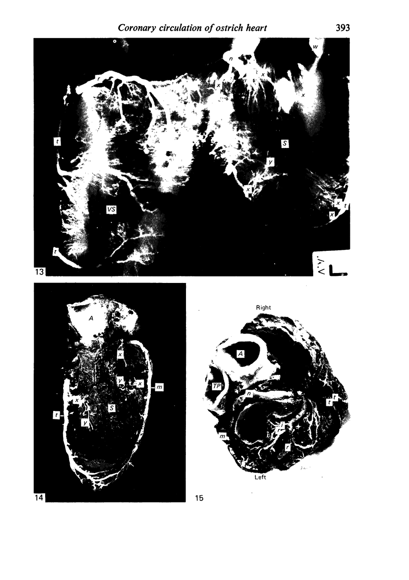
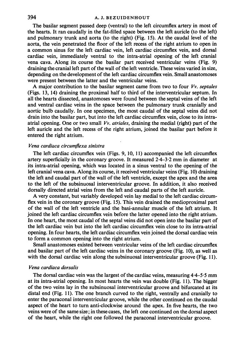
Images in this article
Selected References
These references are in PubMed. This may not be the complete list of references from this article.
- BAROLDI G., MANTERO O., SCOMAZZONI G. The collaterals of the coronary arteries in normal and pathologic hearts. Circ Res. 1956 Mar;4(2):223–229. doi: 10.1161/01.res.4.2.223. [DOI] [PubMed] [Google Scholar]
- LINDSAY F. E., SMITH H. J. CORONARY ARTERIES OF GALLUS DOMESTICUS. Am J Anat. 1965 Jan;116:301–314. doi: 10.1002/aja.1001160115. [DOI] [PubMed] [Google Scholar]
- Lindsay F. E. The cardiac veins of Gallus domesticus. J Anat. 1967 Jun;101(Pt 3):555–568. [PMC free article] [PubMed] [Google Scholar]
- Nerantzis C., Antonakis E., Avgoustakis D. A new corrosion casting technique. Anat Rec. 1978 Jul;191(3):321–325. doi: 10.1002/ar.1091910305. [DOI] [PubMed] [Google Scholar]
- Rigdon R. H., Frölich J. The heart of the duck. Zentralbl Veterinarmed A. 1970 Jan;17(1):85–94. doi: 10.1111/j.1439-0442.1970.tb00777.x. [DOI] [PubMed] [Google Scholar]


