Abstract
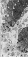
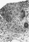
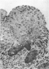
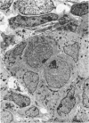
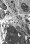
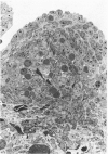
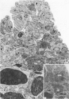
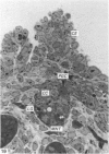
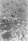
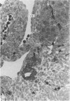
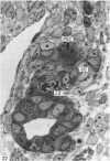
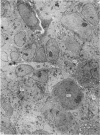
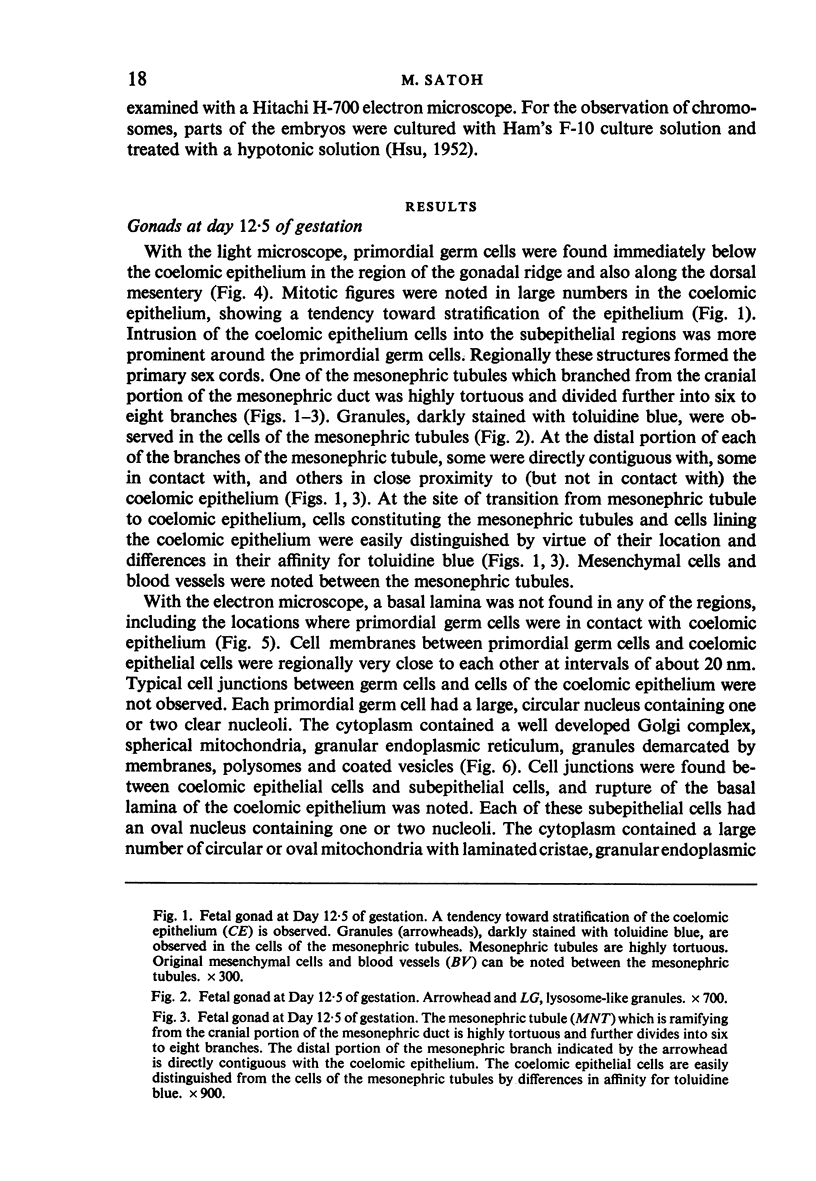
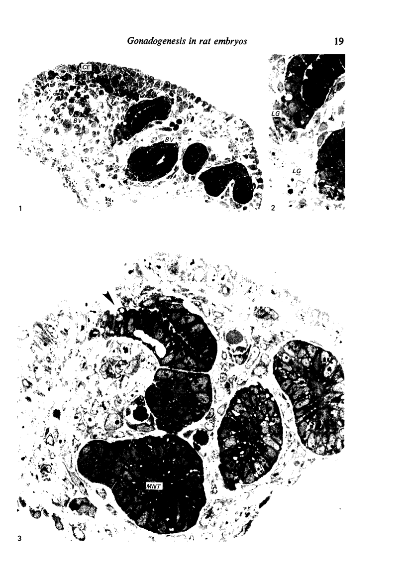
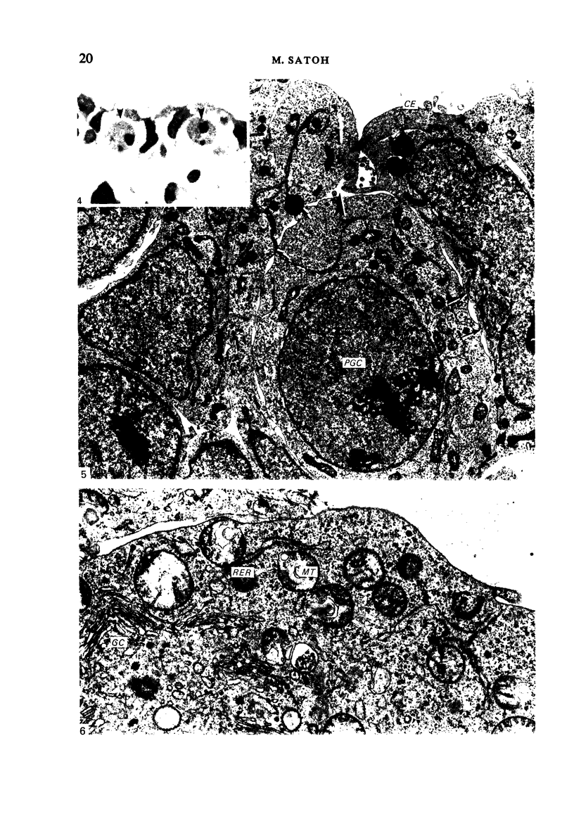
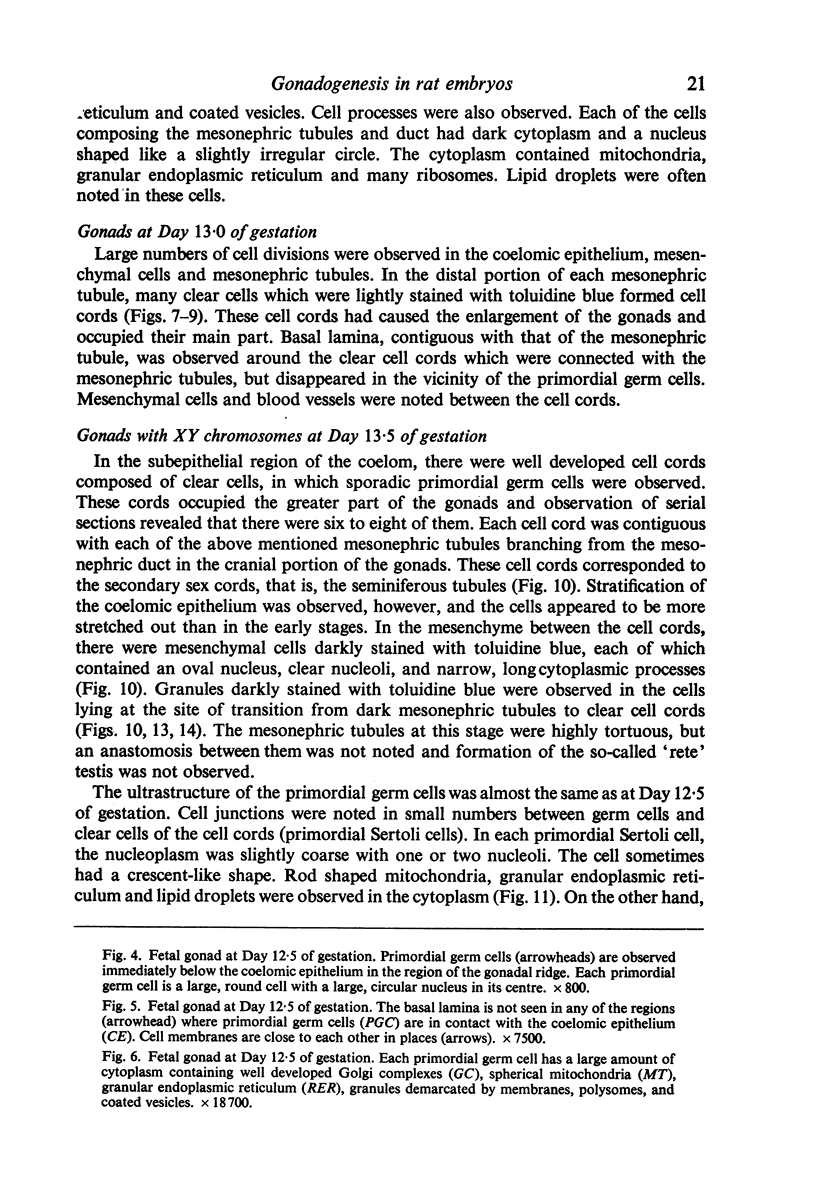
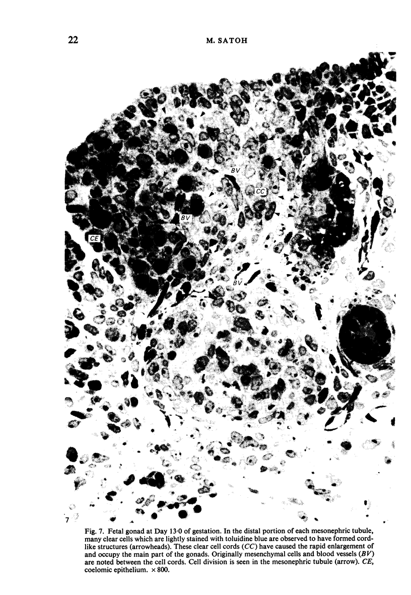
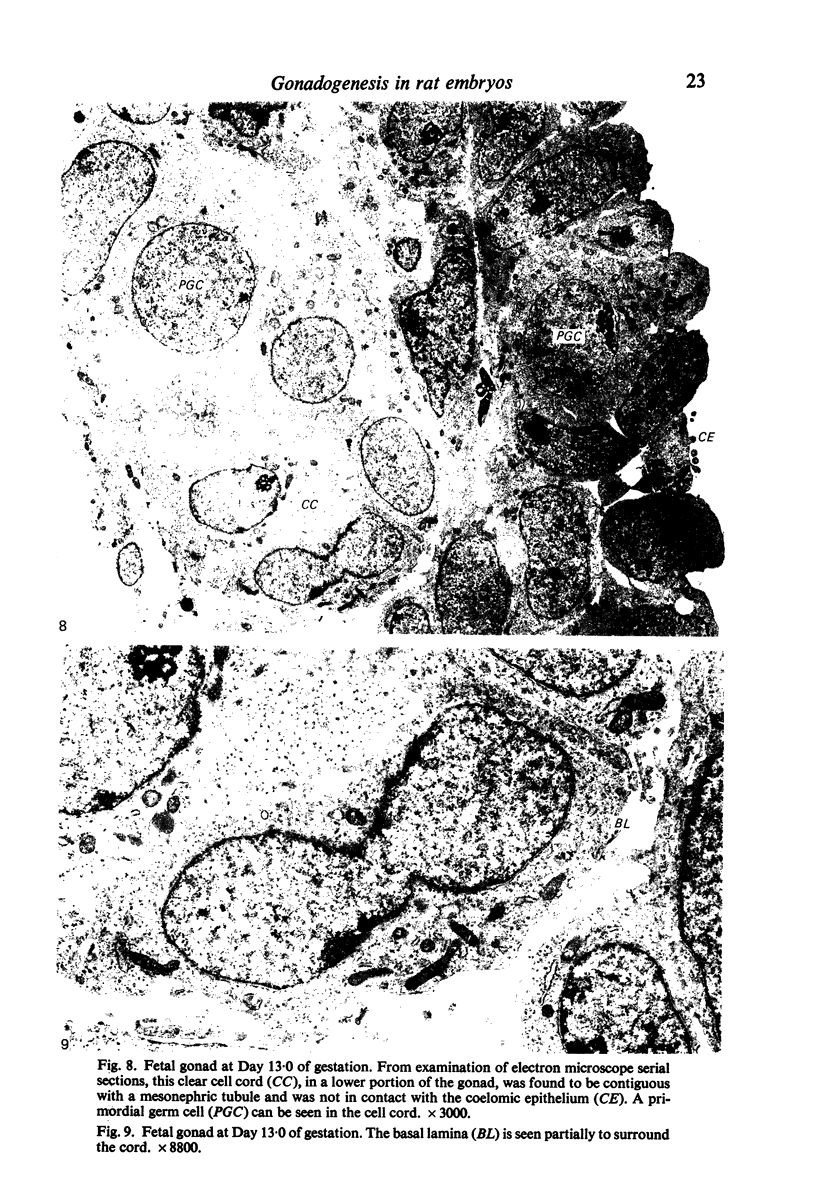
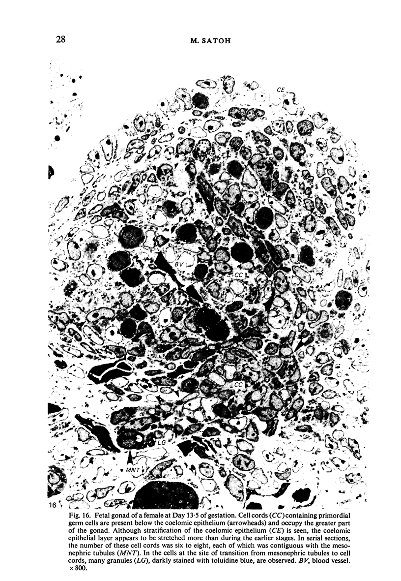
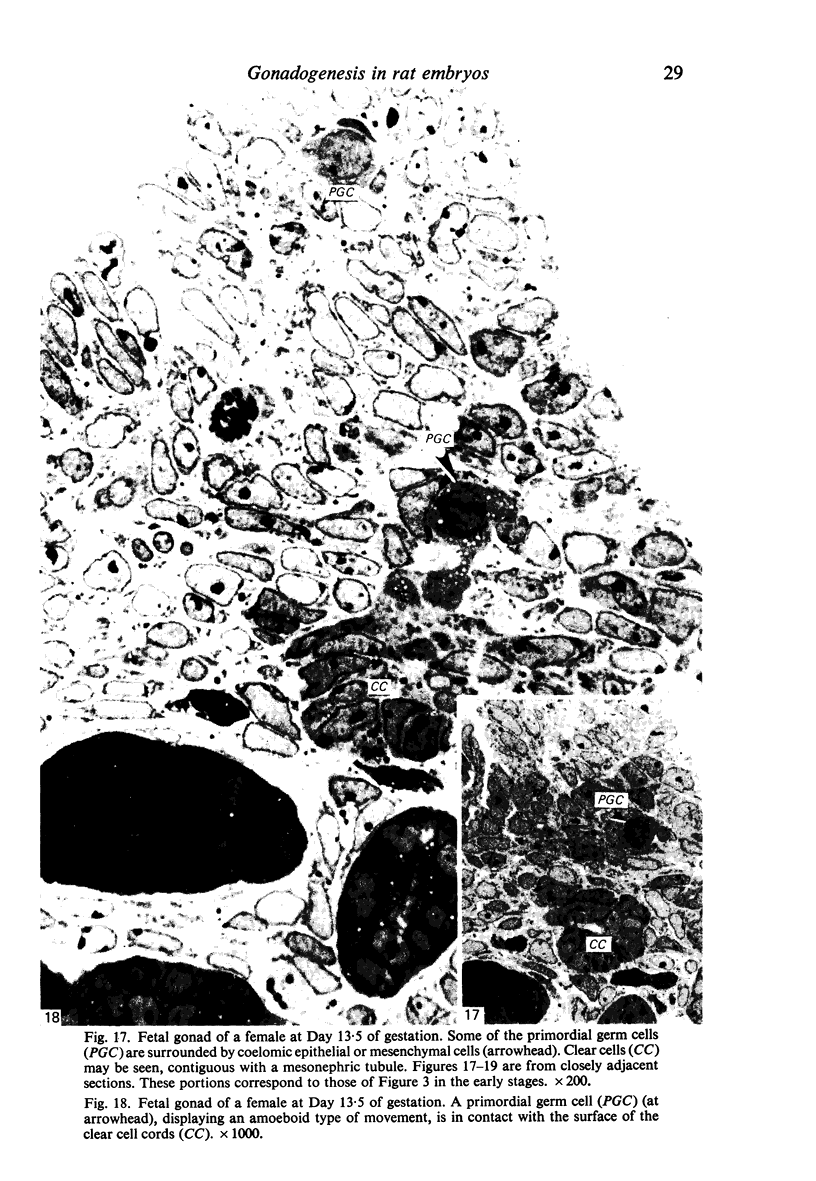
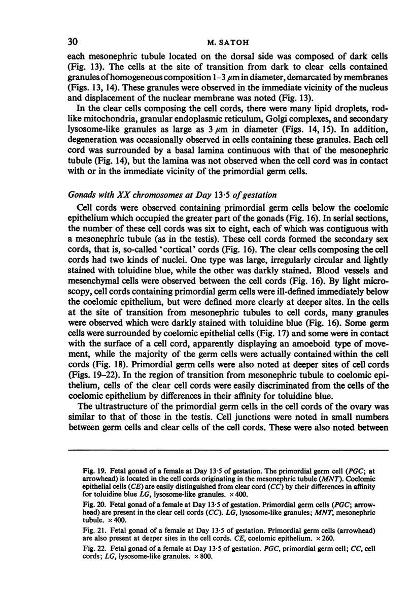
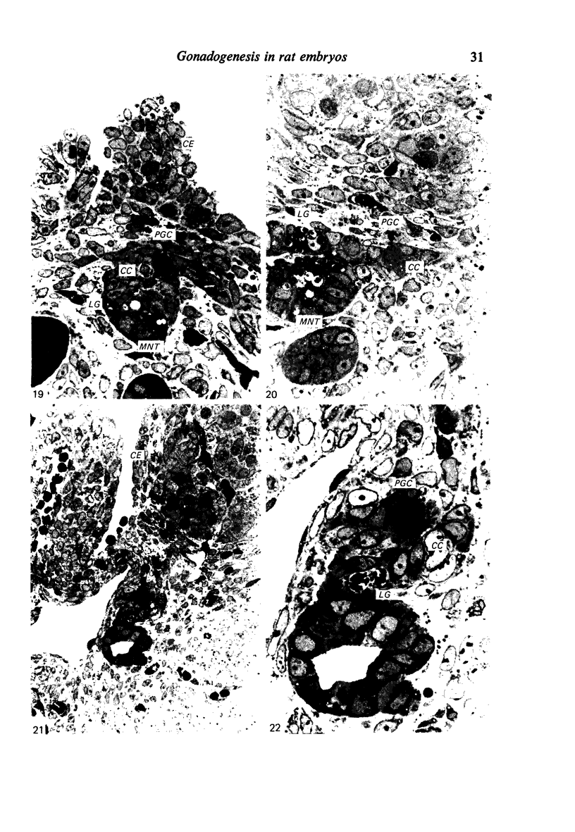
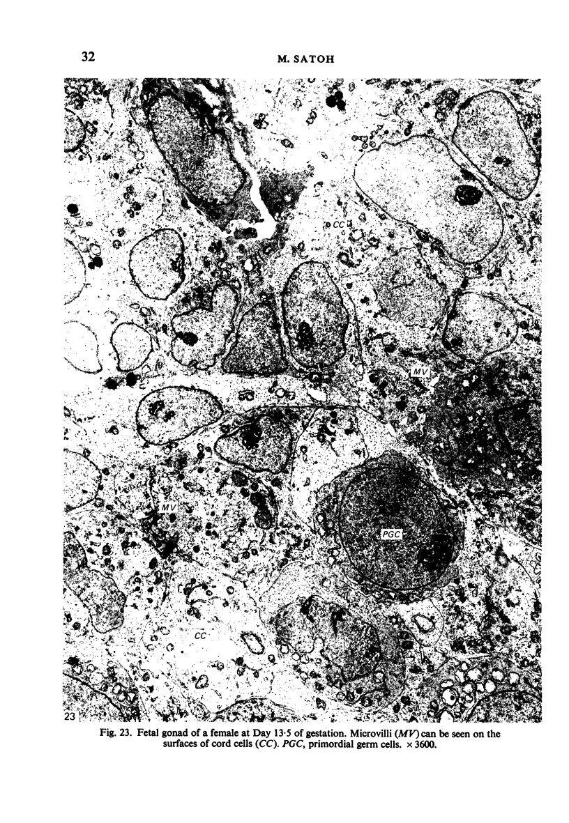
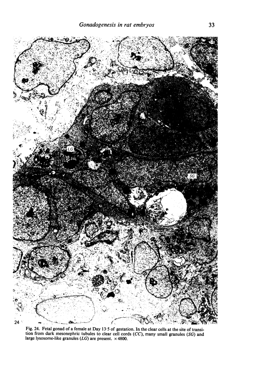
Gonadogenesis was investigated using Wistar rat embryos at 12-14 days after fertilisation. In indifferent gonads, a mesonephric tubule which bifurcates from the mesonephric duct at the upper end of the gonadal anlage ramifies into six to eight branches, the distal portions of which are contiguous, in contact with, or in close proximity to, the coelomic epithelium of the gonadal ridge. After the primordial germ cells reach the gonadal ridge, the overlying epithelium proliferates and clear cells appear in the distal portion of each mesonephric tubule, proliferating and forming cord-like structures. The primordial germ cells appear to enter these cell cords by an amoeboid type of movement. The basal lamina covering the cell cord partially disappears near the germ cells. The germ cells within the cord migrate toward the proximal portion of the cell cord and proliferate in great profusion. From the present observations, it can be concluded that the gonad is mainly formed of clear cell cords originating in mesonephric tubules into which germ cells have entered. The original mesenchymal cells and blood vessels form the interstitial tissue of the gonad. The rete testis and rete ovarii are of mesonephric tubule origin.
Full text
PDF
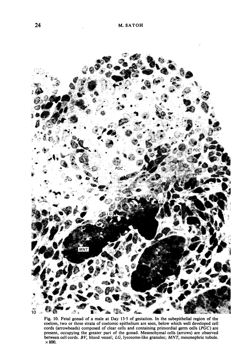
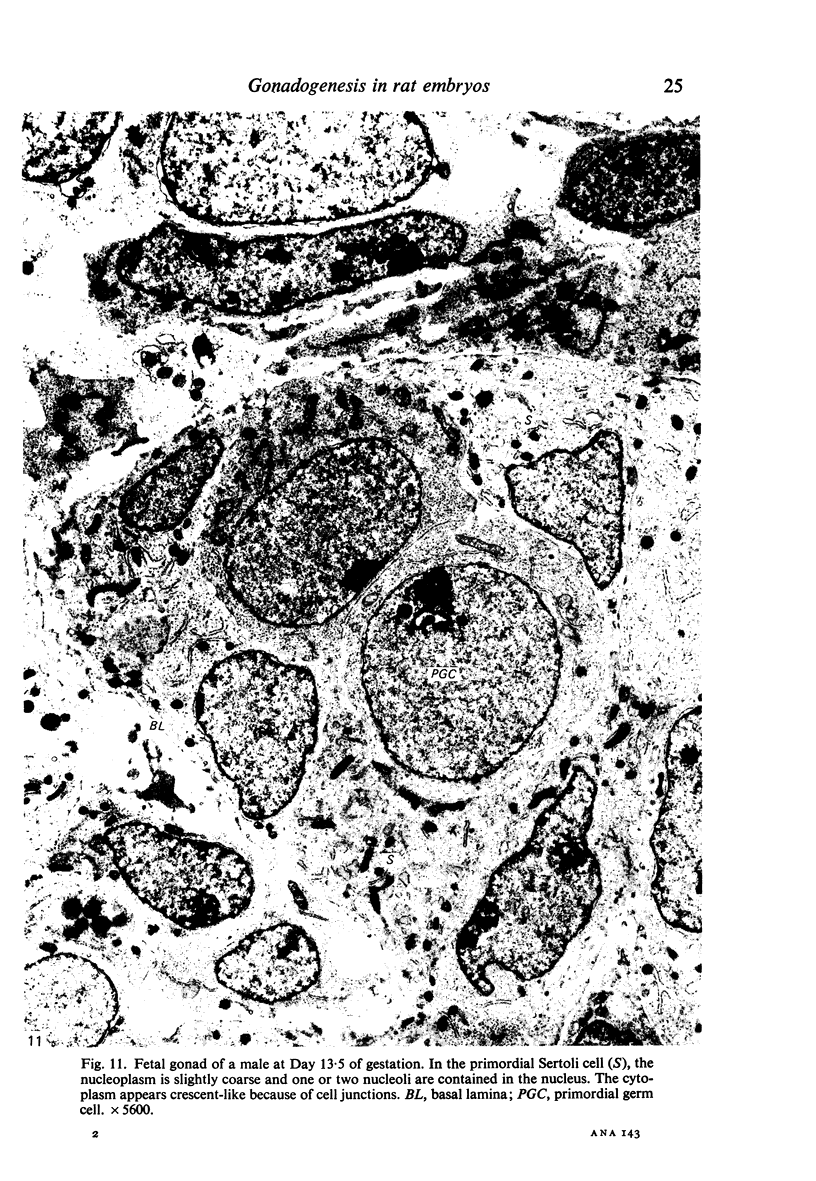
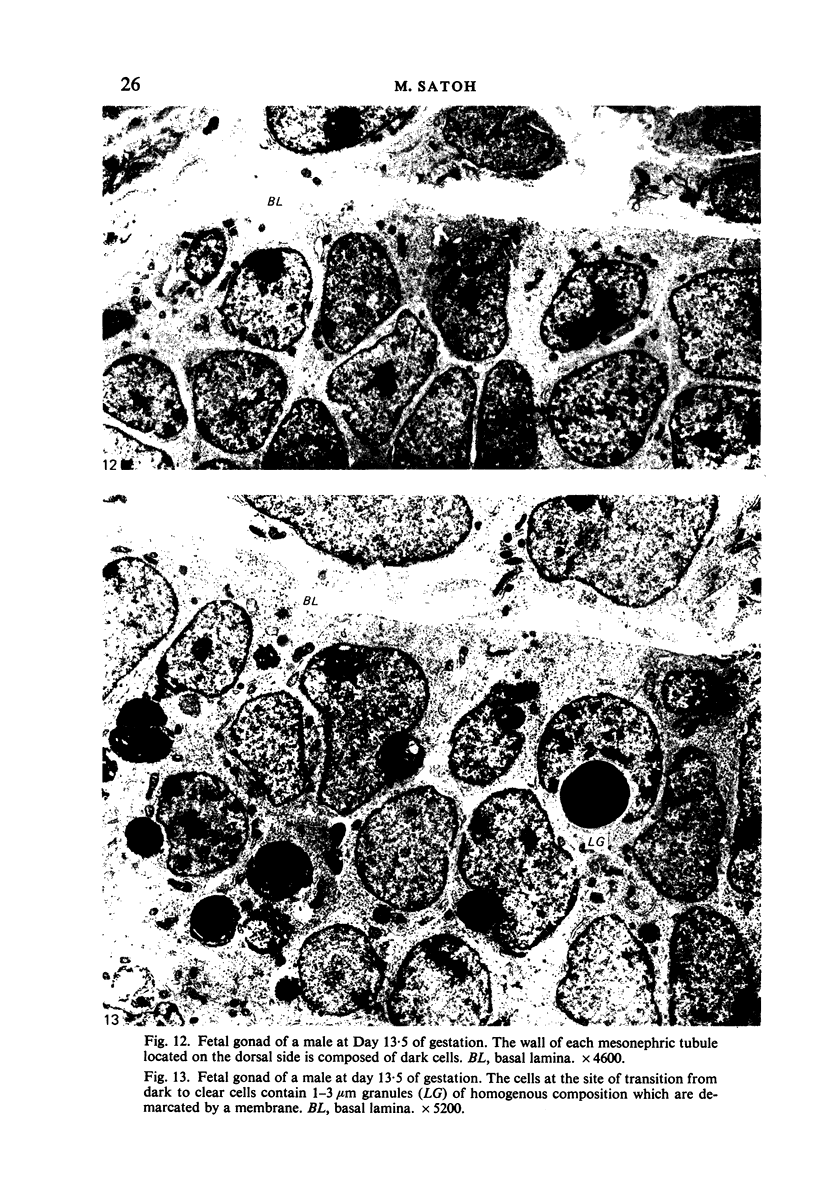
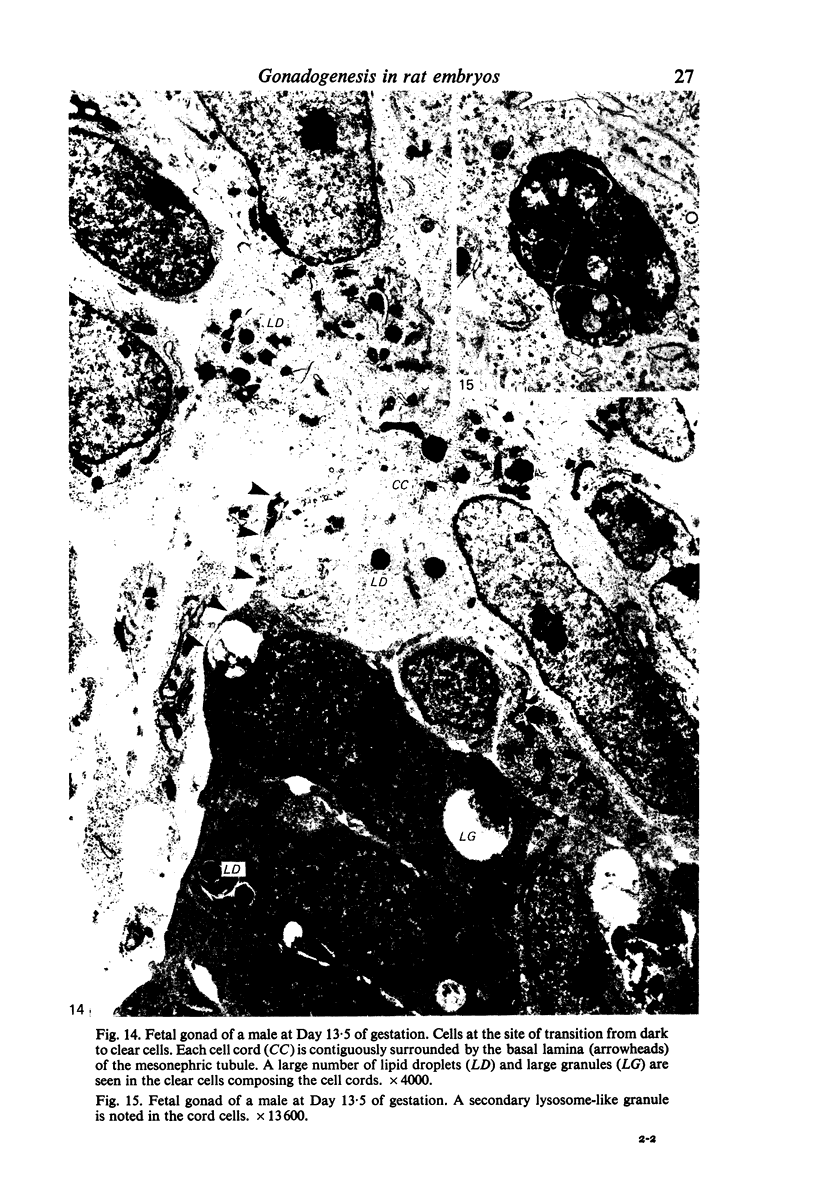
Images in this article
Selected References
These references are in PubMed. This may not be the complete list of references from this article.
- Byskov A. G., Lintern-Moore S. Follicle formation in the immature mouse ovary: the role of the rete ovarii. J Anat. 1973 Nov;116(Pt 2):207–217. [PMC free article] [PubMed] [Google Scholar]
- Gropp A., Ohno S. The presence of a common embryonic blastema for ovarian and testicular parenchymal (follicular, interstitial and tubular) cells in cattle Bos taurus. Z Zellforsch Mikrosk Anat. 1966;74(4):505–528. doi: 10.1007/BF00496841. [DOI] [PubMed] [Google Scholar]
- Merchant H. Rat gonadal and ovarioan organogenesis with and without germ cells. An ultrastructural study. Dev Biol. 1975 May;44(1):1–21. doi: 10.1016/0012-1606(75)90372-3. [DOI] [PubMed] [Google Scholar]
- PINKERTON J. H., McKAY D. G., ADAMS E. C., HERTIG A. T. Development of the human ovary--a study using histochemical technics. Obstet Gynecol. 1961 Aug;18:152–181. [PubMed] [Google Scholar]
- Pelliniemi L. J. Ultrastructure of the indifferent gonad in male and female pig embryos. Tissue Cell. 1976;8(1):163–174. doi: 10.1016/0040-8166(76)90028-8. [DOI] [PubMed] [Google Scholar]
- REYNOLDS E. S. The use of lead citrate at high pH as an electron-opaque stain in electron microscopy. J Cell Biol. 1963 Apr;17:208–212. doi: 10.1083/jcb.17.1.208. [DOI] [PMC free article] [PubMed] [Google Scholar]


