Abstract
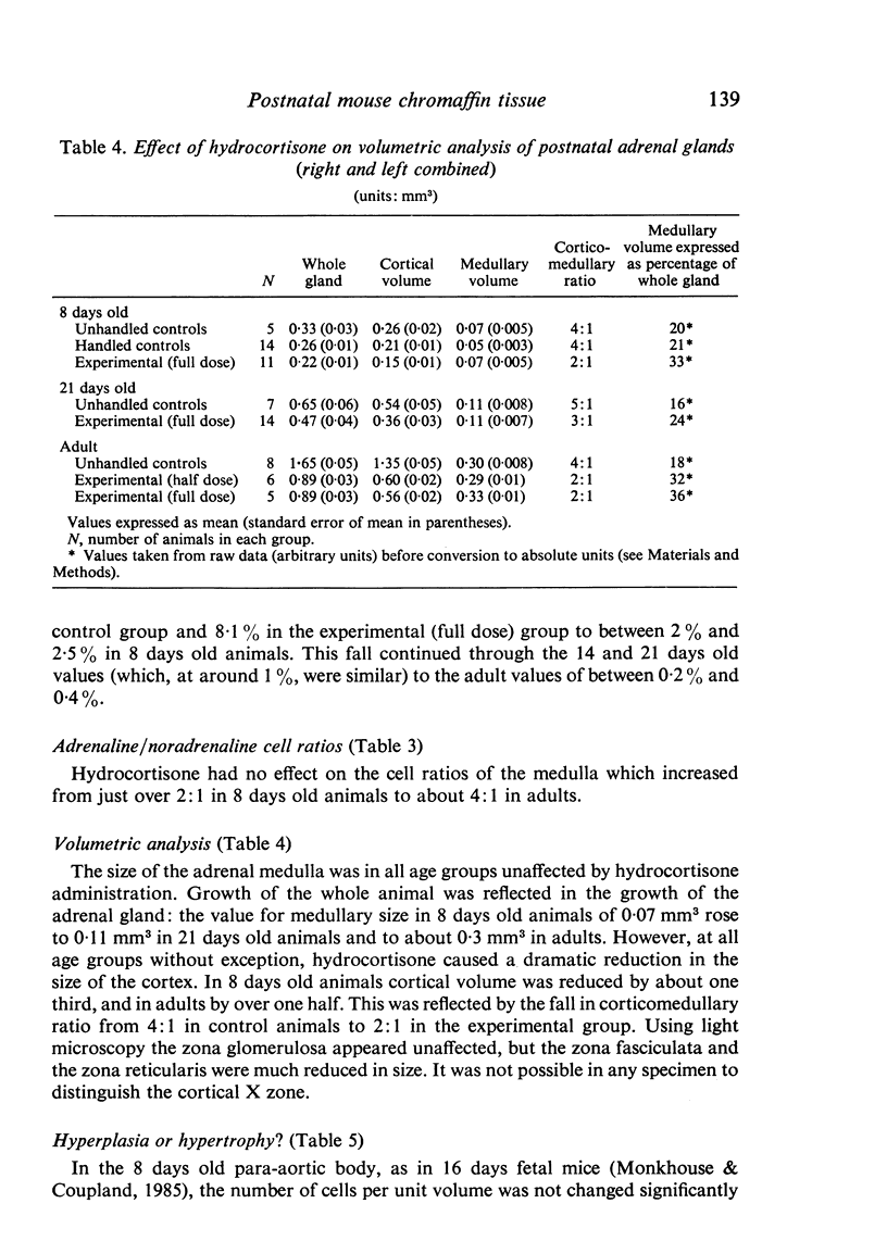
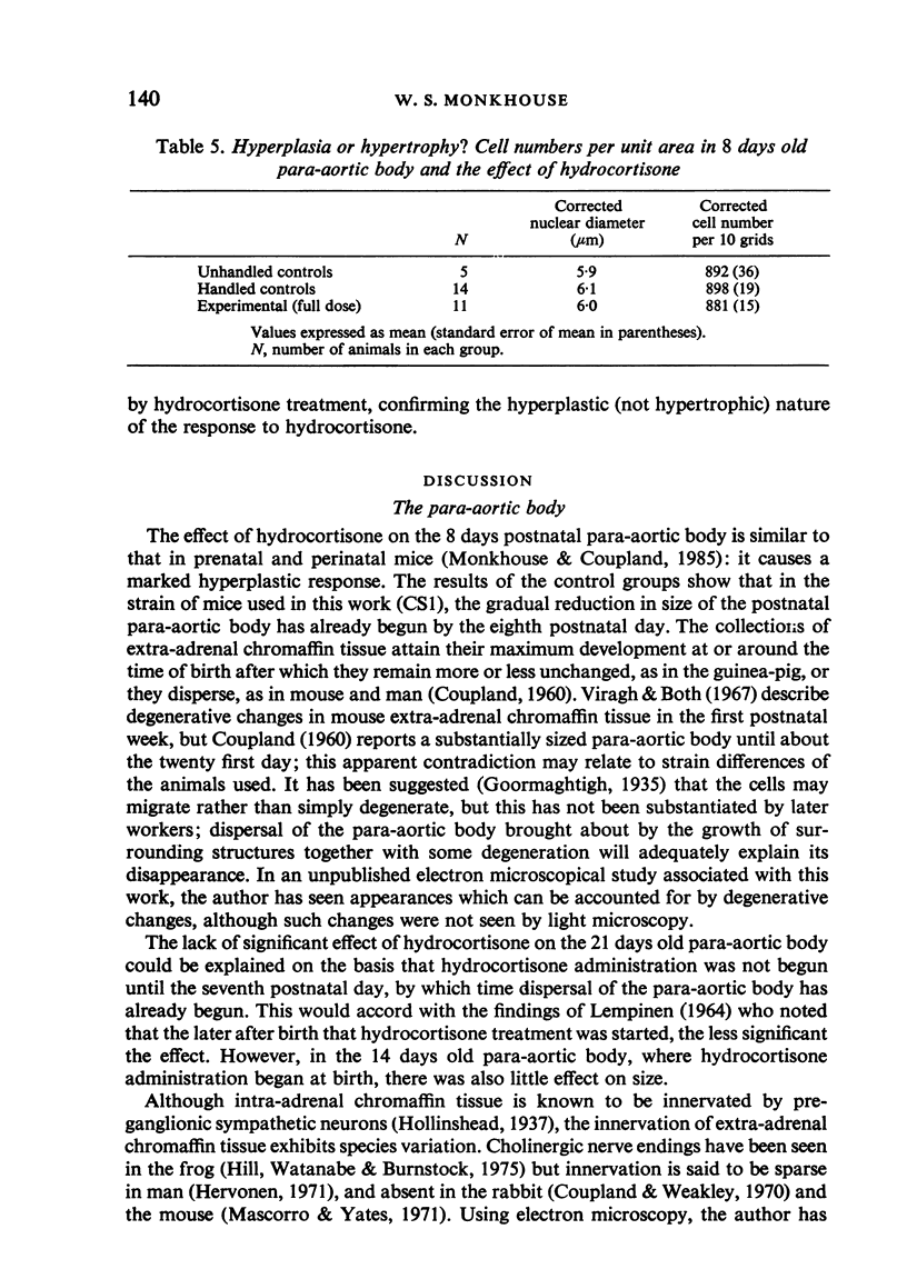
Hydrocortisone administration in vivo to neonatal mice for seven days led to a significant increase in both the size and the labelling index of extra-adrenal chromaffin tissue (as represented by the para-aortic body) of 8 days old mice. In untreated animals at this age, the para-aortic body was in most cases too small to obtain a valid labelling index. In the para-aortic bodies of 14 days old, 21 days old and adult mice, the extra-adrenal chromaffin tissue was too dispersed to obtain values for either volumetric analysis or labelling indices, and hydrocortisone was without significant effect in promoting a hyperplastic response. In the postnatal adrenal medulla at all ages studied, hydrocortisone had no effect on the medullary size or on the labelling indices of either adrenaline- or noradrenaline-storing cells, although it led to a marked diminution of adrenocortical volume. The relative proportion of adrenaline-storing cells increased between the values for 8 days old animals and those for adults; this was unaffected by hydrocortisone. The cortico-medullary ratio remained unchanged from the eighth postnatal day onwards. The results are discussed and related to those of other workers. It is suggested that factors as yet unknown might modulate the response to corticosteroids of developing intra- and extra-adrenal chromaffin tissue.
Full text
PDF
Images in this article
Selected References
These references are in PubMed. This may not be the complete list of references from this article.
- Benedetti A. Mitotic activity of adrenal medullary cells in the mouse at different ages and following unilateral adrenalectomy. Experientia. 1976 Jan 15;32(1):108–109. doi: 10.1007/BF01932650. [DOI] [PubMed] [Google Scholar]
- Brundin T. Studies on the preaortal paraganglia of newborn rabbits. Acta Physiol Scand Suppl. 1966;290:1–54. [PubMed] [Google Scholar]
- COUPLAND R. E. The post-natal distribution of the abdominal chromaffin tissue in the guinea-pig, mouse and white rat. J Anat. 1960 Apr;94:244–256. [PMC free article] [PubMed] [Google Scholar]
- Coupland R. E., Weakley B. S. Electron microscopic observation on the adrenal medulla and extra-adrenal chromaffin tissue of the postnatal rabbit. J Anat. 1970 Mar;106(Pt 2):213–231. [PMC free article] [PubMed] [Google Scholar]
- DIDERHOLM H., HELLMAN B. The cell renewal in the rat adrenals studied with tritiated thymidine. Acta Pathol Microbiol Scand. 1960;49:82–88. doi: 10.1111/j.1699-0463.1960.tb01116.x. [DOI] [PubMed] [Google Scholar]
- Garvey D., Migally N., Sullivan J., Sullivan L. Suppression of adrenal cortical growth and differentiation in fetal rats exposed to dexamethasone. Anat Rec. 1983 Apr;205(4):431–439. doi: 10.1002/ar.1092050408. [DOI] [PubMed] [Google Scholar]
- Hervonen A. Development of catecholamine--storing cells in human fetal paraganglia and adrenal medulla. A histochemical and electron microscopical study. Acta Physiol Scand Suppl. 1971;368:1–94. [PubMed] [Google Scholar]
- Hill C. E., Watanabe H., Burnstock G. Distribution and morphology of amphibian extra-adrenal chromaffin tissue. Cell Tissue Res. 1975 Jul 16;160(3):371–387. doi: 10.1007/BF00222046. [DOI] [PubMed] [Google Scholar]
- Jurecka W., Lassmann H., Hörandner H. The proliferation of adrenal medullary cells in newborn and adult mice. A light and electron microscopic autoradiographic study. Cell Tissue Res. 1978 May 29;189(2):305–312. doi: 10.1007/BF00209279. [DOI] [PubMed] [Google Scholar]
- Lehmann A. R., Stevens S. A rapid procedure for measurement of DNA repair in human fibroblasts and for complementation analysis of xeroderma pigmentosum cells. Mutat Res. 1980 Jan;69(1):177–190. doi: 10.1016/0027-5107(80)90187-6. [DOI] [PubMed] [Google Scholar]
- Malvaldi G., Mencacci P., Viola-Magni M. P. Mitoses in the adrenal medullary cells. Experientia. 1968 May 15;24(5):475–476. doi: 10.1007/BF02144402. [DOI] [PubMed] [Google Scholar]
- Malvaldi G., Viola-Magni M. P. DNA turnover in adrenal medullary cells of different strains of rats and its enhancement after intermittent exposure to cold. Cell Tissue Kinet. 1972 Mar;5(2):103–113. doi: 10.1111/j.1365-2184.1972.tb01007.x. [DOI] [PubMed] [Google Scholar]
- Mascorro J. A., Yates R. D. Ultrastructural studies of the effects of reserpine on mouse abdominal sympathetic paraganglia. Anat Rec. 1971 Jul;170(3):269–279. doi: 10.1002/ar.1091700303. [DOI] [PubMed] [Google Scholar]
- Monkhouse W. S., Coupland R. E. The effect of in vivo hydrocortisone administration on the labelling index and size of the intra- and extra-adrenal chromaffin tissue of the fetal and perinatal mouse. J Anat. 1985 Jun;140(Pt 4):679–696. [PMC free article] [PubMed] [Google Scholar]
- Monkhouse W. S. The effect of section thickness and embedding media on the observed S-phase labelling index of artificially selected cell populations from neonatal mouse liver and spleen. J Anat. 1985 Aug;141:201–209. [PMC free article] [PubMed] [Google Scholar]
- Pelc S. R. Turnover of DNA and function. Nature. 1968 Jul 13;219(5150):162–163. doi: 10.1038/219162a0. [DOI] [PubMed] [Google Scholar]
- Virágh S., Both A. K. The fine structure of abdominal paraganglia in the newborn mouse. Acta Biol Acad Sci Hung. 1967;18(2):161–179. [PubMed] [Google Scholar]


