Abstract
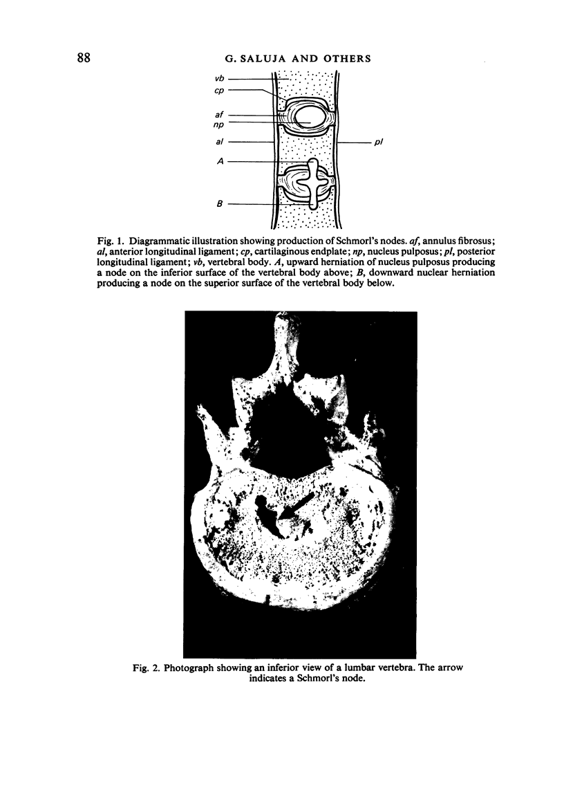
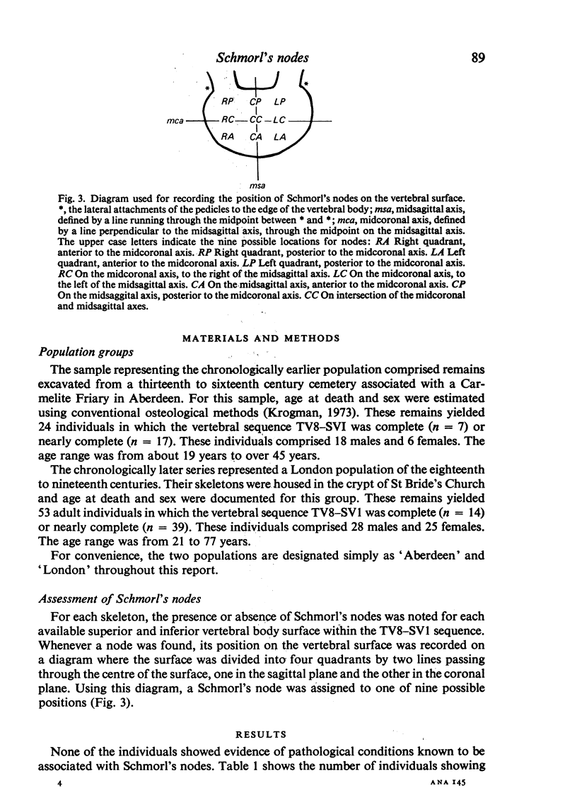
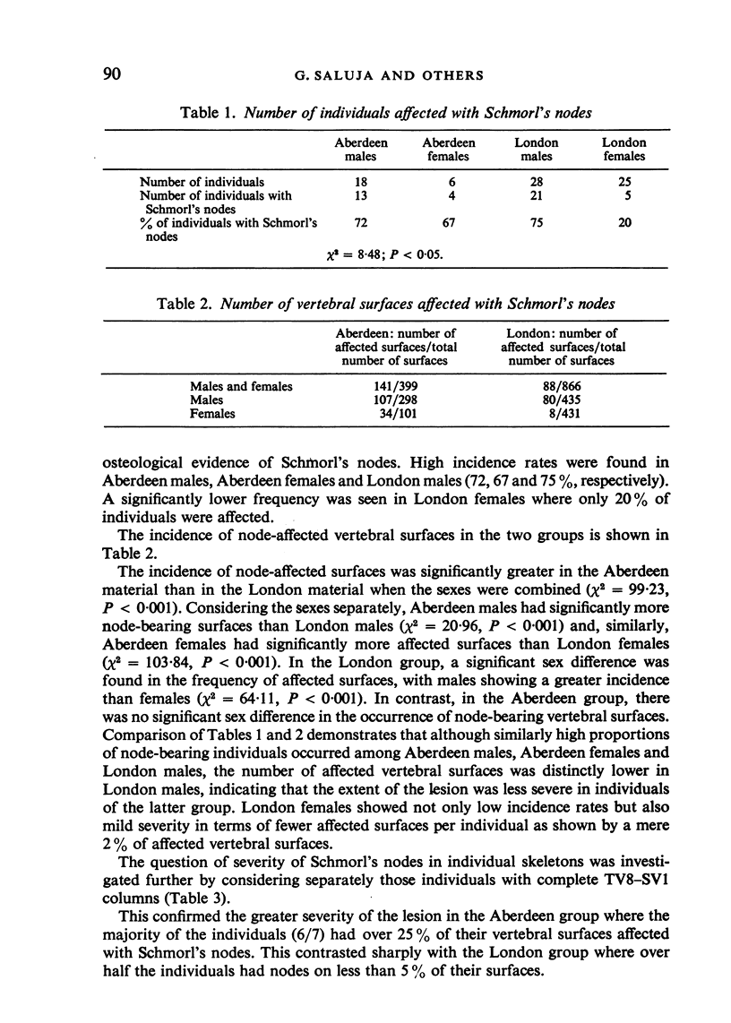
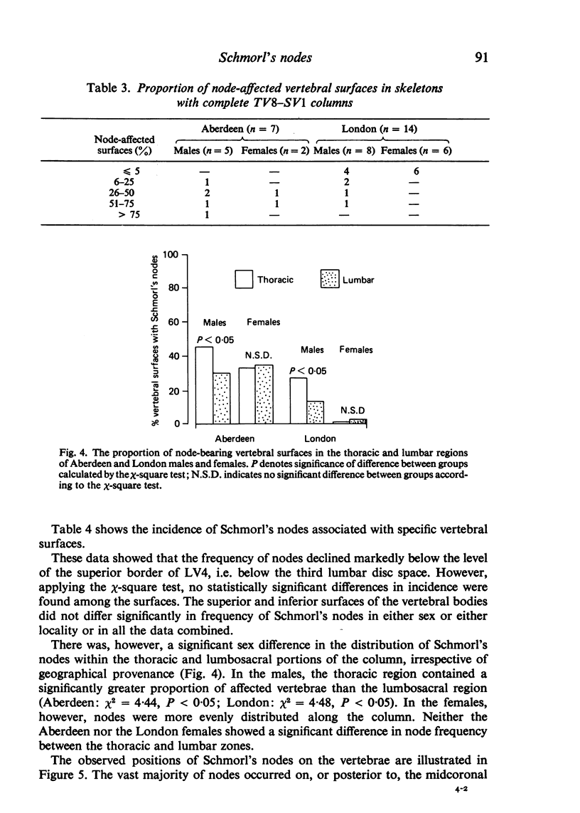
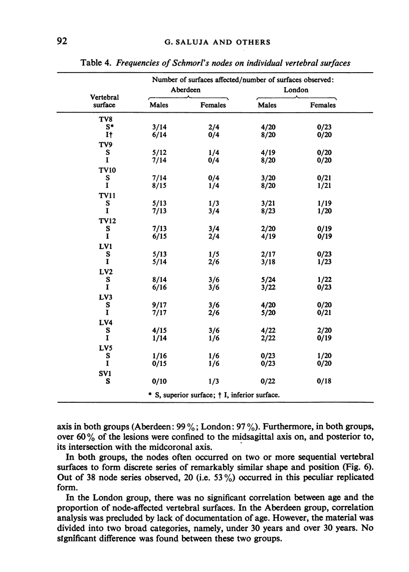
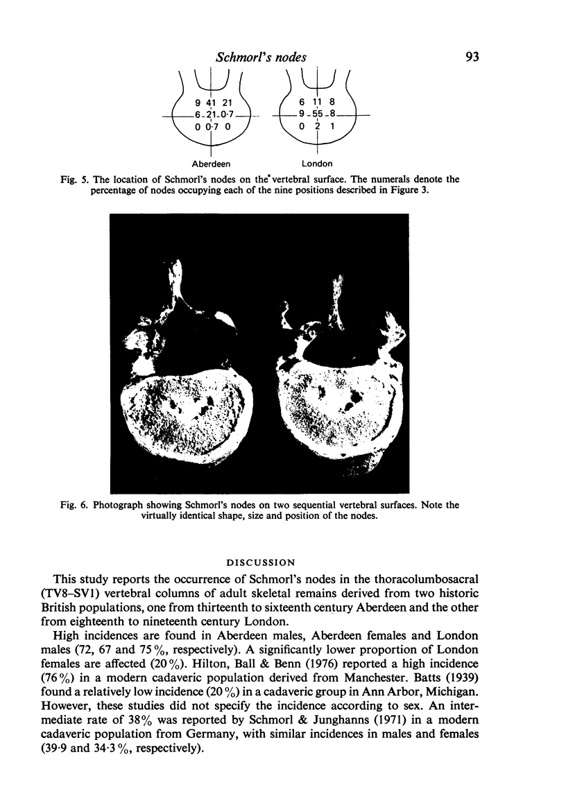
The herniation of the nucleus pulposus into the vertebral body produces ectopic deposit of disc material which are known as Schmorl's nodes. This prolapsed disc tissue leaves characteristic deformations on the surface of the vertebral body and hence the incidence of this lesion can be studied in skeletal remains. This report describes the occurrence of Schmorl's nodes in TV8-SV1 in two historic adult British populations, one from Aberdeen and the other from London. In the Aberdeen group, both males and females showed a high incidence rate and severity of Schmorl's nodes. In the London group, the males had a similarly high affliction whereas the females were nearly free of the condition. The lesion had no significant predilection for any one particular vertebral surface. However, in males in both localities, the frequency of Schmorl's nodes was significantly higher in the thoracic region than in the lumbosacral region. In contrast, both groups of females showed similar node frequency in these two zones. The majority of Schmorl's nodes were localised in the central and central-posterior regions of the vertebral surface. When nodes occurred on successive vertebral surfaces, they often formed sequences showing similar shape and position. The aetiology of Schmorl's nodes is unclear. Various hypothetical causal factors were appraised in relation to the findings of this study. It was suggested that anomalies in vascular and/or notochordal regression may be related to the development of the lesion.
Full text
PDF
Images in this article
Selected References
These references are in PubMed. This may not be the complete list of references from this article.
- Bagnall K. M., Harris P. F., Jones P. R. A radiographic study of the human fetal spine. 2. The sequence of development of ossification centres in the vertebral column. J Anat. 1977 Dec;124(Pt 3):791–802. [PMC free article] [PubMed] [Google Scholar]
- DENT C. E. Idiopathic osteoporosis. Proc R Soc Med. 1955 Jul;48(7):574–578. [PubMed] [Google Scholar]
- Hilton R. C., Ball J., Benn R. T. Vertebral end-plate lesions (Schmorl's nodes) in the dorsolumbar spine. Ann Rheum Dis. 1976 Apr;35(2):127–132. doi: 10.1136/ard.35.2.127. [DOI] [PMC free article] [PubMed] [Google Scholar]
- Maroudas A., Stockwell R. A., Nachemson A., Urban J. Factors involved in the nutrition of the human lumbar intervertebral disc: cellularity and diffusion of glucose in vitro. J Anat. 1975 Sep;120(Pt 1):113–130. [PMC free article] [PubMed] [Google Scholar]
- Nachemson A., Lewin T., Maroudas A., Freeman M. A. In vitro diffusion of dye through the end-plates and the annulus fibrosus of human lumbar inter-vertebral discs. Acta Orthop Scand. 1970;41(6):589–607. doi: 10.3109/17453677008991550. [DOI] [PubMed] [Google Scholar]
- Owsley D. W., Bradtmiller B. Mortality of pregnant females in Arikara villages: osteological evidence. Am J Phys Anthropol. 1983 Jul;61(3):331–336. doi: 10.1002/ajpa.1330610307. [DOI] [PubMed] [Google Scholar]
- Resnick D., Niwayama G. Intravertebral disk herniations: cartilaginous (Schmorl's) nodes. Radiology. 1978 Jan;126(1):57–65. doi: 10.1148/126.1.57. [DOI] [PubMed] [Google Scholar]


