PLOS ONE logoLink to PLOS ONE
. 2024 Dec 23;19(12):e0315868. doi: 10.1371/journal.pone.0315868

Biochemical analysis to study wild-type and polyglutamine-expanded ATXN3 species

Grégoire Quinet 1,2,*, María Cristina Paz-Cabrera 1, Raimundo Freire 1,2,3,*
Editor: Elise A Kikis4
PMCID: PMC11666052  PMID: 39715253

Abstract

Spinocerebellar ataxia type 3 (SCA3) is a cureless neurodegenerative disease recognized as the most prevalent form of dominantly inherited ataxia worldwide. The main hallmark of SCA3 is the expansion of a polyglutamine tract located in the C-terminal of Ataxin-3 (or ATXN3) protein, that triggers the mis-localization and toxic aggregation of ATXN3 in neuronal cells. The propensity of wild type and polyglutamine-expanded ATXN3 proteins to aggregate has been extensively studied over the last decades. In vitro studies with mass spectrometry techniques revealed a time-dependent aggregation of polyglutamine-expanded ATXN3 that occurs in several steps, leading to fibrils formation, a high status of aggregation. For in vivo experiments though, the techniques commonly used to demonstrate aggregation of polyglutamine proteins, such as filter trap assays, SDS-PAGE and SDS-AGE, are unable to unequivocally show all the stages of aggregation of wild type and polyglutamine-expanded ATXN3 proteins. Here we describe a systematic and detailed analysis of different known techniques to detect the various forms of both wild type and pathologic ATXN3 aggregates, and we discuss the power and limitation of each strategy.

Introduction

Polyglutamine (polyQ) disorders are associated with a pathological form of a protein harbouring an interrupted tract of polyQ, resulting from an expanded (CAG)n trinucleotide repeat within the coding region of the associated gene [1]. One of these proteins is Ataxin-3 or ATXN3 (UniProt ID P54252), an evolutionary conserved protein of a theoretical length of 42 kDa from the family of cysteine proteases, that harbours, among others, a Josephin Domain and a polyQ repeat [2, 3]. The length of the polyQ in the normal form of ATXN3 (wild type, ATXN3 WT) ranges from 12 to 44 repeats, while the pathological variant (ATXN3 polyQ) expands from 55 to 87 repetitions [4]. ATXN3 polyQ is the pathogenic protein associated with spinocerebellar ataxia type 3 (SCA3), a dominantly inherited neurodegenerative disorder also called Machado-Joseph disease [5]. As in other polyQ disorders, expansion of the glutamine tract in ATXN3 directly correlates with the severity of the disease [3]. Indeed, ATXN3 polyQ can oligomerize and aggregate leading to toxic intraneuronal inclusions that constitute the pathological hallmark of SCA3 [57].

Although not fully understood, the aggregation features of ATXN3 proteins have been extensively investigated over the past decades. A mass spectrometry study of the aggregation products obtained in vitro with purified recombinant ATXN3 proteins, proposed a four-step mechanism for the aggregation of ATXN3. This mechanism includes an early oligomerization phase, slow for ATXN3 WT but accelerated for ATXN3 polyQ, followed by a late oligomerization phase, that then leads to the formation of a higher order of aggregation, called protofibrils. ATXN3 polyQ proteins finally aggregate irreversibly into SDS-resistant fibrils in a process dependent on large polyQ tract [8]. Several other in vitro studies support this multi-stage mechanism during which both WT and polyQ versions of ATXN3 aggregate, with faster aggregation and additional formation of SDS insoluble aggregates for the pathological variants [911].

Conventional biochemical methods used to study ATXN3 forms in vivo include SDS-Polyacrylamide Electrophoresis (SDS-PAGE) [1215] and filter trap assay (FTA) [12, 1517]. However, these techniques, when used individually, are not able to distinguish the different steps of aggregation of ATXN3 in detail. Furthermore, preparation of the sample containing ATXN3 aggregates before the analysis is a critical step for analysis as conventional lysis methods eliminate the pelleted cellular debris in which insoluble aggregate proteins might remain. Lysis buffers might additionally lack sufficient detergent for effective solubilization therefore affect the detection of late stages ATXN3 aggregates.

Since the degree of aggregation is intimately linked to the SCA3 disease pathogenesis, the capability to study all forms of ATXN3 aggregates through simple and reproducible techniques in vivo is of great interest. Nevertheless, the multi-step aggregation of ATXN3 is time-dependent and can be influenced by several parameters, including the biological model used, the type of ATXN3 isoforms and the amount of expressed proteins [8, 11, 12]. Here we describe a cost-effective and straightforward biochemical method to separate and resolve monomers, oligomers and high molecular weight (HMW) species from WT and pathological ATXN3 variants. By optimizing the time of protein expression, using a modified protein fractionation procedure previously described for Huntingtin polyQ proteins [18], and refining parameters of SDS-PAGE, SDS-Agarose Gel Electrophoresis (SDS-AGE) and FTA assays, this method enables a clear visualization of ATXN3 intermediates in cellular models.

Methods

The protocol described in this article is published on protocols.io dx.doi.org/10.17504/protocols.io.dm6gpz9q8lzp/v1 and is included for printing as S1 File with this article.

Expected results

ATXN3 WT and polyQ expanded variants, fused with a protein tag or a fluorescent protein to ease detection, have been widely used as a molecular tool to express the protein of interest in cellular models for investigating the SCA3 pathogenesis [16, 19, 20]. A straightforward cellular model often chosen to express ATXN3 proteins for their study in vivo is the embryonic kidney HEK-293T cell line [12, 15, 16]. In this study, we developed and optimized protocols applied to HEK-293T cells transiently expressing a GFP-tagged full length human ATXN3 WT with 23 repeats of glutamine [20], as well as ATXN3 with a polyQ tract containing an 80-glutamine tract [19]. To optimize the expression of the protein, we first transfected different amount of the GFP-ATXN3 WT expressing plasmid (S1A Fig). In our hands, expression of ATXN3 was optimal using 3 μg of plasmid, which was subsequently used in all following experiments (S1 File).

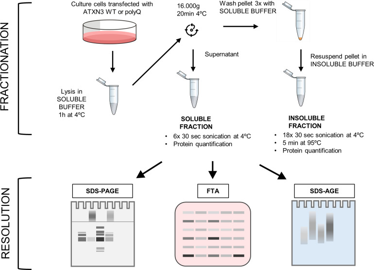
Given the diverse forms and solubilities of ATXN3 species, a biochemical fractionation was performed to isolate the different types of ATXN3 aggregates [21]. For this, a protein fractionation originally developed for the polyQ protein Huntingtin [18] was adapted (Fig 1). First, a “soluble buffer” containing the non-ionic detergent Triton X-100 was used to lyse cells and separate soluble forms of ATXN3 by high-speed centrifugation. Because ATXN3 can bind to chromatin [19], a nuclease was added to the lysis buffer to prevent sedimentation of soluble ATXN3 species with chromatin and remove nucleic acids possibly entangled around aggregates [22]. Non-soluble proteins were then resuspended in a buffer containing 4% SDS (“insoluble buffer”), followed by sonication and boiling of the sample.

Fig 1. Experimental workflow of the fractionation assay.

Fig 1

Cultured cells expressing ATXN3 proteins are washed in PBS and lysed in soluble buffer. After centrifugation, the supernatant is saved as the soluble fraction. Pellets are washed with soluble buffer and resuspended with insoluble buffer, hereafter referred as the insoluble fraction. Fractions are sonicated, incubated for 5 min at 95°C for insoluble fractions, before quantification. Samples are then analysed by Polyacrylamide Gel Electrophoresis (SDS-PAGE), Filter Trap Assay (FTA) and SDS-Agarose Gel Electrophoresis (SDS-AGE) with the optimized parameters described in this article, to allow an optimal resolution and quantification depending on the ATXN3 species of interest.

Separation of proteins into two fractions enables a better resolution of ATXN3 species, providing crucial information about their solubility and thus insights into the progression of ATXN3 aggregation. Monomer and early polymers of ATXN3 are separated in the soluble fraction after centrifugation, while Triton-insoluble ATXN3 forms solubilized in SDS are retained in the second fraction. ATXN3 polyQ SDS-resistant fibrils from the final stage of aggregation were expected to be found in the insoluble fraction since they are highly stable in ionic detergent [9, 23]. We thus tried several buffers containing either SDS, DTT, urea or a combination of these compounds, to resuspend ATXN3 variants from the insoluble pellet. Interestingly, the 7M urea buffer and the mix buffer (4% SDS, 100mM DTT, 7M urea) efficiently solubilized the ATXN3 polyQ HMW forms that remained blocked in the stacking gel when the insoluble fraction was resuspended in 4% SDS only (S1B Fig). Therefore, to preserve ATXN3 polyQ HMW forms for visualization, which are likely ATXN3 polyQ fibrils, the buffer containing 4% SDS is the most suitable (S1B Fig) and is used in the fractionation protocol of soluble and insoluble ATXN3 forms detailed in S1 File.

To subsequently analyse the different ATXN3 species separated by fractionation, SDS-PAGE was used first. The SDS-PAGE settings were optimised for the maximal resolution of all ATXN3 species. Fifteen micrograms of soluble and insoluble fractions were run on an 8% polyacrylamide separating gel with a 4% polyacrylamide stacking gel. ATXN3 SDS-resistant aggregates behave as HMW proteins in SDS-PAGE and remain in the stacking of polyacrylamide gel, as previously described [2325]. The entire gel, including the stacking and the wells was therefore transferred onto a membrane under high transference conditions to allow HMW proteins to transfer. Due to their size and solubility, the ability of these large insoluble aggregates to be charged during the electrophoresis process is limited and could face difficulty to efficiently enter the 4% polyacrylamide stacking gel (S1C Fig). We observed that increasing the electrophoresis voltage improved the migration of the ATXN3 species in the stacking gel (S1C Fig). As a control for the fractionation and analysis by SDS-PAGE, His-tagged ATXN3 WT was expressed, purified and subsequently processed with this protocol. As expected, the ATXN3 WT was only present in the soluble fraction and run as a monomer, demonstrating that the protocol does not result in artifacts, at least with the soluble ATXN3 WT, thereby strongly suggesting that the non-monomeric forms observed after fractionation of cell extracts are the result from aggregation (S1D Fig).

We hypothesized that the signal in the blot from the stacking gel could correlate with the amount of aggregates present in the loaded samples. When loading increasing amounts of the SDS-resistant extracts, a linear correlation with ATXN3 polyQ HMW signal was observed. This indicates that under these SDS-PAGE conditions, the signal of the HMW proteins is quantitative up to the loading of 12 μg of total protein (S1E Fig), although this amount could vary depending on the quantity and the quality of the aggregates formed in the cellular conditions used.

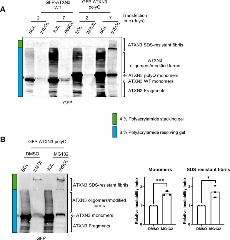
Given that the multi-step aggregation dynamics of ATXN3 polyQ is a time-dependent process influenced by various parameters [8, 11, 12, 26], our analysis was carried out at different times post transfection to optimize the formation of ATXN3 aggregates in our cellular model. Cells transiently expressing GFP-ATXN3 polyQ were harvested at different times from 1 to 7 days post-transfection, subjected to the biochemical fractionation previously mentioned and run on SDS-PAGE (S2A Fig). The GFP signal from the HMW SDS-resistant fibrils was observed in the stacking gel starting from samples collected at 3 days post-transfection, but with a greater amount observed 6 and 7 days after transfection (S2A Fig and Fig 2A). As in our experimental setup no SDS-resistant GFP-ATXN3 polyQ fibrils were observed at 2 days post-transfection, whereas maximal of aggregation was detected at day 7 day post-transfection, subsequent experiments were carried at day 2 and 7 post-transfection as representative time points in the ATXN3 aggregation process. As expected and consistent with previous findings, HMW SDS-resistant aggregates were not detected in extracts of cells transfected with GFP-ATXN3 WT, even after 7 days of transfection (Fig 2A) [11, 25].

Fig 2. SDS-PAGE allows the resolution of ATXN3 monomers, polymers/modified forms and HMW fibrils.

Fig 2

(A) HEK-239T were transfected with GFP-ATXN3 WT or GFP-ATXN3 polyQ plasmids at the indicated times cell were collected and subjected to the fractionation and SDS-PAGE as described in the protocols section. The complete gel including the stacking gel was transferred onto nitrocellulose followed by western blotting using a GFP antibody. In green and blue are indicated parts corresponding to the separating and stacking phases. (B) HEK-239T were transfected with ATXN3 polyQ and 7 days later treated with DMSO or MG132 (5 μM) overnight. Cells were then lysed and processed as in (A) (left panel). Quantification of the insoluble GFP-ATXN3 polyQ, in monomers (middle panel) and HMW aggregates (right panel) (n = 3). The relative ratio between the insoluble and soluble fraction was calculated and called insolubility index. The relative insolubility index of DMSO-treated samples is set to 1. Student’s t‐test was used to determine the statistical significance. Error bars represent the standard deviation. * p<0.5, ** p<0.01 and *** p<0.001.

Interestingly, we observed more ATXN3 aggregation intermediates/oligomers in the soluble fractions of both WT and polyQ GFP-ATXN3 at longer times after transfection (Fig 2A). More ATXN3 WT and polyQ monomers were additionally detected in the insoluble fraction after 7 days of transfection (Fig 2A). This suggests that earlier steps of both WT and polyQ ATXN3 aggregation keep occurring after several days of transfection, possibly due to the new synthesis of ATXN3 proteins. We also noticed that the levels of GFP-ATXN3 polyQ were somewhat higher than those of GFP-ATXN3 WT, especially noticeable at the soluble form. This could be due to differences in the transfection and/or expression process, even though the same amount of plasmid was transfected. Alternatively, this could reflect a higher protein stability of GFP-ATNX3 polyQ compared to ATXN3 WT.

According to the so-called toxic fragment hypothesis, the proteolytic cleavage of ATXN3 by cellular proteases such as calpains plays a crucial role in its toxicity during SCA3 pathogenesis [27]. This hypothesis proposes that the generation of cleaved ATXN3 polyQ proteins initiates the aggregation process associated with cellular dysfunction in the disease [23, 24]. Interestingly, using an anti-GFP antibody, we were able to detect proteins with smaller molecular weight than monomeric GFP-ATXN3 versions of the proteins in the soluble samples after fractionation (indicated as ATXN3 Fragments in Fig 2A and 2B). Size and abundance of these fragments varied between samples transfected for 2 and 7 days, suggesting that the duration of protein expression might have an impact on the proteolysis of ATXN3 proteins (Fig 2A and 2B). As some information could be missed in this analysis using the anti-GFP antibody, the fractionated ATXN3 polyQ samples were studied by western blot using different antibodies. We used an antibody against full length ATXN3, another against the C-terminal part of ATXN3 and finally, an antibody that recognizes polyglutamine tracks (S2B Fig). The patterns resulting from the westerns of the 3 antibodies were comparable to the patterns obtained using anti-GFP. The only exception was that the two ATXN3 antibodies and the polyglutamine antibody recognized some proteins smaller than the GFP ATXN3 polyQ monomer, that were not recognized by the anti-GFP antibody (Fig 2A and S2B Fig). These bands could either correspond to overexpressed GFP-ATXN3 polyQ that was processed to smaller fragments without GFP or to (modified forms of) endogenous ATXN3. Indeed, endogenous ATXN3 migrated with the same mobility as bands observed in the soluble and insoluble fractions of cells expressing GFP-ATXN polyQ when analysed with the different ATXN3 antibodies (S2C Fig). Therefore, the use of different antibodies against ATXN3 is advisable for the detection of protein fragments, and care should be taken when drawing conclusions regarding a model with both endogenous and exogenous proteins. Since the patterns of the blots with the 4 antibodies employed in this study were similar, we decided to use the anti-GFP antibody as it only detects the exogenously expressed polyQ expanded protein of our interest.

Notably, other research groups reported HMW SDS-resistant aggregates after only 2 or 3 days of transfection in cells transiently expressing tagged ATXN3 polyQ [15, 16]. These discrepancies observed in the kinetics of the last stage of ATXN3 polyQ aggregation could be caused by various factors. First, the aggregation of ATXN3 may vary depending on the cellular model used, as a non-exhaustive list of cellular factors such as chaperones, interacting partners, or post-translational modifications has been shown to modulate ATXN3 aggregation [28, 29]. The dynamic of ATXN3 aggregation additionally depends on protein concentration thus the construct used and the efficiency of protein expression will influence the timing of aggregation formation [26]. Also, the length of the polyQ tract carried by the protein influences ATXN3 aggregation dynamics [9, 28].

Several studies have demonstrated that the ubiquitin proteasome system is affected in SCA3 and it is known that proteasome activity regulates the ATXN3 aggregation process [3032]. We therefore investigated the impact of proteasome inhibition on ATXN3 aggregation in our setup. Cells expressing GFP-ATXN3 polyQ for 7 days were treated with MG132 overnight, and samples were subjected to the fractionation and analysis by SDS-PAGE (Fig 2B). The relative insolubility index, determined by quantification of the insoluble vs soluble fractions, demonstrated that proteasome inhibition led to accumulation of ATXN3 insoluble monomers and HMW forms, which is in agreement with previous reports [3032].

Together these results show that cell fractionation followed by SDS-PAGE with adapted settings is an efficient method to study and quantify different ATXN3 variants, including ATXN3 fragments, monomers, polymers, modified forms and HMW SDS-resistant fibrils. However, SDS-PAGE analysis is not suitable to resolve protein species larger than 500 kDa, primarily due to the limitation of polyacrylamide gel pore size, but also, as in our case, due to the poor solubility of HMW SDS-insoluble ATXN3 aggregates that remain in the stacking gel after electrophoresis. We therefore employed the Filter Trap Assay (FTA), a technique proposed for visualizing large protein aggregates. In this assay, vacuum pressure is applied to a cassette forcing samples through a filter membrane where only particles larger than a certain pore size are retained and can be visualized [10, 12, 1517]. We adapted an existing FTA protocol for global aggregated proteins in human cell lines using a 0.2 μm pore size acetate membrane to analyse ATXN3 aggregates (see S1 File for details) [33].

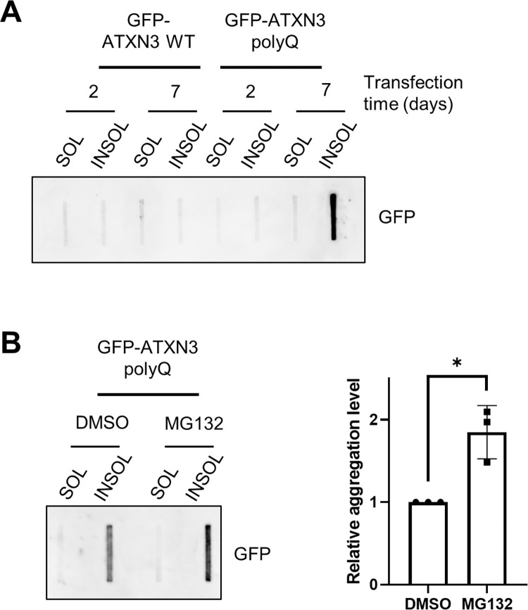
Cell expressing GFP-ATXN3 WT or polyQ for 2 and 7 days were fractionated and analysed by FTA (Fig 3A). Consistent with our observations after SDS-PAGE in Fig 2A and S1D Fig, GFP signal from HMW SDS-resistant forms was only detected in the insoluble fraction of day 7 GFP-ATXN3 polyQ samples (Fig 3A and 3B). Furthermore, positive signal for ATXN3 aggregates was observed when insoluble precipitates were resuspended in 4% SDS, and in a lesser extent with DTT or urea (S3A Fig). The absence of signal in the sample resuspended in the mix buffer (4% SDS; 100 mM DTT; 7M urea) suggests that HMW ATXN3 species were completely solubilized in this condition, as observed in S1B Fig. These results indicate that the trap assay effectively retains the HMW ATXN3 polyQ species. Additionally, when cells were treated with the proteasome inhibitor MG132, more aggregates in the insoluble fraction retained in the FTA filter (Fig 3B), suggesting that proteasome inhibition caused accumulation of ATXN3 polyQ aggregation into HMW fibrils, as observed in Fig 2B and expected from previous studies [3032]. To ensure that FTA assays are quantitative, the filter membrane must remain unsaturated to allow samples to pass through without obstructing the liquid flow [12, 15]. Indeed, a strong linear correlation between the GFP signal detected and the quantity of total protein loaded with FTA was measured (S3B Fig). This result supports the suitability of this assay to visualize and quantify ATXN3 polyQ aggregates of over 0.2 μm (the size of the membrane pores) when up to 40 μg of total protein is loaded, although this amount could vary depending on the aggregate quantity in the samples.

Fig 3. FTA analysis of ATXN3 aggregates.

Fig 3

(A) HEK-239T transfected with the indicated expression plasmids were collected at the indicated times and subjected to the fractionation and FTA protocols described in S1 File. The membrane was subsequently immunoblotted with anti-GFP antibody. (B) HEK-239T cells expressing GFP-ATXN3 polyQ for 7 days were treated with DMSO or 5 μM MG132 overnight, after which cells were collected and analysed as in A (left panel). The relative GFP signal from the insoluble fractions was quantified (n = 3) and normalized to the signal of the DMSO-treated cells (right panel). Student’s t‐test was used to calculate the statistical significance. Error bars represent the standard deviation. * p<0.05.

Although SDS-PAGE and FTA allow a global quantification of SDS-insoluble HMW of ATXN3 polyQ, neither of these assays are suitable for the separation of the HMW ATXN3 species by size. We therefore employed SDS-Agarose Gel Electrophoresis (SDS-AGE), which had been proposed as straightforward technique to separate fibrous protein aggregates, and is routinely used to screen amyloid aggregates [34]. SDS-AGE was used additionally to investigate the large fibrils oligomers of the polyQ protein Huntingtin [35, 36] and more recently also to separate HMW ATXN3 [37, 38]. Similar to SDS-PAGE, this method relies on the ability of the protein substrates to enter the polymerized gel and to distribute according to their molecular weight and their resistance to detergent. However, unlike polyacrylamide gels, the pore size of an agarose gel of 1% has been determined to be around 100 nm [39], theoretically capable to resolve proteins exceeding 1 million kDa [40].

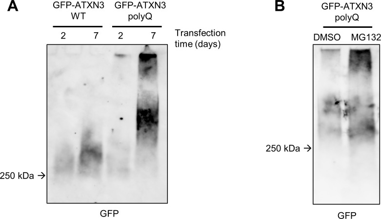
To resolve the HMW ATXN3 species observed in the insoluble fractions after fractionation described in this paper, we adapted a protocol of agarose gel electrophoresis (Fig 4) (see protocol section S1 File) [40, 41]. As in SDS-PAGE and FTA, samples from cells expressing GFP-ATXN3 WT and polyQ were collected and fractionated, after which the insoluble fractions containing the large SDS-resistant ATXN3 species were analysed by SDS-AGE. HMW aggregates were resolved in the agarose gel, as GFP blotting reveals a smear of which the intensity and size can be compared between conditions (Fig 4A and 4B). More HMW ATXN3 aggregates were observed with ATXN3 polyQ at 7 days after transfection (Fig 4A), which is consistent with the previous results observed with SDS-PAGE and FTA (Figs 2A, 3A and S2A Fig). The resolution of HMW species above 250 kDa by SDS-AGE revealed that larger ATXN3 aggregates form 7 days after transfection, mostly in ATXN3 polyQ expressing cells and to a lesser extent in cells expressing the WT version of ATXN3. Furthermore, after MG132 treatment a stronger GFP signal of higher molecular weight species was observed, implying that proteasome inhibition promotes accumulation of such larger aggregates (Fig 4B).

Fig 4. Adapted SDS-AGE for resolving HMW ATXN3 aggregates.

Fig 4

(A) HEK-239T were transfected with the indicated expression plasmids and collected at the specified times before fractionation, SDS-AGE and immunoblotted with an anti-GFP antibody. (B) As in (A) with cells transfected with GFP-ATXN3 polyQ for 7 days and treated with DMSO or 5 μM MG-132 for 16 hours.

Altogether these findings highlight that SDS-AGE is an appropriate method to visualize the different sizes of HMW ATXN3 aggregates. The limitations of SDS-AGE are evident in its challenging quantitative aspect, as the migration of proteins is not well-defined and is influenced by various parameters previously described [42]. However, compared to SDS-PAGE and FTA, SDS-AGE offers additional insights into the distribution of ATXN3 aggregates based on their molecular weight, providing crucial information for researchers investigating the formation of large ATXN3 polyQ fibrils.

It is worth noting that we observe an increase in ATXN3 polyQ and WT aggregation in protein samples from soluble fractions that were heated at more than 40°C or were left at room temperature for more than 24 hours, as a white precipitate was formed that led to a positive signal for HMW proteins when analysed by SDS-PAGE, FTA or SDS-AGE (S3C Fig). This suggests that soluble WT and polyQ ATXN3 monomers/oligomers can artificially aggregate under these conditions, similar to reported for the Huntingtin polyQ protein [18]. It is therefore recommended to avoid several thawing cycles, for example by freezing samples in aliquots, in addition to thawing the fractions on ice to prevent in vitro aggregation after extraction.

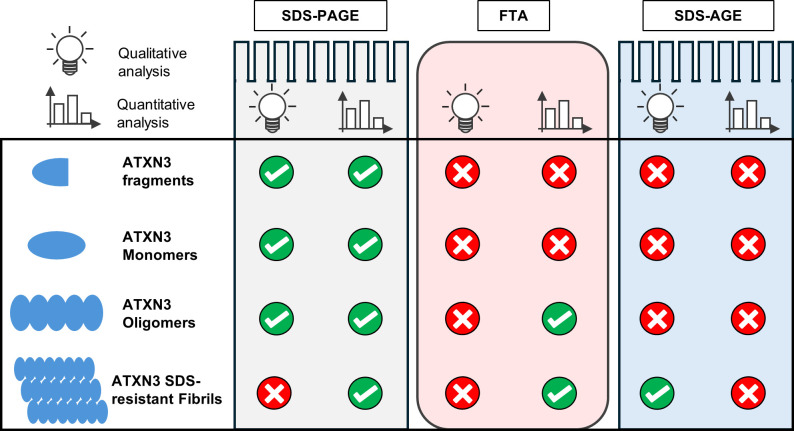
In conclusion, our results show that, when possible, optimization of protein expression time is required to study WT and polyQ-expanded ATXN3 aggregation in vivo. Additionally, an important step in the analysis is the biochemical fractionation that enables an optimized solubilization of ATXN3 species. Among the techniques to visualize ATXN3 intermediates described here, SDS-PAGE can be used as a quantitative method for all kinds of ATXN3 forms, although it is not able to separate HMW SDS-resistant fibrils. FTA is a straightforward and reproducible method, but only allows to quantify ATXN3 aggregates larger than 0.2 μm, while SDS-AGE is well adapted to separate and qualitatively analyse HMW ATXN3 species (Fig 5). Therefore, depending on research context and on the ATXN3 species targeted for analysis, one or a combination of these methods can be used to accurately characterize ATXN3 aggregation (Fig 5). This protocol thus provides adapted and reproducible biochemical methodologies to study the ATXN3 aggregation involved in SCA3 pathogenesis, thereby contributing the evaluation of potential therapeutic outcomes for this still incurable disease.

Fig 5. Analysis of ATXN3 forms by SDS-PAGE, FTA and SDS-AGE.

Fig 5

Table illustrating the different options for qualitative and the quantitative analysis of ATXN3 forms by SDS-PAGE, FTA and SDS-AGE assays. Note that FTA is only suitable for aggregates superior to 0.2 μm.

Supporting information

S1 Fig. SDS-PAGE allows a quantitative study of SDS-resistant ATXN3 aggregates.

(A) HEK-239T were transfected with the indicated plasmid amounts to express GFP-ATXN3 WT. After 48h, cells were lysed and total cell extracts were analysed by SDS-PAGE with the indicated antibody. (B) HEK-239T were transfected with the GFP-ATXN3 polyQ-expressing plasmid and collected 7 days post-transfection. Cells were lysed and fractionated as described in the methods section, except for the last step. After the last wash and centrifugation, the insoluble pellet was resuspended in various buffers: the insoluble buffer containing 4% SDS, a DTT buffer (50mM Tris 7.4, 150 mM NaCl, 100 mM DTT), a Urea buffer (30 mM Tris pH 8.5, 7 M urea, 2 M thiourea) and a mix buffer (30 mM Tris pH 8.5, 7 M urea, 2 M thiourea, 100 mM DTT, 4% SDS). Subsequently, soluble and insoluble fractions were analysed by SDS-PAGE and membranes were immunoblotted for GFP. (C) HEK-239T transfected with GFP-ATXN3 polyQ, were lysed 7 days after transfection, fractionated and run on SDS-PAGE before being immunoblotted with the indicated antibodies. Electrophoresis was performed with 2 different voltages. (D) Purified His-tagged ATXN3-WT protein and extracts from cells expressing GFP-ATXN3 polyQ for 7 days were fractionated using the protocol described in the methods. Soluble and insoluble fractions were analysed by western blot with an antibody raised against the C-terminus of ATXN3 (E) Increasing amounts of insoluble fractions of cells expressing GFP-ATXN3 polyQ for 7 days were analysed by SDS-PAGE. The GFP signal from the stacking gel of 3 replicates was quantified, and a linear correlation was applied between the quantity of protein loaded and the GFP signal. The GFP signal in the stacking gel was measured using ImageJ and quantifications were plotted against the amount of protein loaded.

(TIF)

pone.0315868.s001.tif (2.5MB, tif)
S2 Fig. Detecting ATXN3 aggregate formation by SDS-PAGE using various antibodies.

(A) HEK-239T expressing ATXN3 polyQ were lysed and fractionated at the indicated time points, followed by analysis by SDS-PAGE and immunoblot for GFP. (B) HEK-239T were transfected with GFP-ATXN3 polyQ expressing plasmid and cells were collected at the indicated times. After lysis and fractionation, samples were analysed by western blot using the indicated antibodies. (C) HEK-239T were transfected with siRNAs against luciferase (siLUC) or ATXN3 (siATXN3) for 48 hours, after which whole cell extracts were prepared. The extracts were analysed together with fractionated samples from HEK-239T transfected with GFP-ATXN3 polyQ expressing plasmid for 7 days by SDS-PAGE and western blot using the indicated antibodies. The asterisk marks the mobility of endogenous ATXN3.

(TIF)

pone.0315868.s002.tif (2.2MB, tif)
S3 Fig. FTA as a quantitative method to study of SDS-resistant ATXN3 aggregates.

(A) HEK-239T were transfected with the GFP-ATXN3 polyQ-expressing plasmid and collected 7 days post-transfection, followed by lysis and fractionation as described in the protocol section, except for the last step. After the last wash and centrifugation, the insoluble pellet was resuspended with various buffers, including the insoluble buffer containing 4% SDS, a DTT buffer (50mM Tris 7.4, 150 mM NaCl, 100 mM DTT), an urea buffer (30 mM Tris pH 8.5, 7 M urea, 2 M thiourea) and a mix buffer (30 mM Tris pH 8.5, 7 M urea, 2 M thiourea, 100 mM DTT, 4% SDS). Soluble and insoluble fractions were subsequently analysed by FTA and immunoblotting for GFP. (B) HEK-239T expressing GFP-ATXN3 polyQ for 7 days were lysed, fractionated and analysed by FTA and immunoblot using a GFP antibody. The GFP signal in the insoluble fractions was quantified with ImageJ and plotted against the amount of protein loaded.

(TIF)

pone.0315868.s003.tif (596.3KB, tif)
S4 Fig. Boiling soluble fractions induced GFP signal in the stacking gel of SDS-PAGE.

HEK-239T were transfected with the GFP-ATXN3 polyQ and WT expressing plasmids and collected at day 7 post-transfection followed by lysis, fractionation and analysis by SDS-PAGE as described in the protocol section, except that in this case samples were left untreated or boiled before loading onto the acrylamide gel, as indicated.

(TIF)

pone.0315868.s004.tif (742.3KB, tif)
S1 File

(PDF)

pone.0315868.s005.pdf (437.3KB, pdf)
S1 Raw images

(PDF)

pone.0315868.s006.pdf (13.2MB, pdf)

Acknowledgments

We thank Dr. Veronique Smits for critical reading of the manuscript.

Data Availability

All relevant data are within the manuscript and its Supporting Information files.

Funding Statement

This work was supported by grants PID2022-139691OB-I00 funded by MCIN/AEI/10.13039/501100011033 and ERDF A way of making Europe (European Union) to RF and grant OA23/071 from Fundación DISA to GQ.

References

  • 1.Cummings CJ, Zoghbi HY (2000) Trinucleotide Repeats: Mechanisms and Pathophysiology. Annual Review of Genomics and Human Genetics 1:281–328 doi: 10.1146/annurev.genom.1.1.281 [DOI] [PubMed] [Google Scholar]
  • 2.Burnett B, Li F, Pittman RN (2003) The polyglutamine neurodegenerative protein ataxin-3 binds polyubiquitylated proteins and has ubiquitin protease activity. Human Molecular Genetics 12:3195–3205 doi: 10.1093/hmg/ddg344 [DOI] [PubMed] [Google Scholar]
  • 3.Hernández-Carralero E, Quinet G, Freire R (2024) ATXN3: a multifunctional protein involved in the polyglutamine disease spinocerebellar ataxia type 3. Expert Rev Mol Med 26:e19 doi: 10.1017/erm.2024.10 [DOI] [PMC free article] [PubMed] [Google Scholar]
  • 4.Lima M, Costa MC, Montiel R, et al. (2006) Population Genetics of Wild-Type CAG Repeats in the Machado-Joseph Disease Gene in Portugal. Human Heredity 60:156–163 [DOI] [PubMed] [Google Scholar]
  • 5.Matos CA, de Almeida LP, Nóbrega C (2019) Machado-Joseph disease/spinocerebellar ataxia type 3: lessons from disease pathogenesis and clues into therapy. J Neurochem 148:8–28 doi: 10.1111/jnc.14541 [DOI] [PubMed] [Google Scholar]
  • 6.Seidel K, Siswanto S, Fredrich M, et al. (2016) On the distribution of intranuclear and cytoplasmic aggregates in the brainstem of patients with spinocerebellar ataxia type 2 and 3. Brain Pathol 27:345–355 doi: 10.1111/bpa.12412 [DOI] [PMC free article] [PubMed] [Google Scholar]
  • 7.Costa M do C, Paulson HL (2012) Toward understanding Machado-Joseph Disease. Prog Neurobiol 97:239–257 doi: 10.1016/j.pneurobio.2011.11.006 [DOI] [PMC free article] [PubMed] [Google Scholar]
  • 8.Scarff CA, Almeida B, Fraga J, Macedo-Ribeiro S, Radford SE, Ashcroft AE (2015) Examination of Ataxin-3 (atx-3) Aggregation by Structural Mass Spectrometry Techniques: A Rationale for Expedited Aggregation upon Polyglutamine (polyQ) Expansion. Mol Cell Proteomics 14:1241–1253 doi: 10.1074/mcp.M114.044610 [DOI] [PMC free article] [PubMed] [Google Scholar]
  • 9.Ellisdon AM, Thomas B, Bottomley SP (2006) The Two-stage Pathway of Ataxin-3 Fibrillogenesis Involves a Polyglutamine-independent Step*. Journal of Biological Chemistry 281:16888–16896 doi: 10.1074/jbc.M601470200 [DOI] [PubMed] [Google Scholar]
  • 10.Figueiredo F, Lopes-Marques M, Almeida B, Matscheko N, Martins PM, Silva A, et al. (2022) A Robust Assay to Monitor Ataxin-3 Amyloid Fibril Assembly. Cells 11:1969 doi: 10.3390/cells11121969 [DOI] [PMC free article] [PubMed] [Google Scholar]
  • 11.Saunders HM, Hughes VA, Cappai R, Bottomley SP (2013) Conformational Behavior and Aggregation of Ataxin-3 in SDS. PLOS ONE 8:e69416 doi: 10.1371/journal.pone.0069416 [DOI] [PMC free article] [PubMed] [Google Scholar]
  • 12.Weishäupl D, Schneider J, Peixoto Pinheiro B, Ruess C, Dold SM, von Zweydorf F, et al. (2019) Physiological and pathophysiological characteristics of ataxin-3 isoforms. Journal of Biological Chemistry 294:644–661 doi: 10.1074/jbc.RA118.005801 [DOI] [PMC free article] [PubMed] [Google Scholar]
  • 13.Todi SV, Scaglione KM, Blount JR, Basrur V, Conlon KP, Pastore A, et al. (2010) Activity and Cellular Functions of the Deubiquitinating Enzyme and Polyglutamine Disease Protein Ataxin-3 Are Regulated by Ubiquitination at Lysine 117*. Journal of Biological Chemistry 285:39303–39313 doi: 10.1074/jbc.M110.181610 [DOI] [PMC free article] [PubMed] [Google Scholar]
  • 14.Moore LR, Keller L, Bushart DD, Delatorre RG, Li D, McLoughlin HS, et al. (2019) Antisense oligonucleotide therapy rescues aggresome formation in a novel spinocerebellar ataxia type 3 human embryonic stem cell line. Stem Cell Res 39:101504 doi: 10.1016/j.scr.2019.101504 [DOI] [PMC free article] [PubMed] [Google Scholar]
  • 15.Sena PP, Weber JJ, Bayezit S, et al. (2023) Implications of specific lysine residues within ataxin-3 for the molecular pathogenesis of Machado-Joseph disease. Frontiers in Molecular Neuroscience 16:1133271. doi: 10.3389/fnmol.2023.1133271 [DOI] [PMC free article] [PubMed] [Google Scholar]
  • 16.Breuer P, Haacke A, Evert BO, Wüllner U (2010) Nuclear Aggregation of Polyglutamine-expanded Ataxin-3. J Biol Chem 285:6532–6537 [DOI] [PMC free article] [PubMed] [Google Scholar]
  • 17.Natalello A, Frana AM, Relini A, Apicella A, Invernizzi G, Casari C, et al. (2011) A major role for side-chain polyglutamine hydrogen bonding in irreversible ataxin-3 aggregation. PLoS One 6:e18789 doi: 10.1371/journal.pone.0018789 [DOI] [PMC free article] [PubMed] [Google Scholar]
  • 18.Ochaba J, Morozko EL, O’Rourke JG, Thompson LM (2018) Fractionation for Resolution of Soluble and Insoluble Huntingtin Species. J Vis Exp 57082. [DOI] [PMC free article] [PubMed] [Google Scholar]
  • 19.Hernández-Carralero E, Cabrera E, Rodríguez-Torres G, et al. (2023) ATXN3 controls DNA replication and transcription by regulating chromatin structure. Nucleic Acids Res 51:5396–5413 doi: 10.1093/nar/gkad212 [DOI] [PMC free article] [PubMed] [Google Scholar]
  • 20.Singh AN, Oehler J, Torrecilla I, et al. (2019) The p97–Ataxin 3 complex regulates homeostasis of the DNA damage response E3 ubiquitin ligase RNF8. EMBO J 38:e102361 doi: 10.15252/embj.2019102361 [DOI] [PMC free article] [PubMed] [Google Scholar]
  • 21.Koch P, Breuer P, Peitz M, et al. (2011) Excitation-induced ataxin-3 aggregation in neurons from patients with Machado–Joseph disease. Nature 480:543–546 doi: 10.1038/nature10671 [DOI] [PubMed] [Google Scholar]
  • 22.Mitsui K, Doi H, Nukina N (2006) Proteomics of polyglutamine aggregates. Methods Enzymol 412:63–76 doi: 10.1016/S0076-6879(06)12005-4 [DOI] [PubMed] [Google Scholar]
  • 23.Haacke A, Broadley SA, Boteva R, Tzvetkov N, Hartl FU, Breuer P (2006) Proteolytic cleavage of polyglutamine-expanded ataxin-3 is critical for aggregation and sequestration of non-expanded ataxin-3. Human Molecular Genetics 15:555–568 doi: 10.1093/hmg/ddi472 [DOI] [PubMed] [Google Scholar]
  • 24.Hsu J-Y, Jhang Y-L, Cheng P-H, Chang Y-F, Mao S-H, Yang H-I, et al. (2017) The Truncated C-terminal Fragment of Mutant ATXN3 Disrupts Mitochondria Dynamics in Spinocerebellar Ataxia Type 3 Models. Front Mol Neurosci. doi: 10.3389/fnmol.2017.00196 [DOI] [PMC free article] [PubMed] [Google Scholar]
  • 25.Chai Y, Wu L, Griffin JD, Paulson HL (2001) The Role of Protein Composition in Specifying Nuclear Inclusion Formation in Polyglutamine Disease*. Journal of Biological Chemistry 276:44889–44897 doi: 10.1074/jbc.M106575200 [DOI] [PubMed] [Google Scholar]
  • 26.Ellisdon AM, Pearce MC, Bottomley SP (2007) Mechanisms of Ataxin-3 Misfolding and Fibril Formation: Kinetic Analysis of a Disease-associated Polyglutamine Protein. Journal of Molecular Biology 368:595–605 doi: 10.1016/j.jmb.2007.02.058 [DOI] [PubMed] [Google Scholar]
  • 27.Simões AT, Gonçalves N, Koeppen A, Déglon N, Kügler S, Duarte CB, et al. (2012) Calpastatin-mediated inhibition of calpains in the mouse brain prevents mutant ataxin 3 proteolysis, nuclear localization and aggregation, relieving Machado-Joseph disease. Brain 135:2428–2439 doi: 10.1093/brain/aws177 [DOI] [PubMed] [Google Scholar]
  • 28.Kuiper EFE, de Mattos EP, Jardim LB, Kampinga HH, Bergink S (2017) Chaperones in Polyglutamine Aggregation: Beyond the Q-Stretch. Front Neurosci. doi: 10.3389/fnins.2017.00145 [DOI] [PMC free article] [PubMed] [Google Scholar]
  • 29.Thiruvalluvan A, de Mattos EP, Brunsting JF, et al. (2020) DNAJB6, a Key Factor in Neuronal Sensitivity to Amyloidogenesis. Molecular Cell 78:346–358.e9 doi: 10.1016/j.molcel.2020.02.022 [DOI] [PubMed] [Google Scholar]
  • 30.Wang H-L, Hu S-H, Chou A-H, Wang S-S, Weng Y-H, Yeh T-H (2013) H1152 promotes the degradation of polyglutamine-expanded ataxin-3 or ataxin-7 independently of its ROCK-inhibiting effect and ameliorates mutant ataxin-3-induced neurodegeneration in the SCA3 transgenic mouse. Neuropharmacology 70:1–11 doi: 10.1016/j.neuropharm.2013.01.006 [DOI] [PubMed] [Google Scholar]
  • 31.Schmidt T, Lindenberg KS, Krebs A, Schöls L, Laccone F, Herms J, et al. (2002) Protein surveillance machinery in brains with spinocerebellar ataxia type 3: redistribution and differential recruitment of 26S proteasome subunits and chaperones to neuronal intranuclear inclusions. Ann Neurol 51:302–310 doi: 10.1002/ana.10101 [DOI] [PubMed] [Google Scholar]
  • 32.Chen ZS, Huang X, Talbot K, Chan HYE (2021) A fine balance between Prpf19 and Exoc7 in achieving degradation of aggregated protein and suppression of cell death in spinocerebellar ataxia type 3. Cell Death Dis 12:136 doi: 10.1038/s41419-021-03444-x [DOI] [PMC free article] [PubMed] [Google Scholar]
  • 33.Chhipi-Shrestha JK, Yoshida M, Iwasaki S (2022) Filter trapping protocol to detect aggregated proteins in human cell lines. STAR Protoc 3:101571 doi: 10.1016/j.xpro.2022.101571 [DOI] [PMC free article] [PubMed] [Google Scholar]
  • 34.Halfmann R, Jarosz DF, Jones SK, Chang A, Lancaster AK, Lindquist S (2012) Prions are a common mechanism for phenotypic inheritance in wild yeasts. Nature 482:363–368 doi: 10.1038/nature10875 [DOI] [PMC free article] [PubMed] [Google Scholar]
  • 35.Legleiter J, Mitchell E, Lotz GP, Sapp E, Ng C, DiFiglia M, et al. (2010) Mutant Huntingtin Fragments Form Oligomers in a Polyglutamine Length-dependent Manner in Vitro and in Vivo. J Biol Chem 285:14777–14790 doi: 10.1074/jbc.M109.093708 [DOI] [PMC free article] [PubMed] [Google Scholar]
  • 36.Sontag EM, Lotz GP, Yang G, Sontag CJ, Cummings BJ, Glabe CG, et al. (2012) Detection of Mutant Huntingtin Aggregation Conformers and Modulation of SDS-Soluble Fibrillar Oligomers by Small Molecules. J Huntingtons Dis 1:119–132 doi: 10.3233/JHD-2012-129004 [DOI] [PMC free article] [PubMed] [Google Scholar]
  • 37.Cushman-Nick M, Bonini NM, Shorter J (2013) Hsp104 suppresses polyglutamine-induced degeneration post onset in a drosophila MJD/SCA3 model. PLoS Genet 9:e1003781 doi: 10.1371/journal.pgen.1003781 [DOI] [PMC free article] [PubMed] [Google Scholar]
  • 38.Weber JJ, Golla M, Guaitoli G, et al. (2017) A combinatorial approach to identify calpain cleavage sites in the Machado-Joseph disease protein ataxin-3. Brain 140:1280–1299 doi: 10.1093/brain/awx039 [DOI] [PubMed] [Google Scholar]
  • 39.Holmes DL, Stellwagen NC (1990) The electric field dependence of DNA mobilities in agarose gels: a reinvestigation. Electrophoresis 11:5–15 doi: 10.1002/elps.1150110103 [DOI] [PubMed] [Google Scholar]
  • 40.Krizek DM, Rick ME (2002) Agarose gel electrophoresis of proteins. Curr Protoc Cell Biol Chapter 6:Unit 6.7 doi: 10.1002/0471143030.cb0607s15 [DOI] [PubMed] [Google Scholar]
  • 41.Warren CM, Krzesinski PR, Greaser ML (2003) Vertical agarose gel electrophoresis and electroblotting of high-molecular-weight proteins. Electrophoresis 24:1695–1702 doi: 10.1002/elps.200305392 [DOI] [PubMed] [Google Scholar]
  • 42.Drozdova PB, Barbitoff YA, Belousov MV, Skitchenko RK, Rogoza TM, Leclercq JY, et al. (2020) Estimation of amyloid aggregate sizes with semi-denaturing detergent agarose gel electrophoresis and its limitations. Prion 14:118–128 doi: 10.1080/19336896.2020.1751574 [DOI] [PMC free article] [PubMed] [Google Scholar]

Decision Letter 0

Elise A Kikis

5 Aug 2024

PONE-D-24-24197Systematic biochemical analysis to study wild-type and polyglutamine expanded ATXN3 species in vivo.PLOS ONE

Dear Dr. Freire,

Thank you for submitting your manuscript to PLOS ONE. After careful consideration, we feel that it has merit but does not fully meet PLOS ONE’s publication criteria as it currently stands. Therefore, we invite you to submit a revised version of the manuscript that addresses the points raised during the review process.

Please play close attention to the suggestion to utilize anti-atxn3 antibodies instead of only anti-gfp. A revised manuscript should address this point.

Please submit your revised manuscript by Sep 19 2024 11:59PM. If you will need more time than this to complete your revisions, please reply to this message or contact the journal office at plosone@plos.org. When you're ready to submit your revision, log on to https://www.editorialmanager.com/pone/ and select the 'Submissions Needing Revision' folder to locate your manuscript file.

Please include the following items when submitting your revised manuscript:

  • A rebuttal letter that responds to each point raised by the academic editor and reviewer(s). You should upload this letter as a separate file labeled 'Response to Reviewers'.

  • A marked-up copy of your manuscript that highlights changes made to the original version. You should upload this as a separate file labeled 'Revised Manuscript with Track Changes'.

  • An unmarked version of your revised paper without tracked changes. You should upload this as a separate file labeled 'Manuscript'.

If you would like to make changes to your financial disclosure, please include your updated statement in your cover letter. Guidelines for resubmitting your figure files are available below the reviewer comments at the end of this letter.

If applicable, we recommend that you deposit your laboratory protocols in protocols.io to enhance the reproducibility of your results. Protocols.io assigns your protocol its own identifier (DOI) so that it can be cited independently in the future. For instructions see: https://journals.plos.org/plosone/s/submission-guidelines#loc-laboratory-protocols. Additionally, PLOS ONE offers an option for publishing peer-reviewed Lab Protocol articles, which describe protocols hosted on protocols.io. Read more information on sharing protocols at https://plos.org/protocols?utm_medium=editorial-email&utm_source=authorletters&utm_campaign=protocols.

We look forward to receiving your revised manuscript.

Kind regards,

Elise A. Kikis

Academic Editor

PLOS ONE

Journal requirements: 1. When submitting your revision, we need you to address these additional requirements. Please ensure that your manuscript meets PLOS ONE's style requirements, including those for file naming. The PLOS ONE style templates can be found at https://journals.plos.org/plosone/s/file?id=wjVg/PLOSOne_formatting_sample_main_body.pdf and https://journals.plos.org/plosone/s/file?id=ba62/PLOSOne_formatting_sample_title_authors_affiliations.pdf. 2. PLOS ONE now requires that authors provide the original uncropped and unadjusted images underlying all blot or gel results reported in a submission’s figures or Supporting Information files. This policy and the journal’s other requirements for blot/gel reporting and figure preparation are described in detail at https://journals.plos.org/plosone/s/figures#loc-blot-and-gel-reporting-requirements and https://journals.plos.org/plosone/s/figures#loc-preparing-figures-from-image-files. When you submit your revised manuscript, please ensure that your figures adhere fully to these guidelines and provide the original underlying images for all blot or gel data reported in your submission. See the following link for instructions on providing the original image data: https://journals.plos.org/plosone/s/figures#loc-original-images-for-blots-and-gels.   In your cover letter, please note whether your blot/gel image data are in Supporting Information or posted at a public data repository, provide the repository URL if relevant, and provide specific details as to which raw blot/gel images, if any, are not available. Email us at plosone@plos.org if you have any questions. 3. We note that the grant information you provided in the ‘Funding Information’ and ‘Financial Disclosure’ sections do not match.  When you resubmit, please ensure that you provide the correct grant numbers for the awards you received for your study in the ‘Funding Information’ section. 4. Thank you for stating the following financial disclosure:  [This work was supported by grants PID2022-139691OB-I00 funded by MCIN/AEI/10.13039/501100011033 and ERDF A way of making Europe (European Union) to RF and grant OA23/071 from Fundación DISA to GQ.].  Please state what role the funders took in the study.  If the funders had no role, please state: ""The funders had no role in study design, data collection and analysis, decision to publish, or preparation of the manuscript."" If this statement is not correct you must amend it as needed. Please include this amended Role of Funder statement in your cover letter; we will change the online submission form on your behalf. 5. We note that you have included the phrase “data not shown” in your manuscript. Unfortunately, this does not meet our data sharing requirements. PLOS does not permit references to inaccessible data. We require that authors provide all relevant data within the paper, Supporting Information files, or in an acceptable, public repository. Please add a citation to support this phrase or upload the data that corresponds with these findings to a stable repository (such as Figshare or Dryad) and provide and URLs, DOIs, or accession numbers that may be used to access these data. Or, if the data are not a core part of the research being presented in your study, we ask that you remove the phrase that refers to these data. 6. We are unable to open your Figure file [Fig1.eps, Fig2.eps, Fig3.eps , Fig4.eps, Fig5.eps and supplementary S1, S2]. Please kindly revise as necessary and re-upload.

Additional Editor Comments:

Please address the reviewers’ concerns in a revised manuscript, I encourage you to especially note the suggestion to utilize ATXN3-specific antibodies instead of only anti-gfp antibodies.

[Note: HTML markup is below. Please do not edit.]

Reviewers' comments:

Reviewer's Responses to Questions

Comments to the Author

1. Does the manuscript report a protocol which is of utility to the research community and adds value to the published literature?

Reviewer #1: Yes

Reviewer #2: Yes

**********

2. Has the protocol been described in sufficient detail?

To answer this question, please click the link to protocols.io in the Materials and Methods section of the manuscript (if a link has been provided) or consult the step-by-step protocol in the Supporting Information files.

The step-by-step protocol should contain sufficient detail for another researcher to be able to reproduce all experiments and analyses.

Reviewer #1: Yes

Reviewer #2: Partly

**********

3. Does the protocol describe a validated method?

The manuscript must demonstrate that the protocol achieves its intended purpose: either by containing appropriate validation data, or referencing at least one original research article in which the protocol was used to generate data.

Reviewer #1: Yes

Reviewer #2: No

**********

4. If the manuscript contains new data, have the authors made this data fully available?

The PLOS Data policy requires authors to make all data underlying the findings described in their manuscript fully available without restriction, with rare exception (please refer to the Data Availability Statement in the manuscript PDF file). The data should be provided as part of the manuscript or its supporting information, or deposited to a public repository. For example, in addition to summary statistics, the data points behind means, medians and variance measures should be available. If there are restrictions on publicly sharing data—e.g. participant privacy or use of data from a third party—those must be specified.

Reviewer #1: N/A

Reviewer #2: No

**********

5. Is the article presented in an intelligible fashion and written in standard English?

PLOS ONE does not copyedit accepted manuscripts, so the language in submitted articles must be clear, correct, and unambiguous. Any typographical or grammatical errors should be corrected at revision, so please highlight any specific errors that need correcting in the box below.

Reviewer #1: Yes

Reviewer #2: Yes

**********

6. Review Comments to the Author

Please use the space provided to explain your answers to the questions above. You may also include additional comments for the author, including concerns about dual publication, research ethics, or publication ethics. (Please upload your review as an attachment if it exceeds 20,000 characters)

Reviewer #1: In the manuscript entitled “Systematic biochemical analysis to study wild-type and polyglutamine expanded ATXN3 species in vivo”, Quinet et al. outline a multi-step protocol to evaluate the aggregation stages of the aggregation prone protein ATXN3 in its wild-type and mutant form. To assess aggregation, the authors describe a step by step protocol employing commonly used strategies to evaluate aggregation states. Using their pipeline, researchers are able to identify and evaluate the proposed four-step mechanism for the aggregation of ATXN3 in vivo. This mechanism includes an early and late oligomerization phase followed by protofibril then fibril formation. This reviewer agrees that protocols for characterizing the various stages of aggregation prone proteins from an in vivo system are important for developing strategies to reverse or eliminate aggregation in disease. Recommendations for timing post-transfection are made along with buffer conditions to resolve various species of ATXN3. However, clarification of the timing and quantitation aspect of the pipeline along with additional representative datasets are suggested before publication.

In Figure 2, the authors use SDS-PAGE to resolve ATXN3 states (monomers, polymers/modified forms, and HMW fibrils). Based on the representative dataset, there is a notable difference in the amount of protein in the soluble fraction for the GFP-ATXN3 (WT) and GFP-ATXN3 (polyQ) at day 2 that is not discussed. In line 205, the authors discuss levels of ATXN3 aggregation in the intermediate/oligomer state in the soluble fractions at the longer time point but do not offer an explanation for the different relative amounts at day 2. Is this possibly due to the turnover of GFP-ATXN3 (WT) early (day 2) vs late (day 7)? Is this dependent on the proteosome? The authors state in lines 343-344 that optimization of protein expression time is required to study WT and polyQ-expanded ATXN3 aggregation in vivo. A discussion that includes clarification on this point would strengthen the timing aspect of the study.

In line 245, the authors propose that SDS-PAGE with the adapted settings is a method that can be used to quantify different ATXN3 variants. Furthermore, in line 288, the authors state that SDS-PAGE allows a global quantification of SDS-insoluble HMW of ATXN3 polyQ. And finally, in lines 346-347, the authors conclude that SDS-PAGE can be used as a quantitative method for “all kinds” of ATXN3 forms. However, they do not offer a quantitative analysis for the Figure 2 dataset to support these statements. Adding this analysis would strengthen the quantitative aspect of the study.

Reviewer #2: The manuscript by Freire and coworkers describes several biochemical approaches to investigate ATXN3 with and without polyglutamine expansion in cultured cells. Expanded ATXN3 causes spinocerebellar ataxia type 3 (also known as Machado-Joseph disease), an inherited neurodegenerative disease that is associated with misfolding and aggregation of the mutant protein, and a comprehensive summary of available methods to probe the various states of ATXN3 misfolding would be a valuable addition to the field. The manuscript focuses on three assays, filter trap, SDS-polyacrylamide and SDS-agarose gel electrophoresis, to visualize different species of GFP-tagged ATXN3 protein. These methods have been used extensively by researchers to explore misfolding of ATXN3 and other polyglutamine proteins such as huntingtin over the last 20+ years.

Major comments:

1- My main concern is related to the fact that the authors define monomers, oligomers/polymers/modified forms, and high molecular weight fibrils or aggregates based on electrophoretic shift alone. The results would have been more convincing if the same reference standards (such as purified monomeric ATXN3 and in vitro assembled fibrils) were included to firmly establish the identity of the species and compare the three methods.

2- Another issue is that all three assays employ antibodies that recognize the GFP tag to detect ATXN3 protein. Assuming that the authors transfected commonly used plasmids encoding ATXN3 with an amino-terminal GFP tag (Addgene plasmid #22122 and #22123), such an indirect detection via the GFP tag would miss any fragments containing the carboxy-terminal polyglutamine tract. This is particularly important considering proteolytic processing of ATXN3, which the authors state “plays a crucial role in its toxicity” [p. 7 line 213]. Therefore, all immunoblots should be probed with multiple antibodies targeting other regions in the ATXN3 protein, including antibody clone 1C2, which specifically recognizes the expanded polyglutamine tract (see for example Song et al. 2022 Life Med 1:27-44; and Merry et al. 1998 Hum Mol Genet 7:693-701).

3- It is unclear how the timepoints (1 and 7 days after transfection) were chosen for analysis. HEK293T cells have a doubling time of roughly ~24 hours; waiting one week after the transfection to collect samples seems a bit long. When does misfolding/aggregation start under these conditions? Is it a gradual process?

4- The text states that “multi-step aggregation of ATXN3 is time-dependent and can be influenced by several parameters, including the biological model used, the type of ATXN3 isoforms and the amount of expressed proteins” [p. 4 line 93], yet is remains unclear how parameters were chosen for this particular protocol. This needs to be clarified – ideally with data.

5- The title does not accurately reflect the contents of the manuscript. I recommend removing the terms “Systematic” (see comments above) and “in vivo” (the work was done using protein extracts from HEK293T cells).

Minor comments:

6- The protocol does not include a description of the plasmid DNA used for transfection. This is important, as the extent of protein expression will depend on the promoter used. This information needs to be added.

7- Step 5 of the protocol states “renew medium if it turns yellow”. The methods section should clarify how often the cell culture medium needs to be changed after the transfection.

**********

7. PLOS authors have the option to publish the peer review history of their article (what does this mean?). If published, this will include your full peer review and any attached files.

If you choose “no”, your identity will remain anonymous but your review may still be made public.

Do you want your identity to be public for this peer review? For information about this choice, including consent withdrawal, please see our Privacy Policy.

Reviewer #1: No

Reviewer #2: No

**********

[NOTE: If reviewer comments were submitted as an attachment file, they will be attached to this email and accessible via the submission site. Please log into your account, locate the manuscript record, and check for the action link "View Attachments". If this link does not appear, there are no attachment files.]

While revising your submission, please upload your figure files to the Preflight Analysis and Conversion Engine (PACE) digital diagnostic tool, https://pacev2.apexcovantage.com/. PACE helps ensure that figures meet PLOS requirements. To use PACE, you must first register as a user. Registration is free. Then, login and navigate to the UPLOAD tab, where you will find detailed instructions on how to use the tool. If you encounter any issues or have any questions when using PACE, please email PLOS at figures@plos.org. Please note that Supporting Information files do not need this step.

PLoS One. 2024 Dec 23;19(12):e0315868. doi: 10.1371/journal.pone.0315868.r002

Author response to Decision Letter 0


4 Oct 2024

--

Journal requirements:

1. When submitting your revision, we need you to address these additional requirements.

Please ensure that your manuscript meets PLOS ONE's style requirements, including those for file naming. The PLOS ONE style templates can be found at

https://journals.plos.org/plosone/s/file?id=wjVg/PLOSOne_formatting_sample_main_body.pdf and

https://journals.plos.org/plosone/s/file?id=ba62/PLOSOne_formatting_sample_title_authors_affiliations.pdf.

2. PLOS ONE now requires that authors provide the original uncropped and unadjusted images underlying all blot or gel results reported in a submission’s figures or Supporting Information files. This policy and the journal’s other requirements for blot/gel reporting and figure preparation are described in detail at https://journals.plos.org/plosone/s/figures#loc-blot-and-gel-reporting-requirements and https://journals.plos.org/plosone/s/figures#loc-preparing-figures-from-image-files. When you submit your revised manuscript, please ensure that your figures adhere fully to these guidelines and provide the original underlying images for all blot or gel data reported in your submission. See the following link for instructions on providing the original image data: https://journals.plos.org/plosone/s/figures#loc-original-images-for-blots-and-gels.

In your cover letter, please note whether your blot/gel image data are in Supporting Information or posted at a public data repository, provide the repository URL if relevant, and provide specific details as to which raw blot/gel images, if any, are not available. Email us at plosone@plos.org if you have any questions.

We included all the raw data from the blots presented throughout the manuscript in Supporting Information (S1 raw images file).

3. We note that the grant information you provided in the ‘Funding Information’ and ‘Financial Disclosure’ sections do not match.

When you resubmit, please ensure that you provide the correct grant numbers for the awards you received for your study in the ‘Funding Information’ section.

The work was supported by 2 grants:

1- PID2022-139691OB-I00 from the Agencia Estatal de Investigación (which includes funds from the European Union)

2- OA23/071 from Fundación DISA.

According to the instructions, funding from the Agencia Estatal de Investigación acknowledged in the following way: Funded by MCIN/AEI/10.13039/501100011033 and ERDF A way of making Europe (European Union)”. In the initial submission it was not possible to add all this information in the “Funding information” section, but was added in the “Financial Disclosure”. Since you require the two sections to be the same, we corrected the “Financial Disclosure” to a shorter version. However, is very important that the funding information in the article itself contains the correct, longer version and we therefore left that information in the “Funding” section in the article.

4. Thank you for stating the following financial disclosure:

[This work was supported by grants PID2022-139691OB-I00 funded by MCIN/AEI/10.13039/501100011033 and ERDF A way of making Europe (European Union) to RF and grant OA23/071 from Fundación DISA to GQ.].

Please state what role the funders took in the study. If the funders had no role, please state: ""The funders had no role in study design, data collection and analysis, decision to publish, or preparation of the manuscript.""

If this statement is not correct you must amend it as needed.

Please include this amended Role of Funder statement in your cover letter; we will change the online submission form on your behalf.

The role of the funders in the study (no role) is now added to the funding information of the manuscript.

5. We note that you have included the phrase “data not shown” in your manuscript. Unfortunately, this does not meet our data sharing requirements. PLOS does not permit references to inaccessible data. We require that authors provide all relevant data within the paper, Supporting Information files, or in an acceptable, public repository. Please add a citation to support this phrase or upload the data that corresponds with these findings to a stable repository (such as Figshare or Dryad) and provide and URLs, DOIs, or accession numbers that may be used to access these data. Or, if the data are not a core part of the research being presented in your study, we ask that you remove the phrase that refers to these data.

Data supporting the previous “data not shown” is now provided in S3 FigC. The text in the manuscript has been adapted accordingly (lines 360 to 364).

6. We are unable to open your Figure file [Fig1.eps, Fig2.eps, Fig3.eps , Fig4.eps, Fig5.eps and supplementary S1, S2]. Please kindly revise as necessary and re-upload.

We are now uploading the figure files in .tiff format. Hopefully now all files can be opened.

Thank you for submitting your manuscript to PLOS ONE. After careful consideration, we feel that it has merit but does not fully meet PLOS ONE’s publication criteria as it currently stands. Therefore, we invite you to submit a revised version of the manuscript that addresses the points raised during the review process.

Please play close attention to the suggestion to utilize anti-atxn3 antibodies instead of only anti-gfp. A revised manuscript should address this point.

This revised version of the manuscript contains new experiments suggested by the reviewers, including the use of (3) new, different antibodies to detect ATXN3, as highlighted by the editor. The new manuscript therefore includes more figures/figure panels in addition to changes in the text, following the suggestions of reviewers. We hope this revised version of the manuscript now merits is publication in PLOS ONE.

Reviewer #1: In the manuscript entitled “Systematic biochemical analysis to study wild-type and polyglutamine expanded ATXN3 species in vivo”, Quinet et al. outline a multi-step protocol to evaluate the aggregation stages of the aggregation prone protein ATXN3 in its wild-type and mutant form. To assess aggregation, the authors describe a step by step protocol employing commonly used strategies to evaluate aggregation states. Using their pipeline, researchers are able to identify and evaluate the proposed four-step mechanism for the aggregation of ATXN3 in vivo. This mechanism includes an early and late oligomerization phase followed by protofibril then fibril formation. This reviewer agrees that protocols for characterizing the various stages of aggregation prone proteins from an in vivo system are important for developing strategies to reverse or eliminate aggregation in disease. Recommendations for timing post-transfection are made along with buffer conditions to resolve various species of ATXN3. However, clarification of the timing and quantitation aspect of the pipeline along with additional representative datasets are suggested before publication.

A 7-day time course to study the progression of aggregation in our experimental setup was performed. This experiment made us choose two time points for further experiments in the manuscript: day 2 and day 7 post-transfection, as they represent early and late times of the ATXN3 aggregation process, respectively. We modified the text explaining this (lines 185-189).

In addition, as suggested by reviewer, new quantifications of 3 independent experiments were added in Figure 2B and Figure 3B.

In Figure 2, the authors use SDS-PAGE to resolve ATXN3 states (monomers, polymers/modified forms, and HMW fibrils). Based on the representative dataset, there is a notable difference in the amount of protein in the soluble fraction for the GFP-ATXN3 (WT) and GFP-ATXN3 (polyQ) at day 2 that is not discussed. In line 205, the authors discuss levels of ATXN3 aggregation in the intermediate/oligomer state in the soluble fractions at the longer time point but do not offer an explanation for the different relative amounts at day 2. Is this possibly due to the turnover of GFP-ATXN3 (WT) early (day 2) vs late (day 7)? Is this dependent on the proteosome? The authors state in lines 343-344 that optimization of protein expression time is required to study WT and polyQ-expanded ATXN3 aggregation in vivo. A discussion that includes clarification on this point would strengthen the timing aspect of the study.

The reviewer is right, thank you for pointing this out. We believe this effect is due to differences in transfection efficiency but indeed, also might be due to different protein stability of the two variants. We now discuss these possibilities in the manuscript (lines 214 to 218).

In line 245, the authors propose that SDS-PAGE with the adapted settings is a method that can be used to quantify different ATXN3 variants. Furthermore, in line 288, the authors state that SDS-PAGE allows a global quantification of SDS-insoluble HMW of ATXN3 polyQ. And finally, in lines 346-347, the authors conclude that SDS-PAGE can be used as a quantitative method for “all kinds” of ATXN3 forms. However, they do not offer a quantitative analysis for the Figure 2 dataset to support these statements. Adding this analysis would strengthen the quantitative aspect of the study.

Quantification and statistical analyses of three independent experiments were added in Figure 2B for SDS-PAGE and in Figure 3 for FTA. The text was modified accordingly (lines 264 to 267).

Reviewer #2: The manuscript by Freire and coworkers describes several biochemical approaches to investigate ATXN3 with and without polyglutamine expansion in cultured cells. Expanded ATXN3 causes spinocerebellar ataxia type 3 (also known as Machado-Joseph disease), an inherited neurodegenerative disease that is associated with misfolding and aggregation of the mutant protein, and a comprehensive summary of available methods to probe the various states of ATXN3 misfolding would be a valuable addition to the field. The manuscript focuses on three assays, filter trap, SDS-polyacrylamide and SDS-agarose gel electrophoresis, to visualize different species of GFP-tagged ATXN3 protein. These methods have been used extensively by researchers to explore misfolding of ATXN3 and other polyglutamine proteins such as huntingtin over the last 20+ years.

Major comments:

1- My main concern is related to the fact that the authors define monomers, oligomers/polymers/modified forms, and high molecular weight fibrils or aggregates based on electrophoretic shift alone. The results would have been more convincing if the same reference standards (such as purified monomeric ATXN3 and in vitro assembled fibrils) were included to firmly establish the identity of the species and compare the three methods.

As suggested by the reviewer we expressed and purified a His-tagged ATXN3 WT version of the protein and performed the biochemical fractionation before analysis by SDS-PAGE. The purified ATXN3 WT was only detected in the soluble fraction and ran as a monomer, suggesting that the other ATXN3 forms observed after fractionation and SDS-PAGE analysis from cell extracts are products of different stages of aggregation. Although we failed to clone and express ATXN3 polyQ we believe that the data obtained with purified ATXN3 WT demonstrates that our protocol does not result in extra artifacts. These data was added as S1 FigD with an explanation in the text (and 162 to 167).

2- Another issue is that all three assays employ antibodies that recognize the GFP tag to detect ATXN3 protein. Assuming that the authors transfected commonly used plasmids encoding ATXN3 with an amino-terminal GFP tag (Addgene plasmid #22122 and #22123), such an indirect detection via the GFP tag would miss any fragments containing the carboxy-terminal polyglutamine tract. This is particularly important considering proteolytic processing of ATXN3, which the authors state “plays a crucial role in its toxicity” [p. 7 line 213]. Therefore, all immunoblots should be probed with multiple antibodies targeting other regions in the ATXN3 protein, including antibody clone 1C2, which specifically recognizes the expanded polyglutamine tract (see for example Song et al. 2022 Life Med 1:27-44; and Merry et al. 1998 Hum Mol Genet 7:693-701).

The same soluble and insoluble fractions of ATXN3-PolyQ at 2 and 7 days post transfection were analysed with 3 antibodies: one recognising the full length ATXN3, another recognising the C-terminal region of ATXN3 (amino acids 140-361) and the 5TF1-1C2 monoclonal antibody that recognizes polyQ, suggested by the reviewer. We obtained similar results with all antibodies, validating our results with the anti-GFP antibody (lines 229 to 245). The main difference is that with the new antibodies, extra low molecular weight forms were detected. As some of these bands move exactly as endogenous ATXN3, we are not sure that the antibodies recognize products of degraded GFP-ATXN3 polyQ or they recognize endogenous full length and processed ATXN3. Because of this we decided to use the anti-GFP antibody for the rest of the experiments in the manuscript. This matter is discussed in the revised manuscript (lines 229 to 245).

3- It is unclear how the timepoints (1 and 7 days after transfection) were chosen for analysis. HEK293T cells have a doubling time of roughly ~24 hours; waiting one week after the transfection to collect samples seems a bit long. When does misfolding/aggregation start under these conditions? Is it a gradual process?

As described in various studies (references 8-11) and explained in the introduction (lines 58 to 66, and line 79), aggregation is a time-dependent process. As previous experiments using our experimental setup indicated that the aggregation of ATXN3 polyQ occurs at 5-7 days after transfection, we performed a 7-day time course post transfection.

As shown in S2 FigA, a small fraction of insoluble fibrils can be observed at 3 days after transfection, with amounts increasing at 6-7 days post-transfection. We therefore harvested the cells for all experiments with the different techniques in this manuscript at day 2 (no SDS-resistant HMW ATXN3 polyQ aggregation detected), and at day 7 (maximal SDS-resistant HMW ATXN3 polyQ detected), as representative time points for early and late aggregation stages, respectively.

Similar to our answer to reviewer 1 (comment 1), this choice is better explained in the revised manuscript (lines 185-189).

4- The text states that “multi-step aggregation of ATXN3 is time-dependent and can be influenced by several parameters, including the biological model used, the type of ATXN3 isoforms and the amount of expressed proteins” [p. 4 line 93], yet is remains unclear how parameters were chosen for this particular protocol. This needs to be clarified – ideally with data.

The choice for a simple cellular model is explained in lines 113-114, supported by citations. After preliminary tests (included in the new S1 FigA) we decided to choose to transfect 3 ug of ATXN3 WT plasmid, as in this case the protein was easily detected, but not heavily overexpressed. The same experiments were performed with the ATXN3 polyQ expressing plasmid. Explanation is added to the text (line 117 to 121).

5- The title does not accurately reflect the contents of the manuscript. I recommend removing the terms “Systematic” (see comments above) and “in vivo” (the work was done using protein extracts from HEK293T cells).

As suggested, the title of the manuscript has been adapted.

Minor comments:

6- The protocol does not include a description of the plasmid DNA used for transfection. This is important, as the extent of protein expression will depend on the promoter used. This information needs to be added.

Information regarding the used plasmids is now included.

7- Step 5 of the protocol states “renew medium if it turns yellow”. The methods section should clarify how often the cell culture medium needs to be changed after the transfection.

This sentence was changed (step 5 of the protocol).

Attachment

Submitted filename: Quinet et al Response to Reviewers .pdf

pone.0315868.s007.pdf (94.1KB, pdf)

Decision Letter 1

Elise A Kikis

15 Nov 2024

PONE-D-24-24197R1Biochemical analysis to study wild-type and polyglutamine-

expanded ATXN3 speciesPLOS ONE

Dear Dr. Freire,

Thank you for submitting your manuscript to PLOS ONE. After careful consideration, we feel that it has merit but does not fully meet PLOS ONE’s publication criteria as it currently stands. Therefore, we invite you to submit a revised version of the manuscript that addresses the points raised during the review process.

Please submit your revised manuscript by Dec 30 2024 11:59PM. If you will need more time than this to complete your revisions, please reply to this message or contact the journal office at plosone@plos.org. When you're ready to submit your revision, log on to https://www.editorialmanager.com/pone/ and select the 'Submissions Needing Revision' folder to locate your manuscript file.

Please include the following items when submitting your revised manuscript:

  • A rebuttal letter that responds to each point raised by the academic editor and reviewer(s). You should upload this letter as a separate file labeled 'Response to Reviewers'.

  • A marked-up copy of your manuscript that highlights changes made to the original version. You should upload this as a separate file labeled 'Revised Manuscript with Track Changes'.

  • An unmarked version of your revised paper without tracked changes. You should upload this as a separate file labeled 'Manuscript'.

If you would like to make changes to your financial disclosure, please include your updated statement in your cover letter. Guidelines for resubmitting your figure files are available below the reviewer comments at the end of this letter.

If applicable, we recommend that you deposit your laboratory protocols in protocols.io to enhance the reproducibility of your results. Protocols.io assigns your protocol its own identifier (DOI) so that it can be cited independently in the future. For instructions see: https://journals.plos.org/plosone/s/submission-guidelines#loc-laboratory-protocols. Additionally, PLOS ONE offers an option for publishing peer-reviewed Lab Protocol articles, which describe protocols hosted on protocols.io. Read more information on sharing protocols at https://plos.org/protocols?utm_medium=editorial-email&utm_source=authorletters&utm_campaign=protocols.

We look forward to receiving your revised manuscript.

Kind regards,

Elise A. Kikis

Academic Editor

PLOS ONE

Journal Requirements:

Please review your reference list to ensure that it is complete and correct. If you have cited papers that have been retracted, please include the rationale for doing so in the manuscript text, or remove these references and replace them with relevant current references. Any changes to the reference list should be mentioned in the rebuttal letter that accompanies your revised manuscript. If you need to cite a retracted article, indicate the article’s retracted status in the References list and also include a citation and full reference for the retraction notice.

Additional Editor Comments (if provided):

The manuscript is much improved. Please address the one comment by reviewer #2.

[Note: HTML markup is below. Please do not edit.]

Reviewers' comments:

Reviewer's Responses to Questions

Comments to the Author

1. Does the manuscript report a protocol which is of utility to the research community and adds value to the published literature?

Reviewer #1: Yes

Reviewer #2: Yes

********** 

2. Has the protocol been described in sufficient detail?

To answer this question, please click the link to protocols.io in the Materials and Methods section of the manuscript (if a link has been provided) or consult the step-by-step protocol in the Supporting Information files.

The step-by-step protocol should contain sufficient detail for another researcher to be able to reproduce all experiments and analyses.

Reviewer #1: Yes

Reviewer #2: Yes

********** 

3. Does the protocol describe a validated method?

The manuscript must demonstrate that the protocol achieves its intended purpose: either by containing appropriate validation data, or referencing at least one original research article in which the protocol was used to generate data.

Reviewer #1: Yes

Reviewer #2: Yes

********** 

4. If the manuscript contains new data, have the authors made this data fully available?

The PLOS Data policy requires authors to make all data underlying the findings described in their manuscript fully available without restriction, with rare exception (please refer to the Data Availability Statement in the manuscript PDF file). The data should be provided as part of the manuscript or its supporting information, or deposited to a public repository. For example, in addition to summary statistics, the data points behind means, medians and variance measures should be available. If there are restrictions on publicly sharing data—e.g. participant privacy or use of data from a third party—those must be specified.

Reviewer #1: Yes

Reviewer #2: N/A

********** 

5. Is the article presented in an intelligible fashion and written in standard English?

PLOS ONE does not copyedit accepted manuscripts, so the language in submitted articles must be clear, correct, and unambiguous. Any typographical or grammatical errors should be corrected at revision, so please highlight any specific errors that need correcting in the box below.

Reviewer #1: Yes

Reviewer #2: Yes

********** 

6. Review Comments to the Author

Please use the space provided to explain your answers to the questions above. You may also include additional comments for the author, including concerns about dual publication, research ethics, or publication ethics. (Please upload your review as an attachment if it exceeds 20,000 characters)

Reviewer #1: I have answered all the questions above favorably. Thank you for carefully addressing reviewer comments.

Reviewer #2: Thank you for thoroughly addressing my previous comments. However, there is one point related to the new data that requires clarification. On page 9, line 236 the revised text states that “…two ATXN3 antibodies and the polyglutamine antibody recognized some proteins smaller than the GFP ATXN3 polyQ monomer, that were not recognized by the anti-GFP antibody (Fig 2A, S2 FigB). These bands could either correspond to overexpressed GFP-ATXN3 polyQ that was processed to smaller fragments without GFP or to (modified forms of) endogenous ATXN3.”

It is unclear which smaller bands the authors are referring to in Figure S2B; I assume it is the double-band in the soluble fraction, but this should be specified. Additionally, it seems unlikely that the smaller band corresponds to endogenous ATXN3, given the cross-reactivity with the 1C2 antibody, which recognizes expanded polyglutamine. Aside from this, my concerns have been addressed.

********** 

7. PLOS authors have the option to publish the peer review history of their article (what does this mean?). If published, this will include your full peer review and any attached files.

If you choose “no”, your identity will remain anonymous but your review may still be made public.

Do you want your identity to be public for this peer review? For information about this choice, including consent withdrawal, please see our Privacy Policy.

Reviewer #1: No

Reviewer #2: No

**********

[NOTE: If reviewer comments were submitted as an attachment file, they will be attached to this email and accessible via the submission site. Please log into your account, locate the manuscript record, and check for the action link "View Attachments". If this link does not appear, there are no attachment files.]

While revising your submission, please upload your figure files to the Preflight Analysis and Conversion Engine (PACE) digital diagnostic tool, https://pacev2.apexcovantage.com/. PACE helps ensure that figures meet PLOS requirements. To use PACE, you must first register as a user. Registration is free. Then, login and navigate to the UPLOAD tab, where you will find detailed instructions on how to use the tool. If you encounter any issues or have any questions when using PACE, please email PLOS at figures@plos.org. Please note that Supporting Information files do not need this step.

PLoS One. 2024 Dec 23;19(12):e0315868. doi: 10.1371/journal.pone.0315868.r004

Author response to Decision Letter 1


27 Nov 2024

Journal Requirements:

Please review your reference list to ensure that it is complete and correct. If you have cited papers that have been retracted, please include the rationale for doing so in the manuscript text, or remove these references and replace them with relevant current references. Any changes to the reference list should be mentioned in the rebuttal letter that accompanies your revised manuscript. If you need to cite a retracted article, indicate the article’s retracted status in the References list and also include a citation and full reference for the retraction notice.

Additional Editor Comments (if provided):

The manuscript is much improved. Please address the one comment by reviewer #2.

[Note: HTML markup is below. Please do not edit.]

Reviewers' comments:

Reviewer's Responses to Questions

Reviewers' comments:

Reviewer's Responses to Questions

Comments to the Author

Reviewers' comments:

Reviewer's Responses to Questions

Comments to the Author

1. Does the manuscript report a protocol which is of utility to the research community and adds value to the published literature?

Reviewer #1: Yes

Reviewer #2: Yes

2. Has the protocol been described in sufficient detail?

To answer this question, please click the link to protocols.io in the Materials and Methods section of the manuscript (if a link has been provided) or consult the step-by-step protocol in the Supporting Information files.

The step-by-step protocol should contain sufficient detail for another researcher to be able to reproduce all experiments and analyses.

Reviewer #1: Yes

Reviewer #2: Yes

3. Does the protocol describe a validated method?

The manuscript must demonstrate that the protocol achieves its intended purpose: either by containing appropriate validation data, or referencing at least one original research article in which the protocol was used to generate data.

Reviewer #1: Yes

Reviewer #2: Yes

4. If the manuscript contains new data, have the authors made this data fully available?

The PLOS Data policy requires authors to make all data underlying the findings described in their manuscript fully available without restriction, with rare exception (please refer to the Data Availability Statement in the manuscript PDF file). The data should be provided as part of the manuscript or its supporting information, or deposited to a public repository. For example, in addition to summary statistics, the data points behind means, medians and variance measures should be available. If there are restrictions on publicly sharing data—e.g. participant privacy or use of data from a third party—those must be specified.

Reviewer #1: Yes

Reviewer #2: N/A

5. Is the article presented in an intelligible fashion and written in standard English?

PLOS ONE does not copyedit accepted manuscripts, so the language in submitted articles must be clear, correct, and unambiguous. Any typographical or grammatical errors should be corrected at revision, so please highlight any specific errors that need correcting in the box below.

Reviewer #1: Yes

Reviewer #2: Yes

6. Review Comments to the Author

Please use the space provided to explain your answers to the questions above. You may also include additional comments for the author, including concerns about dual publication, research ethics, or publication ethics. (Please upload your review as an attachment if it exceeds 20,000 characters)

Reviewer #1: I have answered all the questions above favorably. Thank you for carefully addressing reviewer comments.

We thank the reviewer for his/her comments to help to improve our manuscript.

Reviewer #2: Thank you for thoroughly addressing my previous comments. However, there is one point related to the new data that requires clarification. On page 9, line 236 the revised text states that “…two ATXN3 antibodies and the polyglutamine antibody recognized some proteins smaller than the GFP ATXN3 polyQ monomer, that were not recognized by the anti-GFP antibody (Fig 2A, S2 FigB). These bands could either correspond to overexpressed GFP-ATXN3 polyQ that was processed to smaller fragments without GFP or to (modified forms of) endogenous ATXN3.”

It is unclear which smaller bands the authors are referring to in Figure S2B; I assume it is the double-band in the soluble fraction, but this should be specified. Additionally, it seems unlikely that the smaller band corresponds to endogenous ATXN3, given the cross-reactivity with the 1C2 antibody, which recognizes expanded polyglutamine. Aside from this, my concerns have been addressed.

Thanks to the reviewer for his/her comments. To address this last comment, we added new panels in supplemental figure 2 (S2 Fig C) in which we analysed total extracts of control and ATXN3-depleted cells together with the fractionation of GFP-ATXN3 polyQ expressing cells by western blot, using the two ATXN3 antibodies used in S2 Fig B. The double band that corresponds to endogenous ATXN3, was marked with an asterisk in the figure S2 for clarification. This double band migrated at the same mobility as bands present after fractionation of cells expressing GFP-ATXN3 polyQ. Therefore, we find it likely that these bands correspond to the endogenous ATXN3 protein. Accordingly, new text was added (lines 239-242), the S2 figure legend was adapted (lines 423-428) in the main manuscript and information about the siRNA oligonucleotides was included in the S1 file.

7. PLOS authors have the option to publish the peer review history of their article (what does this mean?). If published, this will include your full peer review and any attached files.

If you choose “no”, your identity will remain anonymous but your review may still be made public.

Do you want your identity to be public for this peer review? For information about this choice, including consent withdrawal, please see our Privacy Policy.

Reviewer #1: No

Reviewer #2: No

[NOTE: If reviewer comments were submitted as an attachment file, they will be attached to this email and accessible via the submission site. Please log into your account, locate the manuscript record, and check for the action link "View Attachments". If this link does not appear, there are no attachment files.]

While revising your submission, please upload your figure files to the Preflight Analysis and Conversion Engine (PACE) digital diagnostic tool, https://pacev2.apexcovantage.com/. PACE helps ensure that figures meet PLOS requirements. To use PACE, you must first register as a user. Registration is free. Then, login and navigate to the UPLOAD tab, where you will find detailed instructions on how to use the tool. If you encounter any issues or have any questions when using PACE, please email PLOS at figures@plos.org. Please note that Supporting Information files do not need this step.

Attachment

Submitted filename: Quinet et al response to reviewer.pdf

pone.0315868.s008.pdf (95.6KB, pdf)

Decision Letter 2

Elise A Kikis

3 Dec 2024

Biochemical analysis to study wild-type and polyglutamine-expanded ATXN3 species

PONE-D-24-24197R2

Dear Dr. Freire,

We’re pleased to inform you that your manuscript has been judged scientifically suitable for publication and will be formally accepted for publication once it meets all outstanding technical requirements.

Within one week, you’ll receive an e-mail detailing the required amendments. When these have been addressed, you’ll receive a formal acceptance letter and your manuscript will be scheduled for publication.

An invoice will be generated when your article is formally accepted. Please note, if your institution has a publishing partnership with PLOS and your article meets the relevant criteria, all or part of your publication costs will be covered. Please make sure your user information is up-to-date by logging into Editorial Manager at Editorial Manager® and clicking the ‘Update My Information' link at the top of the page. If you have any questions relating to publication charges, please contact our Author Billing department directly at authorbilling@plos.org.

If your institution or institutions have a press office, please notify them about your upcoming paper to help maximize its impact. If they’ll be preparing press materials, please inform our press team as soon as possible -- no later than 48 hours after receiving the formal acceptance. Your manuscript will remain under strict press embargo until 2 pm Eastern Time on the date of publication. For more information, please contact onepress@plos.org.

Kind regards,

Elise A. Kikis

Academic Editor

PLOS ONE

Additional Editor Comments (optional):

Reviewers' comments:

Acceptance letter

Elise A Kikis

11 Dec 2024

PONE-D-24-24197R2

PLOS ONE

Dear Dr. Freire,

I'm pleased to inform you that your manuscript has been deemed suitable for publication in PLOS ONE. Congratulations! Your manuscript is now being handed over to our production team.

At this stage, our production department will prepare your paper for publication. This includes ensuring the following:

* All references, tables, and figures are properly cited

* All relevant supporting information is included in the manuscript submission,

* There are no issues that prevent the paper from being properly typeset

If revisions are needed, the production department will contact you directly to resolve them. If no revisions are needed, you will receive an email when the publication date has been set. At this time, we do not offer pre-publication proofs to authors during production of the accepted work. Please keep in mind that we are working through a large volume of accepted articles, so please give us a few weeks to review your paper and let you know the next and final steps.

Lastly, if your institution or institutions have a press office, please let them know about your upcoming paper now to help maximize its impact. If they'll be preparing press materials, please inform our press team within the next 48 hours. Your manuscript will remain under strict press embargo until 2 pm Eastern Time on the date of publication. For more information, please contact onepress@plos.org.

If we can help with anything else, please email us at customercare@plos.org.

Thank you for submitting your work to PLOS ONE and supporting open access.

Kind regards,

PLOS ONE Editorial Office Staff

on behalf of

Dr. Elise A. Kikis

Academic Editor

PLOS ONE

Associated Data

    This section collects any data citations, data availability statements, or supplementary materials included in this article.

    Supplementary Materials

    S1 Fig. SDS-PAGE allows a quantitative study of SDS-resistant ATXN3 aggregates.

    (A) HEK-239T were transfected with the indicated plasmid amounts to express GFP-ATXN3 WT. After 48h, cells were lysed and total cell extracts were analysed by SDS-PAGE with the indicated antibody. (B) HEK-239T were transfected with the GFP-ATXN3 polyQ-expressing plasmid and collected 7 days post-transfection. Cells were lysed and fractionated as described in the methods section, except for the last step. After the last wash and centrifugation, the insoluble pellet was resuspended in various buffers: the insoluble buffer containing 4% SDS, a DTT buffer (50mM Tris 7.4, 150 mM NaCl, 100 mM DTT), a Urea buffer (30 mM Tris pH 8.5, 7 M urea, 2 M thiourea) and a mix buffer (30 mM Tris pH 8.5, 7 M urea, 2 M thiourea, 100 mM DTT, 4% SDS). Subsequently, soluble and insoluble fractions were analysed by SDS-PAGE and membranes were immunoblotted for GFP. (C) HEK-239T transfected with GFP-ATXN3 polyQ, were lysed 7 days after transfection, fractionated and run on SDS-PAGE before being immunoblotted with the indicated antibodies. Electrophoresis was performed with 2 different voltages. (D) Purified His-tagged ATXN3-WT protein and extracts from cells expressing GFP-ATXN3 polyQ for 7 days were fractionated using the protocol described in the methods. Soluble and insoluble fractions were analysed by western blot with an antibody raised against the C-terminus of ATXN3 (E) Increasing amounts of insoluble fractions of cells expressing GFP-ATXN3 polyQ for 7 days were analysed by SDS-PAGE. The GFP signal from the stacking gel of 3 replicates was quantified, and a linear correlation was applied between the quantity of protein loaded and the GFP signal. The GFP signal in the stacking gel was measured using ImageJ and quantifications were plotted against the amount of protein loaded.

    (TIF)

    pone.0315868.s001.tif (2.5MB, tif)
    S2 Fig. Detecting ATXN3 aggregate formation by SDS-PAGE using various antibodies.

    (A) HEK-239T expressing ATXN3 polyQ were lysed and fractionated at the indicated time points, followed by analysis by SDS-PAGE and immunoblot for GFP. (B) HEK-239T were transfected with GFP-ATXN3 polyQ expressing plasmid and cells were collected at the indicated times. After lysis and fractionation, samples were analysed by western blot using the indicated antibodies. (C) HEK-239T were transfected with siRNAs against luciferase (siLUC) or ATXN3 (siATXN3) for 48 hours, after which whole cell extracts were prepared. The extracts were analysed together with fractionated samples from HEK-239T transfected with GFP-ATXN3 polyQ expressing plasmid for 7 days by SDS-PAGE and western blot using the indicated antibodies. The asterisk marks the mobility of endogenous ATXN3.

    (TIF)

    pone.0315868.s002.tif (2.2MB, tif)
    S3 Fig. FTA as a quantitative method to study of SDS-resistant ATXN3 aggregates.

    (A) HEK-239T were transfected with the GFP-ATXN3 polyQ-expressing plasmid and collected 7 days post-transfection, followed by lysis and fractionation as described in the protocol section, except for the last step. After the last wash and centrifugation, the insoluble pellet was resuspended with various buffers, including the insoluble buffer containing 4% SDS, a DTT buffer (50mM Tris 7.4, 150 mM NaCl, 100 mM DTT), an urea buffer (30 mM Tris pH 8.5, 7 M urea, 2 M thiourea) and a mix buffer (30 mM Tris pH 8.5, 7 M urea, 2 M thiourea, 100 mM DTT, 4% SDS). Soluble and insoluble fractions were subsequently analysed by FTA and immunoblotting for GFP. (B) HEK-239T expressing GFP-ATXN3 polyQ for 7 days were lysed, fractionated and analysed by FTA and immunoblot using a GFP antibody. The GFP signal in the insoluble fractions was quantified with ImageJ and plotted against the amount of protein loaded.

    (TIF)

    pone.0315868.s003.tif (596.3KB, tif)
    S4 Fig. Boiling soluble fractions induced GFP signal in the stacking gel of SDS-PAGE.

    HEK-239T were transfected with the GFP-ATXN3 polyQ and WT expressing plasmids and collected at day 7 post-transfection followed by lysis, fractionation and analysis by SDS-PAGE as described in the protocol section, except that in this case samples were left untreated or boiled before loading onto the acrylamide gel, as indicated.

    (TIF)

    pone.0315868.s004.tif (742.3KB, tif)
    S1 File

    (PDF)

    pone.0315868.s005.pdf (437.3KB, pdf)
    S1 Raw images

    (PDF)

    pone.0315868.s006.pdf (13.2MB, pdf)
    Attachment

    Submitted filename: Quinet et al Response to Reviewers .pdf

    pone.0315868.s007.pdf (94.1KB, pdf)
    Attachment

    Submitted filename: Quinet et al response to reviewer.pdf

    pone.0315868.s008.pdf (95.6KB, pdf)

    Data Availability Statement

    All relevant data are within the manuscript and its Supporting Information files.


    Articles from PLOS ONE are provided here courtesy of PLOS

    RESOURCES