Abstract
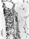
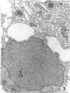
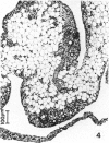
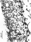
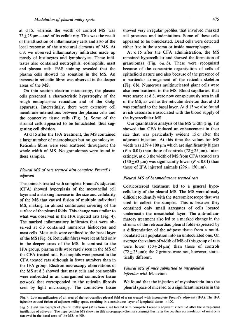
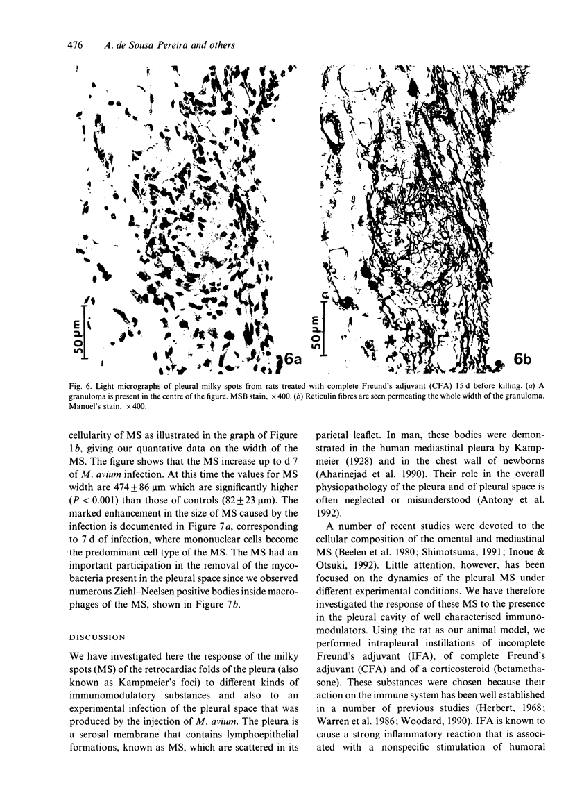
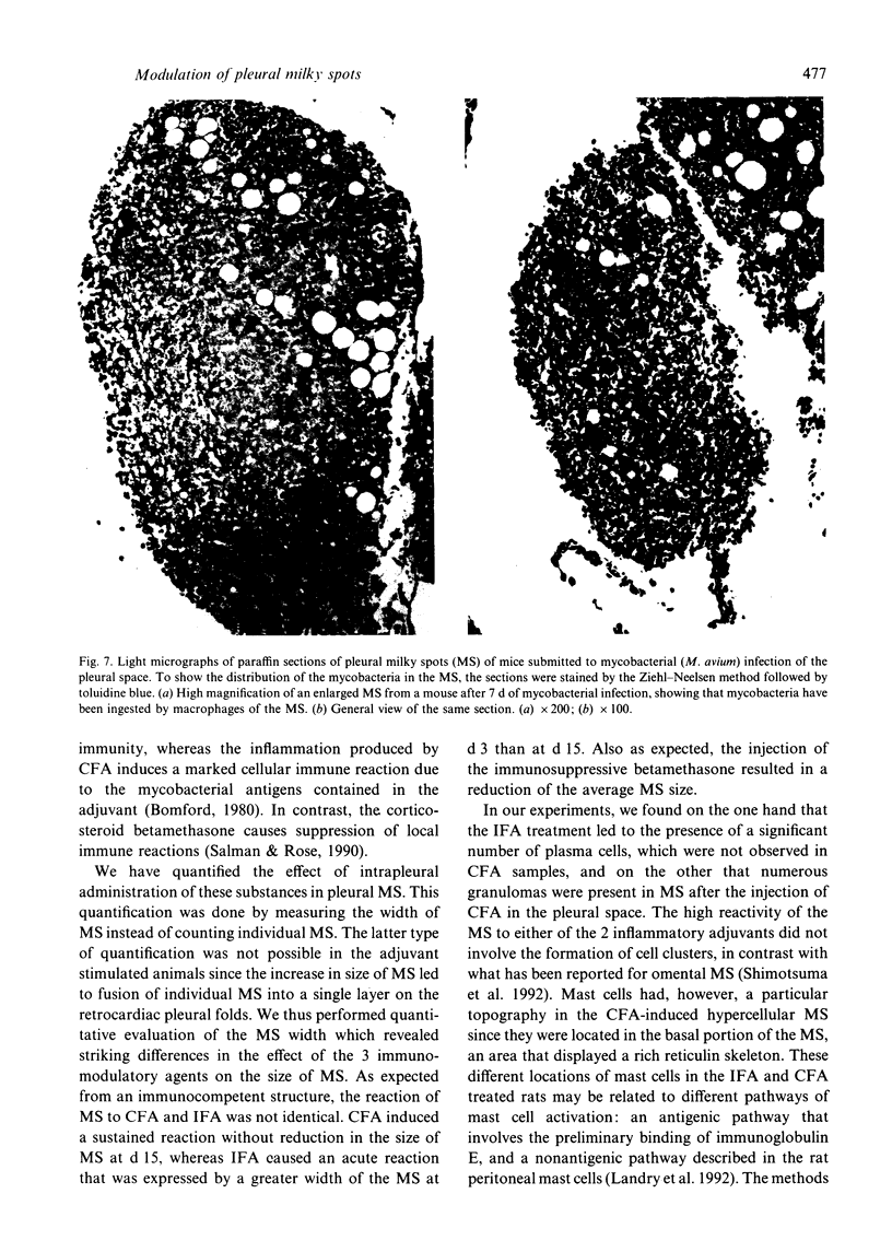
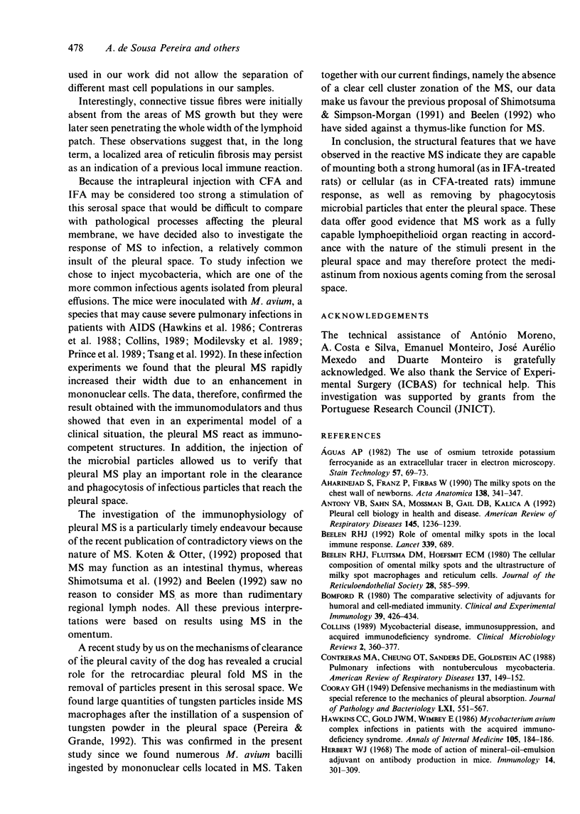
We studied the response of milky spots in the parietal pleura of the rat and mouse to intrapleural instillation of immunomodulatory agents such as complete or incomplete Freund's adjuvants and betamethasone, and also to infection by mycobacteria (M. avium). Both incomplete (mineral oil) and complete (mineral oil plus dead mycobacteria) adjuvants, as well as M. avium infection, induced a striking increase in the size and cellularity of the pleural milky spots whereas betamethasone caused a slight atrophy. The extensive inflammatory infiltrates observed after adjuvant injection differed between milky spots reactive to complete and incomplete Freund's adjuvants. Fifteen days after adjuvant administration, the pleural milky spots of rats were still enlarged and hypercellular but differences were noted in the size of milky spots of the pleura between the 2 adjuvant treatments: animals submitted to injection of complete Freund's adjuvant showed an increase in the size of milky spots from d 3 to d 15, while the size of milky spots of the incomplete Freund's adjuvant treated group showed a decrease in size from d 3 to d 15. The milky spots at d 15 were well organised: reticulin fibres permeated the whole area of the milky spot and the different cell types were evenly distributed. Histiocytes, which were previously confined to the inner layer, were now the main cell type in all areas of milky spots. A moderate number of mast cells and a few eosinophils were also seen. Complete Freund's adjuvant caused the formation of granulomas in the milky spots, a change that was not detected in animals treated with incomplete adjuvant.(ABSTRACT TRUNCATED AT 250 WORDS)
Full text
PDF
Images in this article
Selected References
These references are in PubMed. This may not be the complete list of references from this article.
- Aguas A. P. The use of osmium tetroxide-potassium ferrocyanide as an extracellular tracer in electron microscopy. Stain Technol. 1982 Mar;57(2):69–73. doi: 10.3109/10520298209066530. [DOI] [PubMed] [Google Scholar]
- Aharinejad S., Franz P., Firbas W. The milky spots on the chest wall in newborns. Acta Anat (Basel) 1990;138(4):341–347. doi: 10.1159/000146964. [DOI] [PubMed] [Google Scholar]
- Antony V. B., Sahn S. A., Mossman B., Gail D. B., Kalica A. NHLBI workshop summaries. Pleural cell biology in health and disease. Am Rev Respir Dis. 1992 May;145(5):1236–1239. doi: 10.1164/ajrccm/145.5.1236. [DOI] [PubMed] [Google Scholar]
- Beelen R. H., Fluitsma D. M., Hoefsmit E. C. The cellular composition of omentum milky spots and the ultrastructure of milky spot macrophages and reticulum cells. J Reticuloendothel Soc. 1980 Dec;28(6):585–599. [PubMed] [Google Scholar]
- Beelen R. H. Role of omental milky spots in the local immune response. Lancet. 1992 Mar 14;339(8794):689–689. doi: 10.1016/0140-6736(92)90857-y. [DOI] [PubMed] [Google Scholar]
- Bomford R. The comparative selectivity of adjuvants for humoral and cell-mediated immunity. I. Effect on the antibody response to bovine serum albumin and sheep red blood cells of Freund's incomplete and complete adjuvants, alhydrogel, Corynebacterium parvum, Bordetella pertussis, muramyl dipeptide and saponin. Clin Exp Immunol. 1980 Feb;39(2):426–434. [PMC free article] [PubMed] [Google Scholar]
- Collins F. M. Mycobacterial disease, immunosuppression, and acquired immunodeficiency syndrome. Clin Microbiol Rev. 1989 Oct;2(4):360–377. doi: 10.1128/cmr.2.4.360. [DOI] [PMC free article] [PubMed] [Google Scholar]
- Contreras M. A., Cheung O. T., Sanders D. E., Goldstein R. S. Pulmonary infection with nontuberculous mycobacteria. Am Rev Respir Dis. 1988 Jan;137(1):149–152. doi: 10.1164/ajrccm/137.1.149. [DOI] [PubMed] [Google Scholar]
- Hawkins C. C., Gold J. W., Whimbey E., Kiehn T. E., Brannon P., Cammarata R., Brown A. E., Armstrong D. Mycobacterium avium complex infections in patients with the acquired immunodeficiency syndrome. Ann Intern Med. 1986 Aug;105(2):184–188. doi: 10.7326/0003-4819-105-2-184. [DOI] [PubMed] [Google Scholar]
- Herbert W. J. The mode of action of mineral-oil emulsion adjuvants on antibody production in mice. Immunology. 1968 Mar;14(3):301–318. [PMC free article] [PubMed] [Google Scholar]
- Inoue N., Otsuki Y. Lymphocyte subpopulations in mediastinal milky spots of mice: light- and electron-microscopic immunohistochemical observations. Arch Histol Cytol. 1992 Mar;55(1):89–96. doi: 10.1679/aohc.55.89. [DOI] [PubMed] [Google Scholar]
- Kanazawa K., Roe F. J., Yamamoto T. Milky spots (Taches laiteuses) as structures which trap asbestos in mesothelial layers and their significance in the pathogenesis of mesothelial neoplasia. Int J Cancer. 1979 Jun 15;23(6):858–865. doi: 10.1002/ijc.2910230619. [DOI] [PubMed] [Google Scholar]
- Koten J. W., den Otter W. Are omental milky spots an intestinal thymus? Lancet. 1991 Nov 9;338(8776):1189–1190. doi: 10.1016/0140-6736(91)92043-2. [DOI] [PubMed] [Google Scholar]
- Modilevsky T., Sattler F. R., Barnes P. F. Mycobacterial disease in patients with human immunodeficiency virus infection. Arch Intern Med. 1989 Oct;149(10):2201–2205. [PubMed] [Google Scholar]
- Neiss W. F. Electron staining of the cell surface coat by osmium-low ferrocyanide. Histochemistry. 1984;80(3):231–242. doi: 10.1007/BF00495771. [DOI] [PubMed] [Google Scholar]
- Pereira A. S., Grande N. R. Particle clearance from the canine pleural space into thoracic lymph nodes: an experimental study. Lymphology. 1992 Sep;25(3):120–128. [PubMed] [Google Scholar]
- Prince D. S., Peterson D. D., Steiner R. M., Gottlieb J. E., Scott R., Israel H. L., Figueroa W. G., Fish J. E. Infection with Mycobacterium avium complex in patients without predisposing conditions. N Engl J Med. 1989 Sep 28;321(13):863–868. doi: 10.1056/NEJM198909283211304. [DOI] [PubMed] [Google Scholar]
- REYNOLDS E. S. The use of lead citrate at high pH as an electron-opaque stain in electron microscopy. J Cell Biol. 1963 Apr;17:208–212. doi: 10.1083/jcb.17.1.208. [DOI] [PMC free article] [PubMed] [Google Scholar]
- Shimotsuma M., Simpson-Morgan M. Omental milky spots. Lancet. 1991 Dec 21;338(8782-8783):1596–1596. doi: 10.1016/0140-6736(91)92419-3. [DOI] [PubMed] [Google Scholar]
- Shimotsuma M., Takahashi T., Kawata M., Dux K. Cellular subsets of the milky spots in the human greater omentum. Cell Tissue Res. 1991 Jun;264(3):599–601. doi: 10.1007/BF00319049. [DOI] [PubMed] [Google Scholar]
- Tsang A. Y., Denner J. C., Brennan P. J., McClatchy J. K. Clinical and epidemiological importance of typing of Mycobacterium avium complex isolates. J Clin Microbiol. 1992 Feb;30(2):479–484. doi: 10.1128/jcm.30.2.479-484.1992. [DOI] [PMC free article] [PubMed] [Google Scholar]
- Warren H. S., Vogel F. R., Chedid L. A. Current status of immunological adjuvants. Annu Rev Immunol. 1986;4:369–388. doi: 10.1146/annurev.iy.04.040186.002101. [DOI] [PubMed] [Google Scholar]
- Woodard L. F. Surface chemistry and classification of vaccine adjuvants and vehicles. Adv Biotechnol Processes. 1990;13:281–306. [PubMed] [Google Scholar]


