Abstract
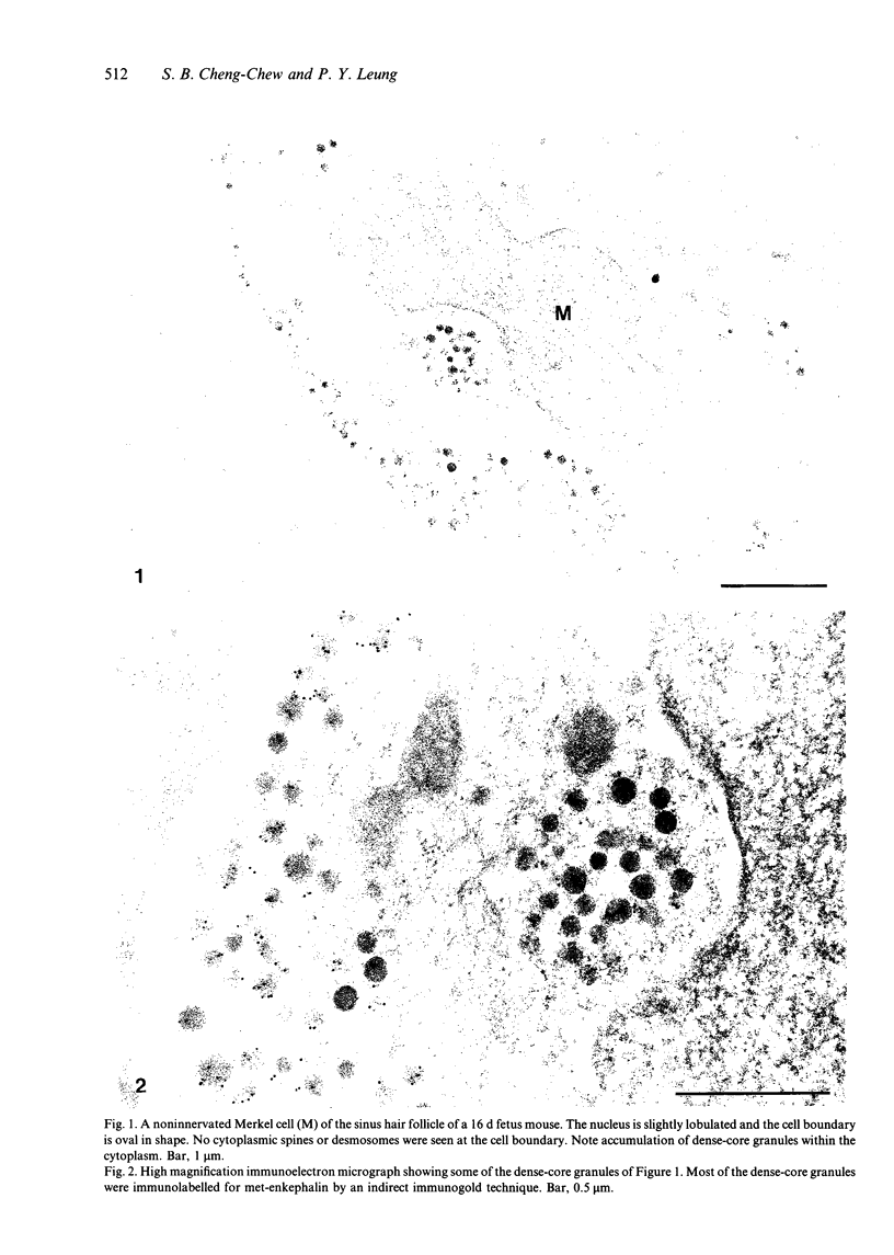
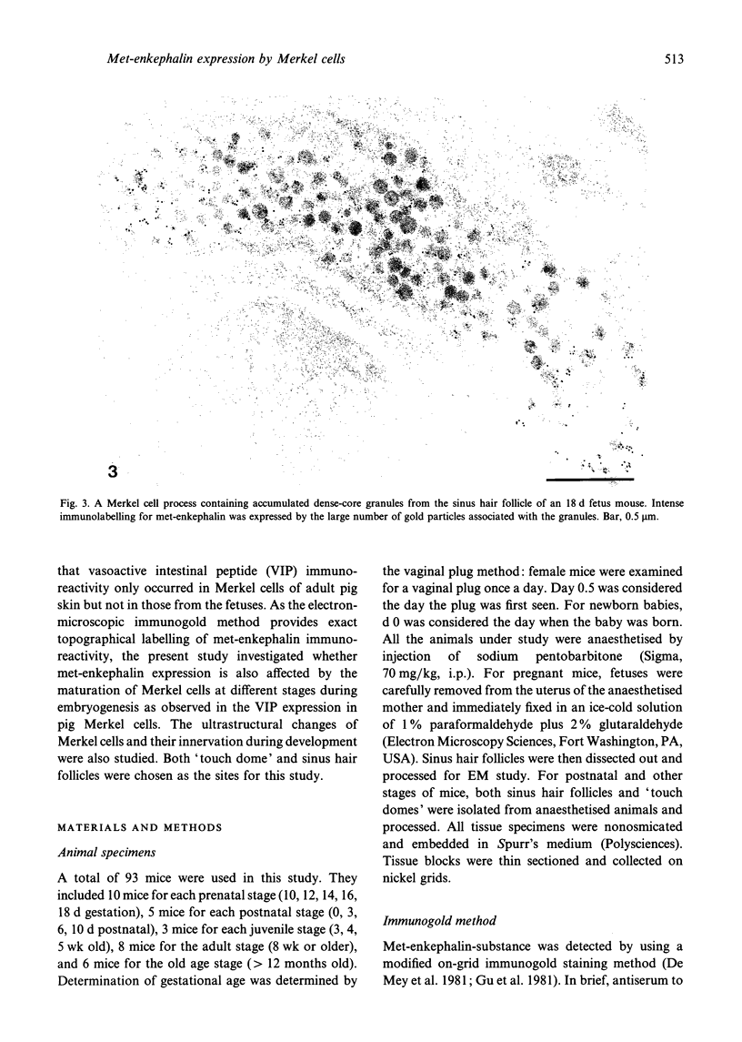
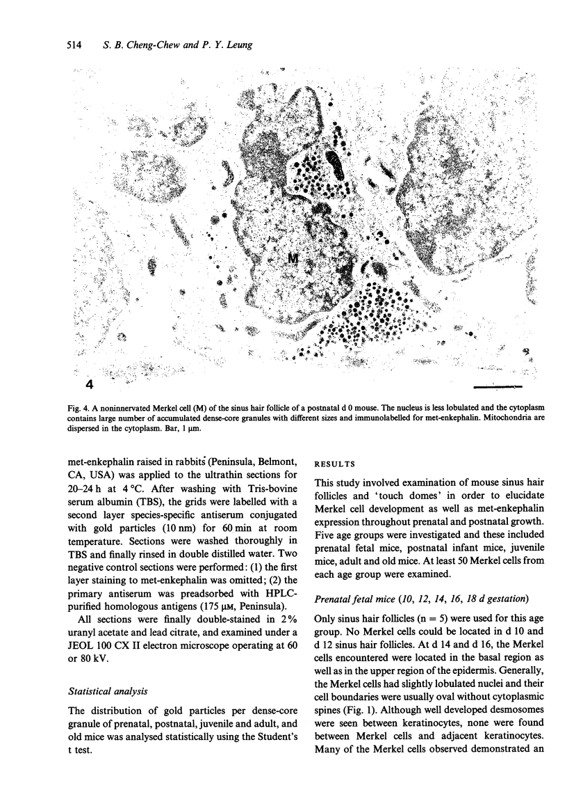
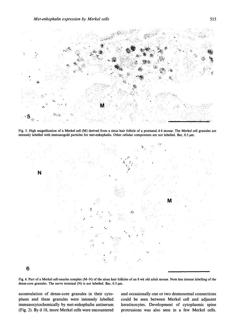
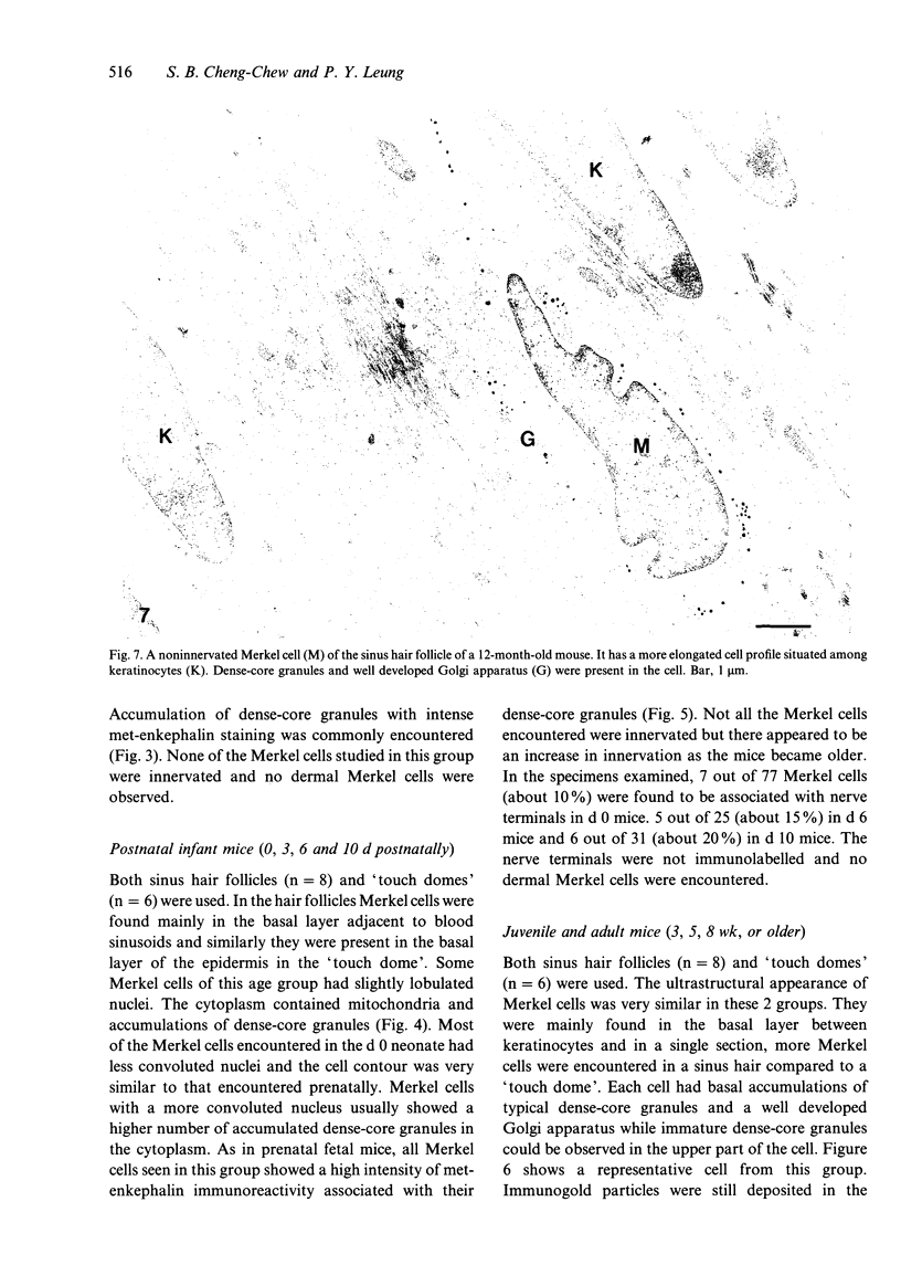
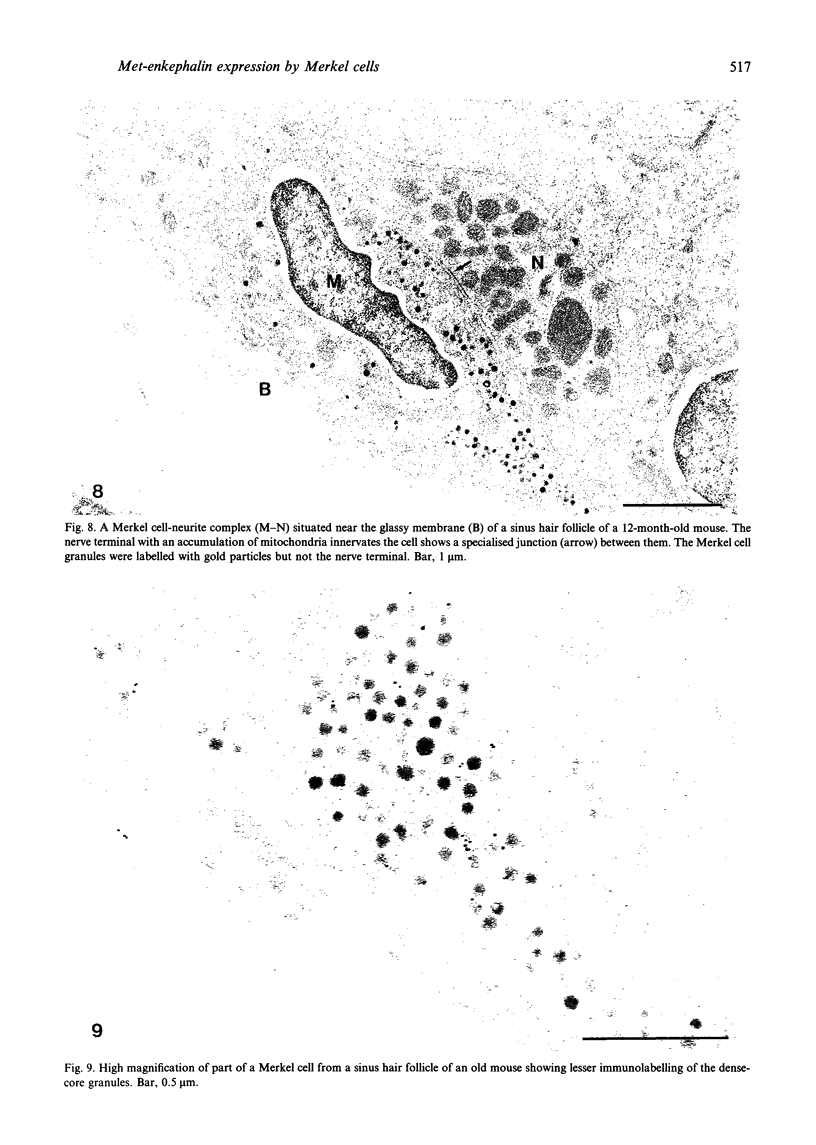
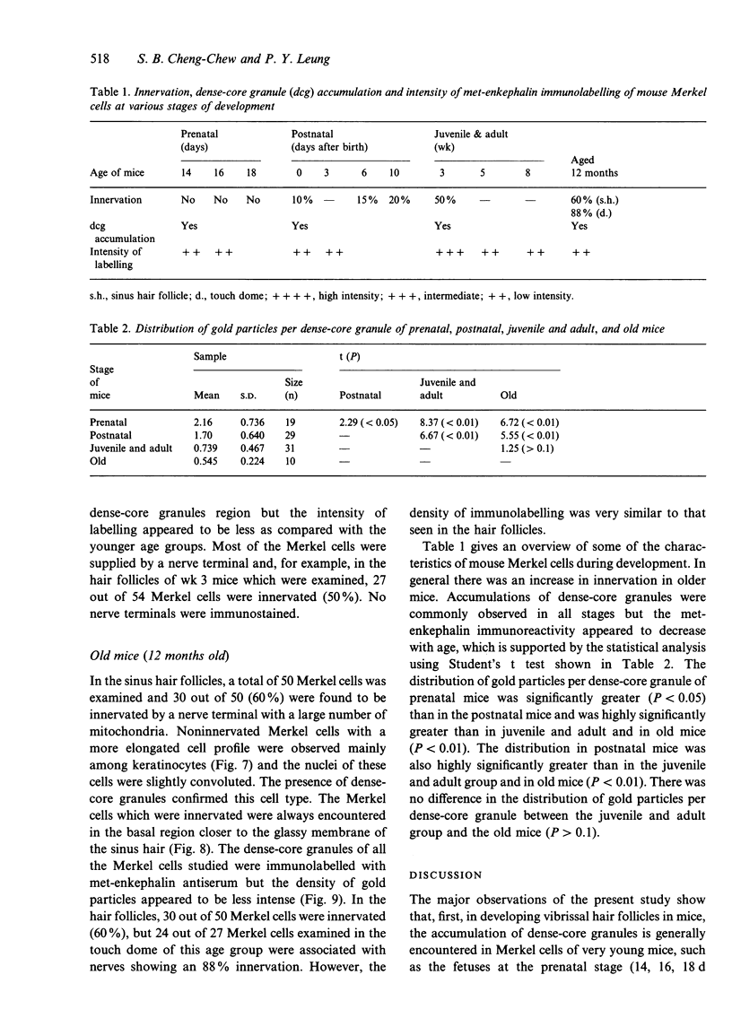
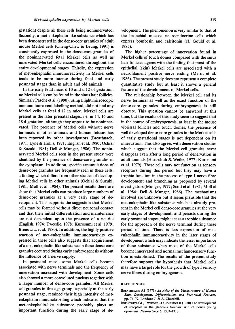
The morphological changes and the expression of met-enkephalin immunoreactivity of Merkel cells during fetal and postnatal development were investigated in touch domes and sinus hair follicles of mice by transmission electron microscopy. In prenatal fetal mice, the Merkel cells were mainly oval in shape and had slightly lobulated nuclei. These fetal Merkel cells (14, 16, 18 d gestation) which were not innervated showed a large number of accumulated dense-core granules in their cytoplasm as compared with the innervated Merkel cells which appeared in adult mice. No Merkel cells could be found in d 10 and d 12 fetuses. Innervation of Merkel cells was found to increase with age. The location of Merkel cells in juvenile, adult and even old mice was very similar, cells being found mainly in the basal layer of the epithelium. Using the electron-microscopic immunogold method, met-enkephalin-like substance was consistently located in the dense-core granule region of both innervated and noninnervated Merkel cells throughout the whole developmental stage. Interestingly, it was also found that the labelling intensity of met-enkephalin immunoreactivity was significantly higher in Merkel cells of younger age groups than in adult and old age groups. None of the nerve terminals associated with Merkel cells were labelled. The present study supports the theory of an epidermal origin of Merkel cells followed by the trophic growth of nerve fibers induced by the peptides.
Full text
PDF
Images in this article
Selected References
These references are in PubMed. This may not be the complete list of references from this article.
- Brenowitz G. L., Tweedle C. D., Johnson J. I. The development of receptors in the glabrous forepaw skin of pouch young opossums. Neuroscience. 1980;5(7):1303–1310. doi: 10.1016/0306-4522(80)90202-x. [DOI] [PubMed] [Google Scholar]
- Cheng Chew S. B., Leung P. Y. Species variability in the expression of met- and leu-enkephalin-like immunoreactivity in mammalian Merkel cell dense-core granules. A light- and electron-microscopic immunohistochemical study. Cell Tissue Res. 1992 Aug;269(2):347–351. doi: 10.1007/BF00319627. [DOI] [PubMed] [Google Scholar]
- Chew S. B., Leung P. Y. Immunocytochemical evidence of a met-enkephalin-like substance in the dense-core granules of mouse Merkel cells. Cell Tissue Res. 1991 Sep;265(3):611–614. doi: 10.1007/BF00340885. [DOI] [PubMed] [Google Scholar]
- Chew S. B., Leung P. Y. Methionine-enkephalin immunoreactivity in Merkel cell dense-core granules of nude mice sinus hair. A post-embedding immunogold electron-microscopic study. In Vivo. 1992 Mar-Apr;6(2):195–198. [PubMed] [Google Scholar]
- De Mey J., Moeremans M., Geuens G., Nuydens R., De Brabander M. High resolution light and electron microscopic localization of tubulin with the IGS (immuno gold staining) method. Cell Biol Int Rep. 1981 Sep;5(9):889–899. doi: 10.1016/0309-1651(81)90204-6. [DOI] [PubMed] [Google Scholar]
- Dell D. A., Munger B. L. The early embryogenesis of papillary (sweat duct) ridges in primate glabrous skin: the dermatotopic map of cutaneous mechanoreceptors and dermatoglyphics. J Comp Neurol. 1986 Feb 22;244(4):511–532. doi: 10.1002/cne.902440408. [DOI] [PubMed] [Google Scholar]
- English K. B., Burgess P. R., Kavka-Van Norman D. Development of rat Merkel cells. J Comp Neurol. 1980 Nov 15;194(2):475–496. doi: 10.1002/cne.901940212. [DOI] [PubMed] [Google Scholar]
- English K. B. Cell types in cutaneous type I mechanoreceptors (Haarscheiben) and their alterations with injury. Am J Anat. 1974 Sep;141(1):105–126. doi: 10.1002/aja.1001410107. [DOI] [PubMed] [Google Scholar]
- Gould V. E., Moll R., Moll I., Lee I., Franke W. W. Neuroendocrine (Merkel) cells of the skin: hyperplasias, dysplasias, and neoplasms. Lab Invest. 1985 Apr;52(4):334–353. [PubMed] [Google Scholar]
- Hartschuh W., Reinecke M., Weihe E., Yanaihara N. VIP-immunoreactivity in the skin of various mammals: immunohistochemical, radioimmunological and experimental evidence for a dual localization in cutaneous nerves and merkel cells. Peptides. 1984 Mar-Apr;5(2):239–245. doi: 10.1016/0196-9781(84)90213-4. [DOI] [PubMed] [Google Scholar]
- Hartschuh W., Weihe E. Multiple messenger candidates and marker substance in the mammalian Merkel cell-axon complex: a light and electron microscopic immunohistochemical study. Prog Brain Res. 1988;74:181–187. doi: 10.1016/s0079-6123(08)63012-5. [DOI] [PubMed] [Google Scholar]
- Hashimoto K. The ultrastructure of the skin of human embryos. X. Merkel tactile cells in the finger and nail. J Anat. 1972 Jan;111(Pt 1):99–120. [PMC free article] [PubMed] [Google Scholar]
- Iggo A., Muir A. R. The structure and function of a slowly adapting touch corpuscle in hairy skin. J Physiol. 1969 Feb;200(3):763–796. doi: 10.1113/jphysiol.1969.sp008721. [DOI] [PMC free article] [PubMed] [Google Scholar]
- Kurosumi K., Kurosumi U., Inoue K. Morphological and morphometric studies with the electron microscope on the Merkel cells and associated nerve terminals of normal and denervated skin. Arch Histol Jpn. 1979 Jul;42(3):243–261. doi: 10.1679/aohc1950.42.243. [DOI] [PubMed] [Google Scholar]
- Lyne A. G., Hollis D. E. Merkel cells in sheep epidermis during fetal development. J Ultrastruct Res. 1971 Mar;34(5):464–472. doi: 10.1016/s0022-5320(71)80059-x. [DOI] [PubMed] [Google Scholar]
- Moll R., Moll I., Franke W. W. Identification of Merkel cells in human skin by specific cytokeratin antibodies: changes of cell density and distribution in fetal and adult plantar epidermis. Differentiation. 1984;28(2):136–154. doi: 10.1111/j.1432-0436.1984.tb00277.x. [DOI] [PubMed] [Google Scholar]
- Munger B. L. Neural-epithelial interactions in sensory receptors. J Invest Dermatol. 1977 Jul;69(1):27–40. doi: 10.1111/1523-1747.ep12497861. [DOI] [PubMed] [Google Scholar]
- Ochiai T., Suzuki H. Fine structural and morphometric studies of the Merkel cell during fetal and postnatal development. J Invest Dermatol. 1981 Dec;77(6):437–443. doi: 10.1111/1523-1747.ep12495677. [DOI] [PubMed] [Google Scholar]
- Pasche F., Mérot Y., Carraux P., Saurat J. H. Relationship between Merkel cells and nerve endings during embryogenesis in the mouse epidermis. J Invest Dermatol. 1990 Sep;95(3):247–251. doi: 10.1111/1523-1747.ep12484847. [DOI] [PubMed] [Google Scholar]
- Pearse A. G. The diffuse neuroendocrine system: peptides, amines, placodes and the APUD theory. Prog Brain Res. 1986;68:25–31. doi: 10.1016/s0079-6123(08)60229-0. [DOI] [PubMed] [Google Scholar]
- Pearse A. G. The neuroendocrine (APUD) cells of the skin. Am J Dermatopathol. 1980 Summer;2(2):121–123. doi: 10.1097/00000372-198000220-00002. [DOI] [PubMed] [Google Scholar]
- Scott S. A., Cooper E., Diamond J. Merkel cells as targets of the mechanosensory nerves in salamander skin. Proc R Soc Lond B Biol Sci. 1981 Mar 27;211(1185):455–470. doi: 10.1098/rspb.1981.0017. [DOI] [PubMed] [Google Scholar]
- Tweedle C. D. Ultrastructure of Merkel cell development in aneurogenic and control amphibian larvae (Ambystoma). Neuroscience. 1978;3(4-5):481–486. doi: 10.1016/0306-4522(78)90052-0. [DOI] [PubMed] [Google Scholar]


