Abstract
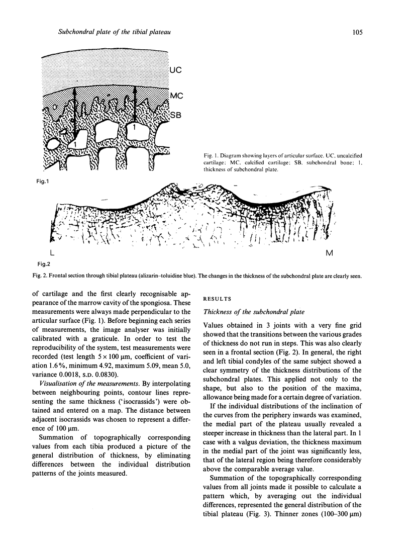
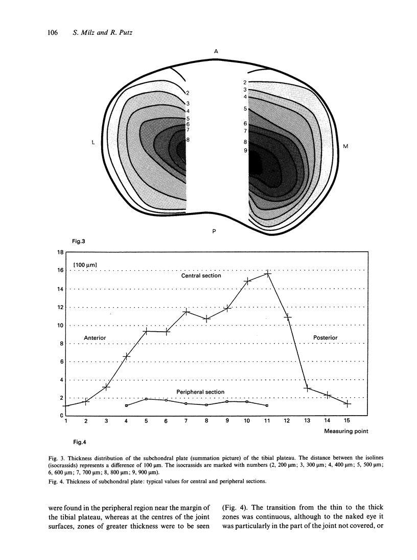
The object of the present investigation was to measure the thickness distribution of the subchondral plate of the tibial plateau. The data were obtained by computerised image analysis of serial sections. The measured values revealed a marked difference in the thickness between the various regions of the joint surface. Thinner zones (100-300 microns) are found in the peripheral region near the margin of the tibial plateau. Thickness maxima (up to 1500 microns and more) are to be seen at the centres of the joint surfaces. The relationship between the thickness distribution of the subchondral plate and information about the stress distribution of this particular joint surface support the conclusion that the morphology of the subchondral plate of the tibial plateau is determined by the function of the joint.
Full text
PDF
Images in this article
Selected References
These references are in PubMed. This may not be the complete list of references from this article.
- Ahmed A. M., Burke D. L. In-vitro measurement of static pressure distribution in synovial joints--Part I: Tibial surface of the knee. J Biomech Eng. 1983 Aug;105(3):216–225. doi: 10.1115/1.3138409. [DOI] [PubMed] [Google Scholar]
- Ateshian G. A., Soslowsky L. J., Mow V. C. Quantitation of articular surface topography and cartilage thickness in knee joints using stereophotogrammetry. J Biomech. 1991;24(8):761–776. doi: 10.1016/0021-9290(91)90340-s. [DOI] [PubMed] [Google Scholar]
- Behrens J. C., Walker P. S., Shoji H. Variations in strength and structure of cancellous bone at the knee. J Biomech. 1974 May;7(3):201–207. doi: 10.1016/0021-9290(74)90010-4. [DOI] [PubMed] [Google Scholar]
- Clark J. M., Huber J. D. The structure of the human subchondral plate. J Bone Joint Surg Br. 1990 Sep;72(5):866–873. doi: 10.1302/0301-620X.72B5.2211774. [DOI] [PubMed] [Google Scholar]
- Duncan H., Jundt J., Riddle J. M., Pitchford W., Christopherson T. The tibial subchondral plate. A scanning electron microscopic study. J Bone Joint Surg Am. 1987 Oct;69(8):1212–1220. [PubMed] [Google Scholar]
- Fukubayashi T., Kurosawa H. The contact area and pressure distribution pattern of the knee. A study of normal and osteoarthrotic knee joints. Acta Orthop Scand. 1980 Dec;51(6):871–879. doi: 10.3109/17453678008990887. [DOI] [PubMed] [Google Scholar]
- Goldstein S. A. The mechanical properties of trabecular bone: dependence on anatomic location and function. J Biomech. 1987;20(11-12):1055–1061. doi: 10.1016/0021-9290(87)90023-6. [DOI] [PubMed] [Google Scholar]
- Hoshino A., Wallace W. A. [Impact absorbing properties of the human knee joint]. Nihon Seikeigeka Gakkai Zasshi. 1989 Jan;63(1):67–74. [PubMed] [Google Scholar]
- Hutzschenreuter P., Brümmer H. Embedding, cutting and grinding methods to produce undecalcified cortical bone sections over the whole bone. Acta Anat (Basel) 1984;118(4):201–204. doi: 10.1159/000145845. [DOI] [PubMed] [Google Scholar]
- Hvid I., Hansen S. L. Subchondral bone strength in arthrosis. Cadaver studies of tibial condyles. Acta Orthop Scand. 1986 Feb;57(1):47–51. doi: 10.3109/17453678608993214. [DOI] [PubMed] [Google Scholar]
- Hvid I., Jensen J., Nielsen S. Bone strength measurements at the proximal tibia. Penetration tests and epiphyseal compressive strength. Int Orthop. 1986;10(4):271–275. doi: 10.1007/BF00454408. [DOI] [PubMed] [Google Scholar]
- Józsa L., Réffy A., Järvinen M., Kannus P., Lehto M., Kvist M. Cortical and trabecular osteopenia after immobilization. A quantitative histological study of the rat knee. Int Orthop. 1988;12(2):169–172. doi: 10.1007/BF00266984. [DOI] [PubMed] [Google Scholar]
- Noble J., Alexander K. Studies of tibial subchondral bone density and its significance. J Bone Joint Surg Am. 1985 Feb;67(2):295–302. [PubMed] [Google Scholar]
- Odgaard A., Pedersen C. M., Bentzen S. M., Jørgensen J., Hvid I. Density changes at the proximal tibia after medial meniscectomy. J Orthop Res. 1989;7(5):744–753. doi: 10.1002/jor.1100070517. [DOI] [PubMed] [Google Scholar]
- Pauwels F. Uber die gestaltende Wirkung der funktionellen Anpassung des Knochens. Anat Anz. 1976;139(3):213–220. [PubMed] [Google Scholar]
- SCHENK R. ZUR HISTOLOGISCHEN VERARBEITUNG VON UNENTKALKTEN KNOCHEN. Acta Anat (Basel) 1965;60:3–19. [PubMed] [Google Scholar]
- Sneppen O., Christensen P., Larsen H., Vang P. S. Mechanical testing of trabecular bone in knee replacement. Int Orthop. 1981;5(4):251–256. doi: 10.1007/BF00271079. [DOI] [PubMed] [Google Scholar]
- Spilker R. L., Donzelli P. S., Mow V. C. A transversely isotropic biphasic finite element model of the meniscus. J Biomech. 1992 Sep;25(9):1027–1045. doi: 10.1016/0021-9290(92)90038-3. [DOI] [PubMed] [Google Scholar]
- Swartz D. E., Wittenberg R. H., Shea M., White A. A., 3rd, Hayes W. C. Physical and mechanical properties of calf lumbosacral trabecular bone. J Biomech. 1991;24(11):1059–1068. doi: 10.1016/0021-9290(91)90022-f. [DOI] [PubMed] [Google Scholar]
- Walker P. S., Hajek J. V. The load-bearing area in the knee joint. J Biomech. 1972 Nov;5(6):581–589. doi: 10.1016/0021-9290(72)90030-9. [DOI] [PubMed] [Google Scholar]


