Abstract
Introduction
Strains of the syphilis spirochete, Treponema pallidum ssp. pallidum, group into one of two deep-branching clades: the Nichols clade or the globally dominant Street Strain 14 (SS14) clade. To date, in-depth proteome-wide analyses have focused on Nichols clade strains.
Methods
The T. pallidum SS14 clade reference strain (SS14) proteome was characterized via protein detection and quantification analyses using mass spectrometry, and comparison was made to the Nichols clade reference strain (Nichols) proteome.
Results
Approximately two thirds of all proteins from T. pallidum SS14 were detected and quantitated, allowing confirmation of expression of 259 proteins for the first time in this strain, including 11 known/putative outer membrane proteins (OMPs). SS14 and Nichols proteome comparative analyses demonstrated similar protein expression/quantification profiles between the two strains, and showed that inter-strain amino acid sequence differences are located primarily within predicted surface-exposed regions in 16 known/putative OMPs.
Discussion
This study provides the first comparative analyses of the proteomes from the T. pallidum SS14 and Nichols strains. The findings inform syphilis vaccine design by confirming the expression of known/predicted OMP vaccine candidates in SS14 treponemes, and via the finding that most inter-strain variable residues found in OMPs are predicted to be located in surface-exposed, host-facing regions of these proteins.
Keywords: Treponema pallidum, syphilis, proteomics, outer membrane proteins, vaccine candidates
Introduction
Treponema pallidum ssp. pallidum (hereafter T. pallidum), the causative agent of infectious and congenital syphilis, continues to be a global public health concern, with rates increasing around the world (Herbert and Middleton, 2012; Savage et al., 2012; Chen et al., 2017; Arrieta and Singh, 2019; Korenromp et al., 2019). Furthermore, syphilis infections increase the risk of transmission and acquisition of other STIs, including HIV (Nusbaum et al., 2004; Douglas, 2009).
Comparative genomics studies have demonstrated that the worldwide distribution of T. pallidum strains consists of two deep-branching phylogenetic clades, with each clade named after their respective reference strain: Nichols and SS14 clades (Mikalova et al., 2010; Smajs et al., 2012; Petrosova et al., 2013; Nechvatal et al., 2014; Arora et al., 2016). The Nichols strain, originally isolated in Washington D.C. in 1912 from the cerebrospinal fluid of a patient with secondary syphilis (Nichols and Hough, 1913), is a laboratory reference strain that has undergone continuous passage in the rabbit animal model (LaFond and Lukehart, 2006), and more recently the in vitro culture system (Edmondson et al., 2018; Edmondson et al., 2021; Edmondson and Norris, 2021), since its isolation. The SS14 strain was first isolated in 1977 in Atlanta, Georgia from a skin lesion of an individual with secondary syphilis, and thus represents a more recently isolated clinical strain that has undergone fewer passages through the animal model or in vitro culture system (Stamm et al., 1983; Stamm et al., 1988). Based on currently available clinical sampling data, strains belonging to the SS14-like clade dominate syphilis infections worldwide (Arora et al., 2016; Beale et al., 2021; Lieberman et al., 2021).
Molecular typing and genomics studies have identified genetic differences between strains from the SS14 and Nichols clades (Petrosova et al., 2013; Nechvatal et al., 2014; Lieberman et al., 2021). Many of these genetic differences are conserved between strains within each clade, and are predicted to result in amino acid sequence differences in the corresponding encoded proteins (Mikalova et al., 2010; Smajs et al., 2012; Petrosova et al., 2013; Nechvatal et al., 2014; Arora et al., 2016; Lieberman et al., 2021). Included in these proteins are known and potential outer membrane proteins (OMPs) (Mikalova et al., 2010; Smajs et al., 2012; Petrosova et al., 2013; Nechvatal et al., 2014; Arora et al., 2016; Lieberman et al., 2021), a protein class that is the focus of current efforts for syphilis vaccine development.
In the present study, the proteome of in vitro-cultured T. pallidum SS14 was characterized using mass spectrometry (MS), providing the detection and quantification of proteins encoded by T. pallidum SS14 (referred to as proteome-wide analyses of SS14). Comparative proteomic analyses of SS14 and Nichols strains revealed shared protein expression and quantification profiles across all detected proteins and also among protein classes of specific interest, and validated inter-strain protein sequence variances identified by genome sequencing. Most of the SS14-Nichols inter-strain protein sequence variances found in the treponemal OMPs were mapped to predicted surface-exposed T. pallidum protein regions. This finding has implications for syphilis vaccine design as the treponemal OMPs are located at the pathogen-host interface and are targeted by the host immune response.
Materials and methods
In vitro culture of Treponema pallidum
Treponema pallidum (SS14 strain; supplied as a frozen stock by Lorenzo Giacani, University of Washington, Seattle, WA (Romeis et al., 2021)) was used for in vitro culture and sub-culture of treponemes in the presence of Sf1Ep (NBL-11) cottontail rabbit epithelial cells (ATCC CCL-68; American Type Culture Collection [ATCC], Rockville, MD, USA), as previously described (Edmondson et al., 2018; Edmondson et al., 2021; Edmondson and Norris, 2021; Houston et al., 2024). This SS14 reference strain was originally isolated by John Clark (Sexually Transmitted Disease Laboratory Program of the Centers for Disease Control) in 1977 in Atlanta, Georgia from an individual with secondary syphilis (Stamm et al., 1983). Trypsin-free dissociation buffer (Edmondson and Norris, 2021) was used to maintain OMP integrity during T. pallidum dissociation steps. The final in vitro-cultured T. pallidum sample contained 2.3 × 10⁹ treponemes suspended in sterile 0.9% saline (0.9% w/v NaCl, pH 7.0).
Treponema pallidum SS14 protein sample preparation: present study
In the present study, the T. pallidum SS14 protein sample was prepared for MS using a previously optimized sample preparation workflow comprised of three main steps: (1) centrifugation steps to remove contaminants, (2) lysis steps to extract T. pallidum proteins, and (3) a final centrifugation step to isolate soluble T. pallidum proteins. In step (1), insoluble rabbit gross cellular debris was removed from in vitro-cultured T. pallidum via slow-speed (220 × g) centrifugation steps. High-speed (17,000 × g) centrifugation steps were then performed to remove soluble contaminants. In step (2), chemical lysis of the T. pallidum pellets was performed via suspension in lysis buffer (50 mM ammonium bicarbonate pH 8.0, 0.9% sodium deoxycholate [Sigma-Aldrich Canada Co., Oakville, ON, Canada]) for 30 min on ice, followed by physical lysis using ultrasonication. In step (3), high speed centrifugation (17,000 × g) was used to remove precipitated, aggregated, or insoluble proteins and cellular debris. Further details are provided in our prior T. pallidum proteomics studies (Houston et al., 2023; Houston et al., 2024).
Treponema pallidum protein sample preparations from previous studies used for data integration
The combined T. pallidum SS14 proteome coverage was determined by integrating MS data from the present study results with the MS data from a previous T. pallidum SS14 genetic engineering study (Romeis et al., 2021). In the previous study by Romeis et al., the authors cultured wild-type and genetically engineered T. pallidum SS14 under in vitro culture conditions and prepared protein samples by pelleting 1 × 10⁹ treponemes using high speed centrifugation, followed by SDS-PAGE analysis, in-gel trypsin digestion, and liquid chromatography-tandem MS (LC-MS/MS) analysis using an LTQ HP1100 mass spectrometer. Additional details on the previous study can be found in Romeis et al. (2021). For the proteome comparative analyses of T. pallidum SS14 and Nichols Strains, we compared the SS14 strain MS data from the present study with MS data derived from our previous T. pallidum Nichols proteome study (Houston et al., 2024). In this previous study, three in vitro-cultured T. pallidum Nichols samples (containing 1.0 × 10⁹, 1.0 × 10⁹, and 1.55 × 10⁹ treponemes) were prepared using the optimized proteomics protocol, analyzed by LC–MS/MS, and analyzed for protein detection and label free quantification (LFQ) analyses as outlined in the current study.
MS sample preparation and LC-MS/MS
MS sample preparation and LC–MS/MS were performed on the T. pallidum protein sample using the method outlined in previous work (Houston et al., 2024) which was comprised of 3 major steps: (1) in-solution trypsin digestion, (2) high-pH reversed phase fractionation, and (3) protein detection via LC–MS/MS. In step (1), in-solution trypsin digestion was performed on the T. pallidum protein sample as previously described (Houston et al., 2024), with the following modifications. Dithiothritol (Sigma-Aldrich Canada Co., Oakville, ON, Canada) was added to a final concentration of 20 mM to reduce disulphide bonds. Cleanup of the trypsin-digested protein sample was performed via centrifugation for 5 min at 17,000 x g followed by lyophilization of the sample supernatant. To reduce sample complexity (step 2), high-pH reversed phase fractionation was used to separate the trypsin-digested T. pallidum sample into 24 fractions based on hydrophobicity, as previously described (Houston et al., 2024), with the following changes. Buffer A was comprised of 10 mM ammonium hydroxide, pH 9.0. Buffer B consisted of 80% acetonitrile and 10 mM ammonium hydroxide, pH 9.0. The 24 concatenated fractions were speed vacuum concentrated, and diluted in 2% acetonitrile/ 0.1% formic acid (100 μL final volume per fraction). In step (3), fractionated samples were subjected to LC–MS/MS for protein detections, as previously described (Houston et al., 2024). Briefly, a 10 μL aliquot of each concatenated fraction was separated by on-line reversed phase liquid chromatography using a Thermo Scientific EASY-nLC 1,000 system with an Acclaim PepMap100 reversed-phase pre-column C18 (100 μm I.D., 2 cm length, 5 μm bead size, 100 Å pore size) (Thermo Fisher Scientific, San Jose, CA), and an AcclaimPepMap100 C-18 reversed phase nano-analytical column (75 μm I.D., 15 cm length, 3 μm bead size, 100 Å pore size) (Thermo Fisher Scientific, San Jose, CA), at a flow rate of 300 nL/min. The chromatography system was coupled on-line with an Orbitrap Fusion Tribrid mass spectrometer (Thermo Fisher Scientific, San Jose, CA, USA) equipped with a Nanospray Flex NG source (Thermo Fisher Scientific). Tryptic peptides were separated using a 120-min gradient of solvent A (A; 2% acetonitrile/0.1% formic acid) and solvent B (B; 90% acetonitrile/0.1% formic acid), comprised of the following steps: (i) 0–100 min, gradient change from 95% A / 5% B to 58% A / 42% B, (ii) 100–115 min, gradient change from 58% A / 42% B to 0% A / 100% B, and (iii) 115–120 min, gradient held at 0% A / 100% B. The Orbitrap Fusion Tribrid mass spectrometer instrument parameters (Fusion Tune 3.5 software) were set as outlined before (Houston et al., 2024).
Protein detection: parameters and data analysis
Data analyses and the parameters used for protein detections were performed as described in our previous studies (Houston et al., 2023; Houston et al., 2024). In brief, tandem mass spectra were extracted using Proteome Discoverer version 3.0. (Thermo Scientific). Charge state deconvolution was not performed. All MS/MS samples were analyzed using Sequest (Thermo Fisher Scientific, San Jose, CA, USA; (version IseNode in Proteome Discoverer 3.0.0.757) containing either (1) a T. pallidum database (T. pallidum proteins, Nichols strain, NCBI reference sequence NC_021490, all proteome annotation revisions from June 17th 2013 – July 4th 2021), or (2) a T. pallidum SS14 database (T. pallidum proteins, SS14 strain, NCBI reference sequence: NC_021508, all proteome annotations from March 12th 2023). Proteome annotations from March 2023 were used in the SS14 database as these were the most up-to-date annotations available on the NCBI website when the present study was performed. Both databases also contained all reviewed (Swiss-Prot) canonical rabbit proteins (UniProt Oryctolagus cuniculus proteome, UP000001811) (Supplementary Table S1). Peptide/protein searches also included common contaminants.1 Protein detections in Scaffold (version Scaffold_5.1.2; Proteome Software Inc., Portland, OR, USA) were accepted if they could be established at 99.9% probability. Protein probabilities were assigned by the Protein Prophet algorithm (Nesvizhskii et al., 2003). Proteome coverage was calculated based on T. pallidum SS14 strain (NCBI reference sequence NC_021508, March 2023 annotation, 987 proteins); 981 proteins from predicted protein-coding genes, and six proteins potentially encoded by “pseudo genes.”
Label free quantification
Label free quantification (LFQ) was performed in Scaffold (version Scaffold_5.2.2) using total spectral counts as a relative quantitative measure of protein abundances in SS14 and Nichols strains. Only protein detections that were derived from the T. pallidum SS14 database search in the present study were used for LFQ analyses. Sample 2 protein detections from the in vitro-cultured T. pallidum Nichols strain (Houston et al., 2024) were used for SS14-Nichols comparative analyses, as this sample contained the highest number of detected treponemal proteins. The default settings for the quantitative analysis setup in Scaffold were used. Normalized total spectral counts were selected for each protein, and proteins were ranked from most abundant (highest spectral count) to least abundant (lowest spectral count). The T. pallidum SS14 and Nichols databases, as described above, were used for searches, peptide detections, and protein quantifications. Scaffold search parameters were as follows: precursor mass tolerance = 10 ppm; fragment mass (MS/MS) tolerance = 0.6 Da; enzyme specificity = trypsin, with a maximum of two missed cleavages allowed; fixed modification = carbamidomethylation (C); variable modifications = acetylation of the peptide N-terminus and oxidation (M).
Identification of SS14 proteome annotation errors (proteins prematurely truncated at the N-termini)
The T. pallidum database containing all proteome annotation revisions from June 17th 2013 to July 4th 2021 was used to identify SS14 proteins that have been annotated in the NCBI proteome with prematurely truncated N-termini, as described previously (Houston et al., 2023; Houston et al., 2024). This was achieved via the detection and identification of SS14 peptides that are not present in the SS14 proteome, but which are annotated as being located within extended N-terminal regions of proteins in previous versions of orthologous proteins from T. pallidum Nichols strain. Previously annotated versions of SS14 orthologs with similar extended N-termini (compared to the 2023 annotations) that also contained the detected SS14 peptides were then identified by searching previous revisions of the NCBI SS14 proteome annotations.
Proteome-wide functional categorization of Treponema pallidum proteins
All treponemal protein sequences were assigned to functional categories based on the “Clusters of Orthologous Genes” (COG) (Tatusov et al., 1997) categories,2 as previously described (Houston et al., 2024).
Data integration: combined Treponema pallidum SS14 proteome coverage
In a previous T. pallidum genetic engineering study, MS was used to confirm the expression of a 31 kDa protein produced from a genetically modified target gene (Romeis et al., 2021) in T. pallidum SS14, via the detection of proteins in the ~20–45 kDa size range. These MS analyses were performed as a genetic engineering confirmatory method (Romeis et al., 2021). For the MS analyses, the authors of the genetic engineering study analyzed a single sample prepared from the wild-type control SS14 strain and a single sample from a genetically engineered SS14 strain (Romeis et al., 2021). A beneficial consequence of these MS findings for the T. pallidum proteomics research field is the potential integration of these data with other T. pallidum MS studies focused on improving the proteome coverage of the SS14 strain. Therefore, to determine the combined T. pallidum SS14 proteome coverage in the present study, we integrated the MS data from the previous T. pallidum SS14 genetic engineering study (Romeis et al., 2021) with the T. pallidum SS14 MS results from the current study. For these MS data integration analyses, all detected T. pallidum proteins from the genetically engineered and wild-type control SS14 strains (Romeis et al., 2021) were used for determining the combined T. pallidum SS14 proteome coverage in the present study.
Data integration: proteome-wide comparative analyses of SS14-Nichols inter-strain protein sequence differences
Proteome-wide comparative analyses of SS14 (NC_021508) and Nichols (NC_021490) protein annotations were conducted to identify treponemal proteins with at least one amino acid difference between the strains. Mass spectrometry-based proteomics analyses were then used to experimentally validate the expression of as many of these differences as possible by checking for detection of peptides containing these variations in our present SS14 study, the previous study that performed MS analysis to confirm genetic engineering of T. pallidum (Romeis et al., 2021), and additional previously performed T. pallidum proteomics studies (Osbak et al., 2016; Houston et al., 2023; Houston et al., 2024). Manual inspection of these peptides was used to confirm inter-species protein differences. AlphaFold 33 (Abramson et al., 2024) was used for structure modeling of predicted/known OMPs with annotated and experimentally validated inter-strain amino acid differences, as detailed in below.
Structure modeling of predicted/known OMPs with annotated inter-strain amino acid differences
Structure modeling was performed using AlphaFold 3 (see footnote 3) (Abramson et al., 2024). The mature amino acid sequences of known/predicted OMPs from SS14 (NCBI reference sequence NC_021508, March 2023 annotation) and the corresponding proteins from Nichols (NCBI reference sequence NC_021490, March 2023 annotation) were generated using SignalP 6.04 (Teufel et al., 2022) and then submitted to the AlphaFold 3 (see footnote 3) server. The coordinate files for the highest confidence structure models were downloaded for visualization, labelling, and comparative analyses in the molecular visualization system, PyMOL5 (Schrodinger, 2015).
Results
Proteome-wide profiling of Treponema pallidum SS14
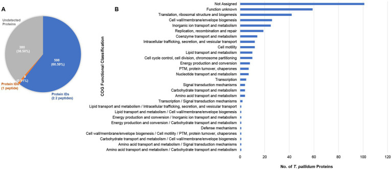
In the present study, 7,163 unique T. pallidum peptides were identified which allowed for the detection of 607 of 987 proteins within treponemes from the SS14 strain, representing 61.5% of the total proteome (Figure 1A, Table 1, and Supplementary Table S2). Among these proteins, nine were identified based on the detection of single peptides, whereas 598 were identified based on the detection of at least two peptides (Figure 1A, Table 1, and Supplementary Table S2). Detailed MS data and Scaffold peptide reports for T. pallidum SS14, including contaminant peptides and proteins, are provided in Supplementary Table S2.
Figure 1.
Proteome coverage of T. pallidum SS14. (A) Pie chart showing the total number of SS14 proteins detected in the present study based on the detection of a single peptide, or at least two peptides. The number of undetected T. pallidum proteins is also shown. Values in parentheses indicate the percentage of T. pallidum proteins found in each of the three categories. (B) Bar chart showing the COG (Clusters of Orthologous Genes) functional classification distribution of 380 T. pallidum proteins that were not detected in the present global proteomics study. COG functional classifications were generated by the eggNOG-mapper tool. Not assigned – proteins where eggNOG-mapper was unable to assign functional classifications. PTM: Post-translational modification.
Table 1.
Summary of T. pallidum SS14 proteins detected.
| Number of detected T. pallidum SS14 proteins | ||
|---|---|---|
| Number of proteins detected | 1 peptide for protein ID | 2 or more peptides for protein ID |
| 607 (61.5% proteome coverage) | 9 (0.91% proteome coverage) | 598 (60.6% proteome coverage) |
| Proteome coverage of T. pallidum SS14: this study and previous study (Romeis et al., 2021) | ||
| Number of proteins detected | 1 peptide for protein ID | 2 or more peptides for protein ID |
| 663 (67.2% proteome coverage) (259 SS14 proteins identified only in the present study) | 7 (0.71% proteome coverage) | 656 (66.6% proteome coverage) (252 SS14 proteins identified only in the present study) |
| Detection of T. pallidum miniproteins of unknown function | ||
| Number of miniproteins detected | 1 peptide for protein ID | 2 or more peptides for protein ID |
| 14 (17.9% miniprotein coverage) (14 SS14 miniproteins uniquely detected in the present study) | 1 (1.3% miniprotein coverage) | 13 (16.7% miniprotein coverage) (13 SS14 miniproteins uniquely detected in the present study) |
| Detection of hypothetical proteins and proteins of unknown function | ||
| Number of proteins detected | 1 peptide for protein ID | 2 or more peptides for protein ID |
| 146/282 total proteins detected (51.8% coverage) (76 SS14 proteins of unknown function uniquely detected in the present study) | 1 (0.4% coverage) | 145 (51.4% coverage) (75 SS14 proteins of unknown function uniquely detected in the present study) |
| Detection of known or predicted OMPs | ||
| Number of proteins detected | 1 peptide for protein ID | 2 or more peptides for protein ID |
| 22 (64.7% known/predicted OMP coverage) (11 SS14 OMPs uniquely detected in the present study) | 0 (0% known/predicted OMP coverage) | 22 (64.7% known/predicted OMP coverage) (11 SS14 OMPs uniquely detected in the present study) |
| Detection of putative pathogenesis-related proteins (PPRPs) | ||
| Number of proteins detected | 1 peptide for protein ID | 2 or more peptides for protein ID |
| 23 (67.6% coverage) (8 PPRPs identified only in the present study) | 0 (0% coverage) | 23 (67.6% coverage) (8 PPRPs identified only in the present study) |
Undetected Treponema pallidum SS14 proteins
The expression of 380 T. pallidum proteins remained undetectable, constituting 38.5% of the treponemal proteome (Figure 1A, Supplementary Tables S2, S3). More than one quarter of these proteins were annotated as proteins of unknown function (Supplementary Tables S2, S3). In line with these results, 42% of undetected proteins were categorized as ‘function unknown’ or were unassignable via COG (Clusters of Orthologous Genes) (Tatusov et al., 1997; Cantalapiedra et al., 2021) analysis (Figure 1B, and Supplementary Table S3). Similar to our previous T. pallidum Nichols proteomics studies (Houston et al., 2023; Houston et al., 2024), proteins classified with ‘translation’ and ‘inorganic ion transport and metabolism’ functions were also among the COG classes with the highest number of undetected proteins (Figure 1B, and Supplementary Table S3).
Data integration: combined coverage of the Treponema pallidum SS14 proteome
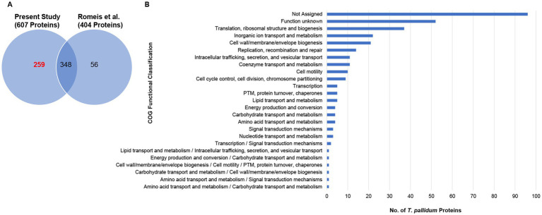
In a previous study that performed mass spectrometry analysis to confirm genetic engineering of T. pallidum, 404 proteins were detected from in vitro-grown T. pallidum SS14 (Romeis et al., 2021) that are also annotated in the reference proteome used here (Figure 2A, and Supplementary Table S4). Our analysis from the current study detected an additional 259 proteins from T. pallidum SS14 (Figure 2A, Table 1, and Supplementary Table S4). Data integration of our MS findings with the MS-related results from the genetic engineering study performed by Romeis et al. (2021) extended the SS14 proteome coverage to 663 proteins, or 67.2% (Figure 2A, Table 1, and Supplementary Table S4).
Figure 2.
Combined proteome coverage of T. pallidum SS14. (A) Venn diagram showing the total number of shared and exclusive protein detections corresponding to T. pallidum proteins detected in the present study and in a previous SS14 genetic engineering study (Romeis et al., 2021). The total number of T. pallidum proteins detected only in the current study (based on the detection of at least one tryptic peptide per identification) is highlighted in red text. (B) Bar chart showing the COG functional classification distribution of 324 treponemal proteins not detected in the present study or in the previous SS14 genetic engineering study (Romeis et al., 2021). COG functional classifications were generated by the eggNOG-mapper tool. Not assigned – proteins that were unable to be assigned functional categories by eggNOG-mapper. PTM: Post-translational modification.
Data integration: undetected proteins from the combined SS14 proteome coverages
After data integration of the MS-related results from the prior study performed by Romeis et al. (2021) with our current results, 324 (32.8%) T. pallidum proteins from the SS14 strain remained undetected (Supplementary Table S5). Almost one third of the undetected proteins have no clearly assigned functions in the proteome (Supplementary Table S5). Consistent with this observation, almost half of the 324 undetected proteins could not be classified into functional categories through COG analysis (Tatusov et al., 1997; Cantalapiedra et al., 2021) (Figure 2B and Supplementary Table S5). One third of the undetected proteins were miniproteins, defined as being comprised of 150 amino acids or less (Houston et al., 2022), with an average size of 99 amino acids (Supplementary Table S5). The high number of undetected miniproteins is likely due to their physicochemical properties that limit their detection via MS (Houston et al., 2023; Houston et al., 2024).
Expression-based comparative analyses of Treponema pallidum SS14 and Nichols strains
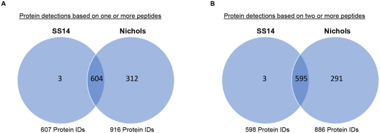
Six hundred four treponemal proteins were detected in both the present SS14 study and in our previous Nichols strain study (Houston et al., 2024) based on the detection of at least one peptide (Figure 3A and Supplementary Table S6). When protein identifications were based on the detection of at least two peptides, five hundred ninety-five treponemal proteins were detected in both T. pallidum strains (Figure 3B and Supplementary Table S6). Only three proteins were detected exclusively in strain SS14: TPASS_20517 (crossover junction endodeoxyribonuclease, RuvC), TPASS_20849 (50S ribosomal protein L35), and TPASS_ RS02370 (PQQ-like beta-propeller repeat protein) (Figure 3 and Supplementary Table S6). The 291 treponemal proteins that were detected only in the Nichols strain, based on the detection of at least two peptides, are listed in Supplementary Table S6 with annotated functions listed in Supplementary Table S2.
Figure 3.
Comparison of T. pallidum SS14 and Nichols proteome-wide expression profiles. Venn diagrams showing the number of shared and exclusive protein detections from SS14 and Nichols strains (Houston et al., 2024) based on the detection of (A) at least one tryptic peptide or (B) two or more tryptic peptides.
Quantitation-based comparative analyses of SS14 and Nichols strains
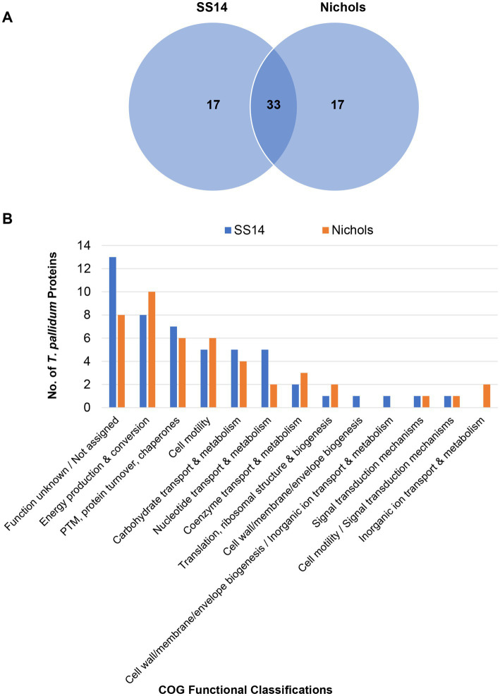
Label-free quantification (LFQ) analysis, based on total spectral counts, allowed for the relative quantification of 598 T. pallidum proteins within the SS14 strain and 914 T. pallidum proteins within the Nichols strain from our previous study (Houston et al., 2024) (Supplementary Table S7). High abundance proteins (proteins with LFQ intensity values greater than the mean average) from both strains are listed in Supplementary Table S8. Ninety-five percent of all high abundance proteins in the SS14 strain were also detected as high abundance proteins in treponemes from the Nichols strain (Supplementary Table S8), and 33 of the top 50 proteins with the highest expression levels were found in both strains (Supplementary Table S9 and Figure 4A). Most high abundance proteins were assigned functions related to ‘homeostasis’, ‘metabolism’, and ‘protein translation’ in both strains (Supplementary Tables S8, S9; Figure 4B; Supplementary Figure S1).
Figure 4.
Comparative analyses of the top 50 highest abundant proteins from T. pallidum SS14 and Nichols. (A) Venn diagram showing the total number of shared and exclusive protein detections corresponding to the top 50 highest abundant proteins from both T. pallidum strains. (B) Bar chart depicting the COG-based functional classification distribution of the top 50 highest abundant proteins from both T. pallidum strains. COG functional classifications were generated by the eggNOG-mapper tool. Not assigned – proteins where eggNOG-mapper was unable to assign functional classifications. PTM: Post-translational modification.
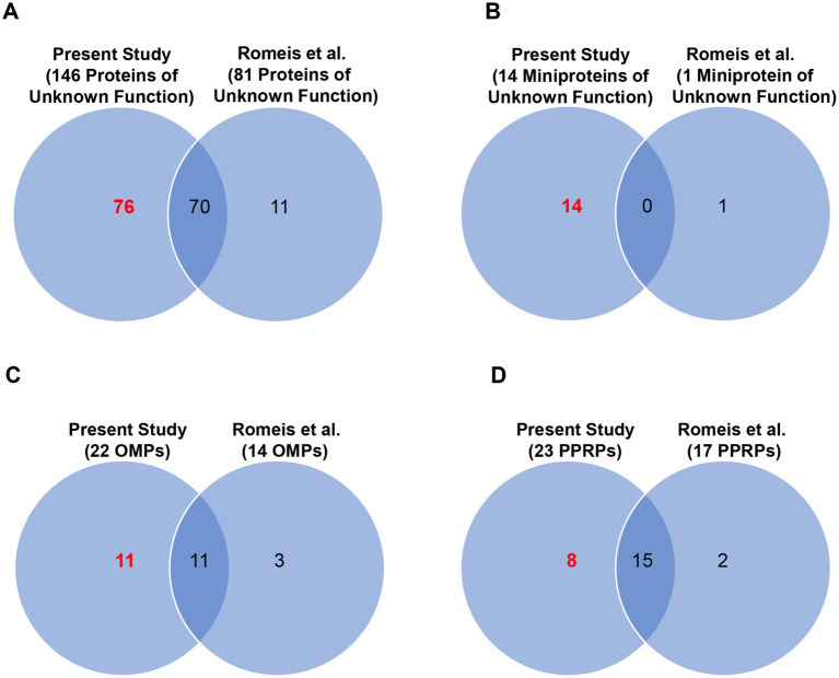
We detected expression of 146 of the 282 proteins from T. pallidum SS14 that have no clearly assigned functions in the proteome, including 76 proteins of unknown function whose expression was detected for the first time in this strain (Figure 5A; Table 1; Supplementary Table S10). All 146 proteins of unknown function detected in T. pallidum SS14 were also detected in the Nichols strain (Houston et al., 2024) (Supplementary Tables S6, S11). Additionally, proteins with no assigned function that are annotated as either “hypothetical proteins” or DUF (domain of unknown function)-containing proteins were found to comprise less than 10% of the high abundance proteins from both strains (Supplementary Table S8). In the SS14 strain, we also detected expression of 14 of the 78 miniproteins of unknown function (Figure 5B; Table 1; Supplementary Table S12), all of which were also detected in the Nichols strain (Houston et al., 2024) (Supplementary Tables S6, S11). Only one miniprotein of unknown function, TPASS_20777/TPANIC_0777, was detected with high expression levels in both strains (Supplementary Table S8).
Figure 5.
Combined proteome coverage for T. pallidum SS14 protein classes of interest. Venn diagrams showing the total number of shared and exclusive protein detections corresponding to (A) T. pallidum proteins of unknown function (no size restriction), (B) T. pallidum miniproteins of unknown function, (C) known/predicted OMPs, and (D) PPRPs, detected in a previous genetic engineering study (Romeis et al., 2021) and in the present MS-based proteomics studies. The total number of T. pallidum proteins of interest detected only in the current study (based on the detection of at least one tryptic peptide per identification) is highlighted in red text.
Previously, we compiled a list of 34 known and putative OMPs from the Nichols strain from literature reports (Houston et al., 2023). Here we detected 22 of the 34 proteins from SS14, including 11 proteins detected for the first time in this strain (Figure 5C, Tables 1, 2). One putative OMP was detected in both T. pallidum strains as a high abundance protein: the “hypothetical protein” TPASS_20855/TPANIC_0855 (Supplementary Table S8). Only one other potential OMP from the Nichols strain was detected as a high abundance protein: the UPF0164 family protein, TPANIC_0858 (Supplementary Table S8). The LFQ rankings for known/putative OMPs ranged from 78 to 579 out of a total of 598 SS14 strain proteins and 71–890 out of a total of 914 Nichols strain proteins (Supplementary Figure S2; Supplementary Table S7).
Table 2.
MS-based detection of predicted/known OMPs from T. pallidum SS14.
| OMP locus tag | NCBI functional annotation | MS detection |
|---|---|---|
| TPASS_RS05540 (WP_014505382) | Major outer sheath N-terminal domain-containing protein (TprA, N- and C termini truncated) | ND |
| TPASS_20011 | Major outer sheath C-terminal domain-containing protein (TprB) | Present, Romeis |
| TPASS_20117 | Major outer sheath N-terminal domain-containing protein (TprC) | Present |
| TPASS_20126 | Hypothetical protein | ND |
| TPASS_20131 | Major outer sheath N-terminal domain-containing protein (TprD) | Present |
| TPASS_20155 | M23 family metallopeptidase | ND |
| TPASS_20313 | Major outer sheath N-terminal domain-containing protein (TprE) | Present |
| TPASS_20316 | Major outer sheath N-terminal domain-containing protein (TprF) | ND |
| TPASS_20324 | Translocation/assembly module TamB domain-containing protein | Present |
| TPASS_20326 | Outer membrane protein assembly factor BamA | Present, Romeis |
| TPASS_20421 | Tetratricopeptide repeat protein | Present |
| TPASS_20479 | DUF2715 domain-containing protein | Present |
| TPASS_20483 | Fibronectin type III domain-containing protein | ND |
| TPASS_20515 | LPS-assembly protein LptD | Present |
| TPASS_20548 | UPF0164 family protein | Romeis |
| TPASS_20557 | DUF1007 family protein | Romeis |
| TPASS_20620 | Major outer sheath N-terminal domain-containing protein (TprI) | ND |
| TPASS_20621 | Major outer sheath N-terminal domain-containing protein (TprJ) | Present |
| TPASS_20698 | DUF2715 domain-containing protein | ND |
| TPASS_20733 | Outer membrane beta-barrel protein | ND |
| TPASS_20751 | Vascular adhesin/metalloprotease pallilysin | Present, Romeis |
| TPASS_20855 | Hypothetical protein | Present |
| TPASS_20856 | UPF0164 family protein | Romeis |
| TPASS_20858 | UPF0164 family protein | Present, Romeis |
| TPASS_20859 | UPF0164 family protein | Present |
| TPASS_20865 | UPF0164 family protein | Present |
| TPASS_20897 | MSP porin (TprK) | Present, Romeis |
| TPASS_20923 | PEGA domain-containing protein | ND |
| TPASS_20952 | Alpha/beta fold hydrolase | Present, Romeis |
| TPASS_20966 | Hypothetical protein | Present, Romeis |
| TPASS_20967 | Hypothetical protein | Present, Romeis |
| TPASS_20968 | Hypothetical protein | Present, Romeis |
| TPASS_RS04790 | hypothetical protein | Present, Romeis |
| TPASS_21031 | Major outer sheath N-terminal domain-containing protein (TprL) | Present, Romeis |
ND, Not detected; Present: Protein detected in the present Study. Romeis: Protein detected in a previous genetic engineering study by Romeis et al. (2021).
We also detected 23 predicted pathogenesis-related proteins (PPRPs) (Cejkova et al., 2012; Petrosova et al., 2012; Petrosova et al., 2013; Houston et al., 2018) from T. pallidum SS14, including eight proteins detected for the first time in this strain (Figure 5D; Tables 1, 3). Most PPRPs from both strains were found to be of low relative abundance, with only five PPRPs from SS14 and seven PPRPs from Nichols detected as high abundance proteins (Supplementary Table S8). In addition, four of the high abundance proteins were shared between SS14 and Nichols (Supplementary Table S8). The single, highly expressed PPRP from SS14 that was not detected as highly abundant in the Nichols strain was TPASS_20594 (DUF2147-containing protein) (Supplementary Table S8). This protein was previously identified as a structural ortholog of a Helicobacter pylori protein that functions in host colonization and persistence (Houston et al., 2018).
Table 3.
MS-based detection of PPRPs from T. pallidum SS14.
| PPRP locus tag | NCBI functional annotation | MS detection |
|---|---|---|
| TPASS_20020 | vWA domain-containing protein | Present, Romeis |
| TPASS_20027 | Hemolysin family protein | ND |
| TPASS_20028 | Hemolysin family protein | ND |
| TPASS_20126 | Hypothetical protein | ND |
| TPASS_20134 | Hypothetical protein | Present, Romeis |
| TPASS_20225 | Leucine-rich repeat domain-containing protein | Present, Romeis |
| TPASS_20246 | VWA domain-containing protein | ND |
| TPASS_20262 | Crp/Fnr family transcriptional regulator | Present |
| TPASS_20399 | Flagellar basal-body MS-ring/collar protein FliF | Present, Romeis |
| TPASS_20401 | Flagellar assembly protein FliH | Present, Romeis |
| TPASS_20402 | Flagellar protein export ATPase FliI | Present, Romeis |
| TPASS_20421 | Tetratricopeptide repeat protein | Present |
| TPASS_20544 | SpnA family nuclease | Present, Romeis |
| TPASS_20579 | Hypothetical protein | ND |
| TPASS_20594 | DUF2147 domain-containing protein | Present |
| TPASS_20598 | Hypothetical protein | Present |
| TPASS_20625 | Hypothetical protein | Present |
| TPASS_20649 | Hemolysin family protein | Present, Romeis |
| TPASS_20714 | Flagellar biosynthesis protein FlhA | Present |
| TPASS_20715 | Flagellar biosynthesis protein FlhB | Present |
| TPASS_20733 | Outer membrane beta-barrel protein | ND |
| TPASS_20783 | Hypothetical protein | ND |
| TPASS_20789 | Outer membrane lipoprotein-sorting protein | Present, Romeis |
| TPASS_20854 | SpoIIE family protein phosphatase | Present |
| TPASS_20862 | FKBP-type peptidyl-prolyl cis-trans isomerase | Present, Romeis |
| TPASS_20911 | FlhB-like flagellar biosynthesis protein | ND |
| TPASS_20928 | Hypothetical protein | Present, Romeis |
| TPASS_20936 | Hemolysin family protein | Romeis |
| TPASS_20966 | Hypothetical protein | Present, Romeis |
| TPASS_20967 | Hypothetical protein | Present, Romeis |
| TPASS_20968 | Hypothetical protein | Present, Romeis |
| TPASS_RS04790 | Hypothetical protein | Present, Romeis |
| TPASS_21033 | Patatin-like phospholipase family protein | Romeis |
| TPASS_21037 | Hemolysin III family protein | ND |
ND, Not detected; Present: Protein detected in the present Study. Romeis: Protein detected in a previous genetic engineering study by Romeis et al. (2021).
Identification of proteome annotation errors in Treponema pallidum SS14
In previous proteomics studies, we identified 23 protein annotation errors from T. pallidum Nichols; seven proteins that were incorrectly deleted from the proteome (“deletion errors”), nine proteins that were annotated with prematurely truncated or incorrect N-termini (“N-terminal errors”), and seven proteins for which we detected expression but whose ORFs had been annotated as “pseudo” genes/non-coding ORFs (“pseudo errors”) (Houston et al., 2023; Houston et al., 2024). Here, we identified nine “N-terminal errors,” three “pseudo errors,” and no “deletion errors” in proteins from T. pallidum SS14 (Table 4, Supplementary Table S13, and Supplementary Figure S3). Similar to the Nichols strain (Houston et al., 2023; Houston et al., 2024), most of the errors found in the SS14 proteome were associated with proteins of unknown function (Table 4 and Supplementary Table S13). Only two of the 12 SS14 proteome annotation errors were previously identified in the Nichols proteome (Houston et al., 2023; Houston et al., 2024) (Table 4 and Supplementary Table S13). This low overlap in inter-strain proteome annotation errors is partially due to the fact that 12 of the 23 proteins from the Nichols strain with annotation errors (Houston et al., 2024) were either not detected in the present SS14 study, or the proteins were not present in the SS14 proteome annotation. These findings indicate a SS14 proteome annotation error rate of at least 1.2% which is comparable to the Nichols proteome annotation error rate of 2.3% (Houston et al., 2024), when adjusted for proteome coverage.
Table 4.
Proteome annotation errors identified in T. pallidum SS14.
| Locus tag | Functional annotation | NCBI proteome annotation errors (NCBI reference sequence NC_021508, March 2023 annotation) |
|---|---|---|
| TPASS_20185 | Signal peptidase I | Incorrectly truncated N-terminus |
| TPASS_20491 | Endolytic transglycosylase MltG | Incorrectly truncated N-terminus |
| TPASS_20496 | Tetratricopeptide repeat protein | Incorrectly truncated N-terminus |
| TPASS_20535 | Hypothetical protein | Incorrectly truncated N-terminus |
| TPASS_20648 | Tetratricopeptide repeat protein | Incorrectly truncated N-terminus |
| TPASS_20675 | TraB/GumN family protein | Incorrectly truncated N-terminus |
| TPASS_20776 | ComF family protein | Incorrectly truncated N-terminus |
| TPASS_20938 | Hypothetical protein | Incorrectly truncated N-terminus |
| TPASS_20978 | Signal peptidase II | Incorrectly truncated N-terminus |
| TPASS_RS05380 (equivalent to TPANIC_0868) | Flagellin | “Pseudo” (non-coding annotation) |
| TPASS_20897 | MSP porin (TprK) | “Pseudo” (non-coding annotation) |
| TPASS_RS04790 (equivalent to TPANIC_0969) | Hypothetical protein | “Pseudo” (non-coding annotation) |
Data integration analyses: identification of SS14-Nichols inter-strain protein sequence differences
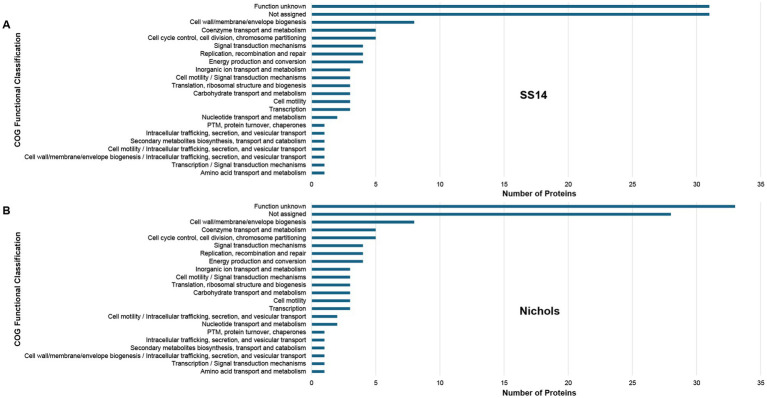
Using proteome-wide annotation comparative analyses, we identified 119 proteins with at least one amino acid difference between the corresponding proteins from SS14 and Nichols strains (Supplementary Figure S4 and Supplementary Table S14). Half of these 119 proteins were identified as proteins of unknown function (Figure 6 and Supplementary Table S14), 10 were previously identified as PPRPs (Cejkova et al., 2012; Petrosova et al., 2012; Petrosova et al., 2013; Houston et al., 2018) (Table 3 and Supplementary Table S14), and seven were identified as miniproteins of unknown function. Two of the miniproteins were previously identified as potential antimicrobial peptides (AMPs) in the Nichols strain (Houston et al., 2022) (Supplementary Tables S12, S14). Furthermore, 19 known/putative OMPs were found to be annotated with at least one amino acid difference between the two strains (Table 5).
Figure 6.
Functional classification of 119 variable proteins from T. pallidum SS14 and Nichols strains. Bar charts showing the COG functional classification distribution of (A) 119 treponemal proteins from T. pallidum SS14, and (B) the corresponding 119 T. pallidum Nichols proteins, that are annotated with at least one amino acid difference between the two strains. COG functional classifications were generated by the eggNOG-mapper tool. Not assigned – proteins that were unable to be assigned functional categories by eggNOG-mapper. PTM: Post-translational modification. Experimental data was derived from the present and previous studies (Osbak et al., 2016; Romeis et al., 2021; Houston et al., 2023; Houston et al., 2024).
Table 5.
SS14 and Nichols OMP inter-strain sequence differences and experimental validations.
| Tp SS14 Locus Tag | Annotated Function | Annotated Inter-Strain Variable Amino Acids (IVAA) | IVAA Detecteda |
|---|---|---|---|
| TPASS_RS05540 | Major outer sheath N-terminal domain-containing protein, TprA | 42 amino acid N-terminal truncation | No |
| TPASS_20117 | Major outer sheath N-terminal domain-containing protein, TprC | 10 amino acid substitutions | 2/10 |
| TPASS_20131 | Major outer sheath N-terminal domain-containing protein, TprD | 117 amino acid substitutions 4 amino acid insertions 6 amino acid gaps |
0/127 |
| TPASS_20313 | Major outer sheath N-terminal domain-containing protein, TprE | 1 amino acid substitution | No |
| TPASS_20316 | Major outer sheath N-terminal domain-containing protein, TprF | 5 amino acid N-terminal extension | No |
| TPASS_20326 | Outer membrane protein assembly factor BamA | 9 amino acid substitutions | 5/9 |
| TPASS_20479 | DUF2715 domain-containing protein | 17 amino acid N-terminal truncation | No |
| TPASS_20515 | LPS-assembly protein LptD | 10 amino acid substitutions | 0/10 |
| TPASS_20548 | UPF0164 family protein | 25 amino acid substitutions 1 amino acid insertion |
0/26 |
| TPASS_20620 | Major outer sheath N-terminal domain-containing protein, TprI | 8 amino acid substitutions | No |
| TPASS_20621 | Major outer sheath N-terminal domain-containing protein TprJ | 2 amino acid substitutions | 0/2 |
| TPASS_20858 | UPF0164 family protein | 1 amino acid substitution | 1/1 |
| TPASS_20865 | UPF0164 family protein | 2 amino acid substitutions 1 amino acid insertion |
1/3 (1 amino acid substitution) |
| TPASS_20897 | MSP porin, TprK | 101 amino acid substitutions | 0/101 |
| TPASS_20952 | Alpha/beta fold hydrolase | 1 amino acid substitution | 0/1 |
| TPASS_20966 | Hypothetical protein | 3 amino acid substitutions | 0/3 |
| TPASS_20968 | Hypothetical protein | 1 amino acid substitution | 1/1 |
| TPASS_RS04790 | Hypothetical protein | 8 amino acid N-terminal extensionb 6 amino acid substitutions 9 amino acid gaps |
No |
| TPASS_21031 | Major outer sheath N-terminal domain-containing protein, TprL | 25 amino acid substitutions | 4/25 |
Number of IVAAs that were confirmed in SS14 and Nichols strains via the detection of corresponding peptides in both strains (present study and Osbak et al., 2016; Romeis et al., 2021; Houston et al., 2023; Houston et al., 2024).
SS14 protein annotated as a Pseudo ORF (N-terminal extension does not contain a start site residue).
IVAA, Annotated Inter-Strain Variable Amino Acids.
No, peptides with IVAAs were not detected.
IVAA descriptions relate to the SS14 proteins in comparison to the corresponding Nichols strain proteins.
Data integration analyses: experimental validation of SS14-Nichols inter-strain protein sequence differences
Data integration analyses using proteome-wide protein expression comparative analyses, which incorporated experimental data from the present study and previous studies (Osbak et al., 2016; Romeis Lieberman et al., 2021; Houston et al., 2023; Houston et al., 2024), confirmed the expression of inter-strain protein sequence differences in 32 of the 119 proteins (Supplementary Figure S4 and Supplementary Table S14). These analyses also confirmed amino acid sequence differences for six of the 19 known/putative T. pallidum OMPs that are annotated with at least one amino acid difference between the two strains (Table 5). Protein tertiary structure modeling using AlphaFold 3 (see footnote 3) (Abramson et al., 2024) demonstrated that most of these inter-strain OMP differences, including the six experimentally validated amino acid differences, were predicted to occur within surface-exposed regions of the OMPs that are located at the interface between the bacterium and the host (LaFond and Lukehart, 2006) (Supplementary Figure S5).
Discussion
The Nichols and SS14 strains of T. pallidum are both highly virulent. However, SS14 strains exhibit a higher frequency of macrolide resistance (Stamm et al., 1988; Stamm and Bergen, 2000), and based on currently available clinical sampling data are the more prevalent infecting strain worldwide (Arora et al., 2016; Beale et al., 2021; Lieberman et al., 2021; Sena et al., 2024). Although previous proteomics studies have detected 95% of all proteins in Nichols/Nichols-like strains (McGill et al., 2010; Osbak et al., 2016; Houston et al., 2023; Houston et al., 2024), in-depth proteome-wide analyses of SS14 strains had not been performed prior to this study.
In the present study, we characterized the proteome of T. pallidum SS14 via the detection of 61.5% of proteins from this strain. Quantitative comparative analyses revealed similar protein abundance profiles between the SS14 and Nichols strains, for both the total proteome coverage and across specific protein functional classes of interest. These included OMPs and potential pathogenesis related proteins, which in agreement with previous T. pallidum studies (Izard et al., 2009; Liu et al., 2010; Radolf and Kumar, 2017; Houston et al., 2023; Houston et al., 2024) generally exhibited low-level expression. This low abundance of known and putative OMPs may partially contribute to the ability of both T. pallidum strains to evade the immune response and establish and maintain persistent infection (Cameron, 2005). Given that the SS14 and Nichols mass spectrometry analyses were temporally independent, statistical analyses of the LFQ results could not be performed and analyses were limited to relative quantification comparisons based solely on LFQ rankings.
Bacterial OMPs, located at the interface between the bacterium and the host, are targets of protective antibodies during infection (LaFond and Lukehart, 2006) and therefore have been identified as leading syphilis vaccine candidates (Giacani et al., 2010; Lithgow et al., 2017; Haynes et al., 2021; Delgado et al., 2022). The analyses of T. pallidum proteomes can aid in syphilis vaccine development by confirming inter-strain protein expression of vaccine candidates, including OMPs, since universal expression among strains is a necessary feature of a vaccine candidate. The present study detected expression of 11 OMPs, including several vaccine candidates, for the first time in T. pallidum SS14. Proteomic analyses can also confirm annotated protein sequence variations among strains, including within treponemal OMPs. A previous T. pallidum genome sequencing study reported that inter-strain sequence variability occurs mostly within the predicted surface-exposed loops of four known/putative OMPs being investigated as vaccine candidates (TP_0326, TP_0548, TP_0966, and TP_0967) (Lieberman et al., 2021). In the present study, comparative proteomic analyses showed that an additional 16 known/putative OMPs have inter-strain amino acid sequence differences, with the differences being confirmed in six known/putative T. pallidum OMPs via the analyses of MS data from the present and past studies (Osbak et al., 2016; Romeis et al., 2021; Houston et al., 2023; Houston et al., 2024). Furthermore, protein tertiary structure modeling showed that these inter-strain amino acid differences were found to be located primarily within predicted surface-exposed regions in the 16 additional OMPs. Several of these treponemal OMPs with surface-exposed inter-strain variable amino acids play key roles in T. pallidum pathogenesis, and have been identified as current syphilis vaccine candidates, including TPASS_20326/TPANIC_0326 (BamA) (Cameron et al., 2000; Luthra et al., 2015), TPASS_20858/TPANIC_0858 (UPF0164 family protein) (Hawley et al., 2021; Delgado et al., 2022), TPASS_20897/TPANIC_0897 (TprK) (Centurion-Lara et al., 2000; Giacani et al., 2010; Parveen et al., 2019), and TPASS_21031/TPANIC_1031 (TprL) (Haynes Lieberman et al., 2021). In addition, potential B cell epitopes (BCEs) have been identified in the literature that include some of the surface-exposed inter-strain variable amino acids reported here: these include BCE3 and BCE8 of TPANIC_0515 (Hawley et al., 2021), and BCE1 and BCE3 of TPANIC_0548 (Hawley et al., 2021). The detection and localization of these inter-strain variable residues within OMPs can aid subunit-based vaccine design by allowing identification of surface-exposed regions of the proteins that are conserved across circulating T. pallidum strains.
The present study has limitations. A relatively high number of undetected proteins were observed in the SS14 strain compared to the Nichols strain (Houston et al., 2024), which limited the comparative analyses that could be performed. Possible explanations for the higher number of undetected proteins in SS14 include: (1) the presence of more post-translational modifications (PTMs) within proteins found in the SS14 strain which could hinder MS detection due to the generation of tryptic peptides with observed masses that differ from the expected masses; or (2) lower protein expression levels within treponemes from the SS14 strain. Another limitation, which is inherent to MS analyses, is that lack of protein detection does not equate to lack of protein expression; instead, proteins may not be detected due to incompatibility with MS methodology. Therefore, it is possible that the undetected proteins in this study may indeed be expressed by T. pallidum. However, it should be noted that identical sample preparation methods were used to prepare samples for proteomic analyses for both the Nichols and SS14 strains, suggesting the lower protein detection coverage within treponemes from the SS14 strain occurred due to biological rather than technical reasons. The use of the optimized proteomics protocol from the previously performed Nichols proteomic study may represent a further limitation of the study, since this resulted in a single sample being analyzed by MS for SS-14 compared to the three samples analyzed for Nichols (Houston et al., 2024). A final limitation is that treponemes used in the present study were cultured under in vitro conditions, and thus protein expression may not be fully representative of circulating strains.
In conclusion, the present study reveals the protein repertoire expressed by the SS14 T. pallidum strain during in vitro growth, and provides the first comparative analyses of protein expression and quantitation profiles of the reference strains from each of the two T. pallidum phylogenetic lineages. This knowledge can be used to inform syphilis vaccine design by confirming the expression of leading vaccine candidates in a clinical T. pallidum strain (SS14), and via the mapping and confirmation of annotated inter-strain amino acid sequence variances in leading vaccine candidates from SS14 and Nichols strains. The data presented in the study are deposited in the MassIVE repository, accession number MSV000095818 (ProteomeXchange identifier PXD055753).
Acknowledgments
We gratefully acknowledge the University of Victoria-Genome BC Proteomics Centre for providing MS analytical services. We also thank Dr. Lorenzo Giacani at the University of Washington for supplying T. pallidum strain SS14 and for providing guidance on the in vitro culturing of T. pallidum strain SS14.
Funding Statement
The author(s) declare that financial support was received for the research, authorship, and/or publication of this article. This work was supported by grants R37AI051334, U19AI144133 and U01AI182035 (CEC) from the National Institute of Allergy and Infectious Diseases (NIAID) at the National Institutes of Health (NIH), as well as an award from Open Philanthropy (52345).
Footnotes
Data availability statement
The datasets presented in this study can be found in online repositories. The names of the repository/repositories and accession number(s) can be found in the article/Supplementary material.
Ethics statement
Ethical approval was not required for the studies on animals in accordance with the local legislation and institutional requirements because only commercially available established cell lines were used.
Author contributions
SH: Conceptualization, Data curation, Formal analysis, Investigation, Methodology, Visualization, Writing – original draft, Writing – review & editing. SM: Data curation, Investigation, Methodology, Writing – review & editing. AG: Investigation, Methodology, Writing – review & editing. CC: Conceptualization, Funding acquisition, Project administration, Resources, Supervision, Writing – review & editing.
Conflict of interest
The authors declare that the research was conducted in the absence of any commercial or financial relationships that could be construed as a potential conflict of interest.
Generative AI statement
The author(s) declare that no Generative AI was used in the preparation of this manuscript.
Publisher’s note
All claims expressed in this article are solely those of the authors and do not necessarily represent those of their affiliated organizations, or those of the publisher, the editors and the reviewers. Any product that may be evaluated in this article, or claim that may be made by its manufacturer, is not guaranteed or endorsed by the publisher.
Supplementary material
The Supplementary material for this article can be found online at: https://www.frontiersin.org/articles/10.3389/fmicb.2024.1505893/full#supplementary-material
References
- Abramson J., Adler J., Dunger J., Evans R., Green T., Pritzel A., et al. (2024). Accurate structure prediction of biomolecular interactions with AlphaFold 3. Nature 630, 493–500. doi: 10.1038/s41586-024-07487-w, PMID: [DOI] [PMC free article] [PubMed] [Google Scholar]
- Arora N., Schuenemann V. J., Jager G., Peltzer A., Seitz A., Herbig A., et al. (2016). Origin of modern syphilis and emergence of a pandemic Treponema pallidum cluster. Nat. Microbiol. 2:16245. doi: 10.1038/nmicrobiol.2016.245, PMID: [DOI] [PubMed] [Google Scholar]
- Arrieta A. C., Singh J. (2019). Congenital syphilis. N. Engl. J. Med. 381:2157. doi: 10.1056/NEJMicm1904420 [DOI] [PubMed] [Google Scholar]
- Beale M. A., Marks M., Cole M. J., Lee M. K., Pitt R., Ruis C., et al. (2021). Global phylogeny of Treponema pallidum lineages reveals recent expansion and spread of contemporary syphilis. Nat. Microbiol. 6, 1549–1560. doi: 10.1038/s41564-021-01000-z, PMID: [DOI] [PMC free article] [PubMed] [Google Scholar]
- Cameron C. E. (2005). “T. pallidum outer membrane and outer membrane proteins” in Pathogenic Treponema, molecular and cellular biology. eds. Radolf J. D., Lukehart S. A. (Norfolk, England: Caister Academic Press; ), 237–266. [Google Scholar]
- Cameron C. E., Lukehart S. A., Castro C., Molini B., Godornes C., Van Voorhis W. C. (2000). Opsonic potential, protective capacity, and sequence conservation of the Treponema pallidum subspecies pallidum Tp92. J. Infect. Dis. 181, 1401–1413. doi: 10.1086/315399, PMID: [DOI] [PubMed] [Google Scholar]
- Cantalapiedra C. P., Hernandez-Plaza A., Letunic I., Bork P., Huerta-Cepas J. (2021). eggNOG-mapper v2: functional annotation, orthology assignments, and domain prediction at the metagenomic scale. Mol. Biol. Evol. 38, 5825–5829. doi: 10.1093/molbev/msab293, PMID: [DOI] [PMC free article] [PubMed] [Google Scholar]
- Cejkova D., Zobanikova M., Chen L., Pospisilova P., Strouhal M., Qin X., et al. (2012). Whole genome sequences of three Treponema pallidum ssp. pertenue strains: yaws and syphilis treponemes differ in less than 0.2% of the genome sequence. PLoS Negl. Trop. Dis. 6:e1471. doi: 10.1371/journal.pntd.0001471, PMID: [DOI] [PMC free article] [PubMed] [Google Scholar]
- Centurion-Lara A., Gordones C., Castro C., Van Voorhis W. C., Lukehart S. A. (2000). The tprK gene is heterogeneous among Treponema pallidum strains and has multiple alleles. Infect. Immun. 68, 824–831. doi: 10.1128/IAI.68.2.824-831.2000, PMID: [DOI] [PMC free article] [PubMed] [Google Scholar]
- Chen G., Cao Y., Yao Y., Li M., Tang W., Li J., et al. (2017). Syphilis incidence among men who have sex with men in China: results from a meta-analysis. Int. J. STD AIDS 28, 170–178. doi: 10.1177/0956462416638224 [DOI] [PMC free article] [PubMed] [Google Scholar]
- Delgado K. N., Montezuma-Rusca J. M., Orbe I. C., Caimano M. J., La Vake C. J., Luthra A., et al. (2022). Extracellular loops of the Treponema pallidum FadL orthologs TP0856 and TP0858 elicit IgG antibodies and IgG(+)-specific B-cells in the rabbit model of experimental syphilis. MBio 13:e0163922. doi: 10.1128/mbio.01639-22, PMID: [DOI] [PMC free article] [PubMed] [Google Scholar]
- Douglas J. M., Jr. (2009). Penicillin treatment of syphilis: clearing away the shadow on the land. JAMA 301, 769–771. doi: 10.1001/jama.2009.143 [DOI] [PubMed] [Google Scholar]
- Edmondson D. G., Delay B. D., Kowis L. E., Norris S. J. (2021). Parameters affecting continuous in vitro culture of Treponema pallidum strains. mBio 12, e03536–e03520. doi: 10.1128/mBio.03536-20 [DOI] [PMC free article] [PubMed] [Google Scholar]
- Edmondson D. G., Hu B., Norris S. J. (2018). Long-term in vitro culture of the syphilis spirochete Treponema pallidum subsp pallidum. mBio 9, e01153–e01118. doi: 10.1128/mBio.01153-18, PMID: [DOI] [PMC free article] [PubMed] [Google Scholar]
- Edmondson D. G., Norris S. J. (2021). In vitro cultivation of the syphilis spirochete Treponema pallidum. Curr. Protoc. 1:e44. doi: 10.1002/cpz1.44, PMID: [DOI] [PMC free article] [PubMed] [Google Scholar]
- Giacani L., Molini B. J., Kim E. Y., Godornes B. C., Leader B. T., Tantalo L. C., et al. (2010). Antigenic variation in Treponema pallidum: TprK sequence diversity accumulates in response to immune pressure during experimental syphilis. J. Immunol. 184, 3822–3829. doi: 10.4049/jimmunol.0902788, PMID: [DOI] [PMC free article] [PubMed] [Google Scholar]
- Hawley K. L., Montezuma-Rusca J. M., Delgado K. N., Singh N., Uversky V. N., Caimano M. J., et al. (2021). Structural modeling of the Treponema pallidum outer membrane protein repertoire: a road map for deconvolution of syphilis pathogenesis and development of a syphilis vaccine. J. Bacteriol. 203:e0008221. doi: 10.1128/JB.00082-21, PMID: [DOI] [PMC free article] [PubMed] [Google Scholar]
- Haynes A. M., Fernandez M., Romeis E., Mitja O., Konda K. A., Vargas S. K., et al. (2021). Transcriptional and immunological analysis of the putative outer membrane protein and vaccine candidate TprL of Treponema pallidum. PLoS Negl. Trop. Dis. 15:e0008812. doi: 10.1371/journal.pntd.0008812, PMID: [DOI] [PMC free article] [PubMed] [Google Scholar]
- Herbert L. J., Middleton S. I. (2012). An estimate of syphilis incidence in Eastern Europe. J. Glob. Health 2:010402. doi: 10.7189/jogh.02.010402, PMID: [DOI] [PMC free article] [PubMed] [Google Scholar]
- Houston S., Gomez A., Geppert A., Eshghi A., Smith D. S., Waugh S., et al. (2023). Deep proteome coverage advances knowledge of Treponema pallidum protein expression profiles during infection. Sci. Rep. 13:18259. doi: 10.1038/s41598-023-45219-8, PMID: [DOI] [PMC free article] [PubMed] [Google Scholar]
- Houston S., Gomez A., Geppert A., Goodyear M. C., Cameron C. E. (2024). In-depth proteome coverage of in vitro-cultured Treponema pallidum and quantitative comparison analyses with in vivo-grown treponemes. J. Proteome Res. 23, 1725–1743. doi: 10.1021/acs.jproteome.3c00891 [DOI] [PMC free article] [PubMed] [Google Scholar]
- Houston S., Lithgow K. V., Osbak K. K., Kenyon C. R., Cameron C. E. (2018). Functional insights from proteome-wide structural modeling of Treponema pallidum subspecies pallidum, the causative agent of syphilis. BMC Struct. Biol. 18:7. doi: 10.1186/s12900-018-0086-3, PMID: [DOI] [PMC free article] [PubMed] [Google Scholar]
- Houston S., Schovanek E., Conway K. M. E., Mustafa S., Gomez A., Ramaswamy R., et al. (2022). Identification and functional characterization of peptides with antimicrobial activity from the syphilis spirochete, Treponema pallidum. Front. Microbiol. 13:888525. doi: 10.3389/fmicb.2022.888525, PMID: [DOI] [PMC free article] [PubMed] [Google Scholar]
- Izard J., Renken C., Hsieh C. E., Desrosiers D. C., Dunham-Ems S., La V. C., et al. (2009). Cryo-electron tomography elucidates the molecular architecture of Treponema pallidum, the syphilis spirochete. J. Bacteriol. 191, 7566–7580. doi: 10.1128/JB.01031-09, PMID: [DOI] [PMC free article] [PubMed] [Google Scholar]
- Korenromp E. L., Rowley J., Alonso M., Mello M. B., Wijesooriya N. S., Mahiane S. G., et al. (2019). Global burden of maternal and congenital syphilis and associated adverse birth outcomes - estimates for 2016 and progress since 2012. PLoS One 14:e0211720. doi: 10.1371/journal.pone.0211720 [DOI] [PMC free article] [PubMed] [Google Scholar]
- Lafond R. E., Lukehart S. A. (2006). Biological basis for syphilis. Clin. Microbiol. Rev. 19, 29–49. doi: 10.1128/CMR.19.1.29-49.2006, PMID: [DOI] [PMC free article] [PubMed] [Google Scholar]
- Lieberman N. A. P., Lin M. J., Xie H., Shrestha L., Nguyen T., Huang M.-L., et al. (2021). Treponema pallidum genome sequencing from six continents reveals variability in vaccine candidate genes and dominance of Nichols clade strains in Madagascar. PLoS Negl. Trop. Dis. 15:e0010063. doi: 10.1371/journal.pntd.0010063, PMID: [DOI] [PMC free article] [PubMed] [Google Scholar]
- Lithgow K. V., Hof R., Wetherell C., Phillips D., Houston S., Cameron C. E. (2017). A defined syphilis vaccine candidate inhibits dissemination of Treponema pallidum subspecies pallidum. Nat. Commun. 8:14273. doi: 10.1038/ncomms14273, PMID: [DOI] [PMC free article] [PubMed] [Google Scholar]
- Liu J., Howell J. K., Bradley S. D., Zheng Y., Zhou Z. H., Norris S. J. (2010). Cellular architecture of Treponema pallidum: novel flagellum, periplasmic cone, and cell envelope as revealed by cryo electron tomography. J. Mol. Biol. 403, 546–561. doi: 10.1016/j.jmb.2010.09.020, PMID: [DOI] [PMC free article] [PubMed] [Google Scholar]
- Luthra A., Anand A., Hawley K. L., Ledoyt M., La Vake C. J., Caimano M. J., et al. (2015). A homology model reveals novel structural features and an immunodominant surface loop/opsonic target in the Treponema pallidum BamA ortholog TP_0326. J. Bacteriol. 197, 1906–1920. doi: 10.1128/JB.00086-15, PMID: [DOI] [PMC free article] [PubMed] [Google Scholar]
- Mcgill M. A., Edmondson D. G., Carroll J. A., Cook R. G., Orkiszewski R. S., Norris S. J. (2010). Characterization and serologic analysis of the Treponema pallidum proteome. Infect. Immun. 78, 2631–2643. doi: 10.1128/IAI.00173-10, PMID: [DOI] [PMC free article] [PubMed] [Google Scholar]
- Mikalova L., Strouhal M., Cejkova D., Zobanikova M., Pospisilova P., Norris S. J., et al. (2010). Genome analysis of Treponema pallidum subsp. pallidum and subsp. pertenue strains: most of the genetic differences are localized in six regions. PLoS One 5:e15713. doi: 10.1371/journal.pone.0015713, PMID: [DOI] [PMC free article] [PubMed] [Google Scholar]
- Nechvatal L., Petrosova H., Grillova L., Pospisilova P., Mikalova L., Strnadel R., et al. (2014). Syphilis-causing strains belong to separate SS14-like or Nichols-like groups as defined by multilocus analysis of 19 Treponema pallidum strains. Int. J. Med. Microbiol. 304, 645–653. doi: 10.1016/j.ijmm.2014.04.007, PMID: [DOI] [PubMed] [Google Scholar]
- Nesvizhskii A. I., Keller A., Kolker E., Aebersold R. (2003). A statistical model for identifying proteins by tandem mass spectrometry. Anal. Chem. 75, 4646–4658. doi: 10.1021/ac0341261 [DOI] [PubMed] [Google Scholar]
- Nichols H. J., Hough W. H. (1913). Demonstration of Spirochaeta pallida in the cerebrospinal fluid from a patient with nervous relapse following the use of Salvarsan. JAMA J. Am. Med. Assoc. 60, 108–110. doi: 10.1001/jama.1913.04340020016005 [DOI] [Google Scholar]
- Nusbaum M. R., Wallace R. R., Slatt L. M., Kondrad E. C. (2004). Sexually transmitted infections and increased risk of co-infection with human immunodeficiency virus. J. Am. Osteopath. Assoc. 104, 527–535. doi: 10.7556/jaoa.2004.104.12.527, PMID: [DOI] [PubMed] [Google Scholar]
- Osbak K. K., Houston S., Lithgow K. V., Meehan C. J., Strouhal M., Smajs D., et al. (2016). Characterizing the syphilis-causing Treponema pallidum ssp. pallidum proteome using complementary mass spectrometry. PLoS Negl. Trop. Dis. 10:e0004988. doi: 10.1371/journal.pntd.0004988, PMID: [DOI] [PMC free article] [PubMed] [Google Scholar]
- Parveen N., Fernandez M. C., Haynes A. M., Zhang R. L., Godornes B. C., Centurion-Lara A., et al. (2019). Non-pathogenic Borrelia burgdorferi expressing Treponema pallidum TprK and Tp0435 antigens as a novel approach to evaluate syphilis vaccine candidates. Vaccine 37, 1807–1818. doi: 10.1016/j.vaccine.2019.02.022, PMID: [DOI] [PMC free article] [PubMed] [Google Scholar]
- Petrosova H., Pospisilova P., Strouhal M., Cejkova D., Zobanikova M., Mikalova L., et al. (2013). Resequencing of Treponema pallidum ssp. pallidum strains Nichols and SS14: correction of sequencing errors resulted in increased separation of syphilis treponeme subclusters. PLoS One 8:e74319. doi: 10.1371/journal.pone.0074319, PMID: [DOI] [PMC free article] [PubMed] [Google Scholar]
- Petrosova H., Zobanikova M., Cejkova D., Mikalova L., Pospisilova P., Strouhal M., et al. (2012). Whole genome sequence of Treponema pallidum ssp. pallidum, strain Mexico a, suggests recombination between yaws and syphilis strains. PLoS Negl. Trop. Dis. 6:e1832. doi: 10.1371/journal.pntd.0001832, PMID: [DOI] [PMC free article] [PubMed] [Google Scholar]
- Radolf J. D., Kumar S. (2017). The Treponema pallidum outer membrane. Curr. Top. Microbiol. Immunol. 415, 1–38. doi: 10.1007/82_2017_44, PMID: [DOI] [PMC free article] [PubMed] [Google Scholar]
- Romeis E., Tantalo L., Lieberman N., Phung Q., Greninger A., Giacani L. (2021). Genetic engineering of Treponema pallidum subsp. pallidum, the syphilis spirochete. PLoS Pathog. 17:e1009612. doi: 10.1371/journal.ppat.1009612, PMID: [DOI] [PMC free article] [PubMed] [Google Scholar]
- Savage E. J., Marsh K., Duffell S., Ison C. A., Zaman A., Hughes G. (2012). Rapid increase in gonorrhoea and syphilis diagnoses in England in 2011. Euro Surveill. 17:20224. doi: 10.2807/ese.17.29.20224-en [DOI] [PubMed] [Google Scholar]
- Schrodinger L. L. C. (2015). The PyMOL molecular graphics system. New York, NY: Schrodinger, LLC. [Google Scholar]
- Sena A. C., Matoga M. M., Yang L., Lopez-Medina E., Aghakhanian F., Chen J. S., et al. (2024). Clinical and genomic diversity of Treponema pallidum subspecies pallidum to inform vaccine research: an international, molecular epidemiology study. Lancet Microbe 5:100871. doi: 10.1016/S2666-5247(24)00087-9, PMID: [DOI] [PMC free article] [PubMed] [Google Scholar]
- Smajs D., Norris S. J., Weinstock G. M. (2012). Genetic diversity in Treponema pallidum: implications for pathogenesis, evolution and molecular diagnostics of syphilis and yaws. Infect. Genet. Evol. 12, 191–202. doi: 10.1016/j.meegid.2011.12.001, PMID: [DOI] [PMC free article] [PubMed] [Google Scholar]
- Stamm L. V., Bergen H. L. (2000). A point mutation associated with bacterial macrolide resistance is present in both 23S rRNA genes of an erythromycin-resistant Treponema pallidum clinical isolate. Antimicrob. Agents Chemother. 44, 806–807. doi: 10.1128/AAC.44.3.806-807.2000, PMID: [DOI] [PMC free article] [PubMed] [Google Scholar]
- Stamm L. V., Kerner T. C., Jr., Bankaitis V. A., Bassford P. J., Jr. (1983). Identification and preliminary characterization of Treponema pallidum protein antigens expressed in Escherichia coli. Infect. Immun. 41, 709–721. doi: 10.1128/iai.41.2.709-721.1983, PMID: [DOI] [PMC free article] [PubMed] [Google Scholar]
- Stamm L. V., Stapleton J. T., Bassford P. J., Jr. (1988). In vitro assay to demonstrate high-level erythromycin resistance of a clinical isolate of Treponema pallidum. Antimicrob. Agents Chemother. 32, 164–169. doi: 10.1128/AAC.32.2.164, PMID: [DOI] [PMC free article] [PubMed] [Google Scholar]
- Tatusov R. L., Koonin E. V., Lipman D. J. (1997). A genomic perspective on protein families. Science 278, 631–637. doi: 10.1126/science.278.5338.631 [DOI] [PubMed] [Google Scholar]
- Teufel F., Almagro Armenteros J. J., Johansen A. R., Gislason M. H., Pihl S. I., Tsirigos K. D., et al. (2022). SignalP 6.0 predicts all five types of signal peptides using protein language models. Nat. Biotechnol. 40, 1023–1025. doi: 10.1038/s41587-021-01156-3 [DOI] [PMC free article] [PubMed] [Google Scholar]
Associated Data
This section collects any data citations, data availability statements, or supplementary materials included in this article.
Supplementary Materials
Data Availability Statement
The datasets presented in this study can be found in online repositories. The names of the repository/repositories and accession number(s) can be found in the article/Supplementary material.


