

Abstract
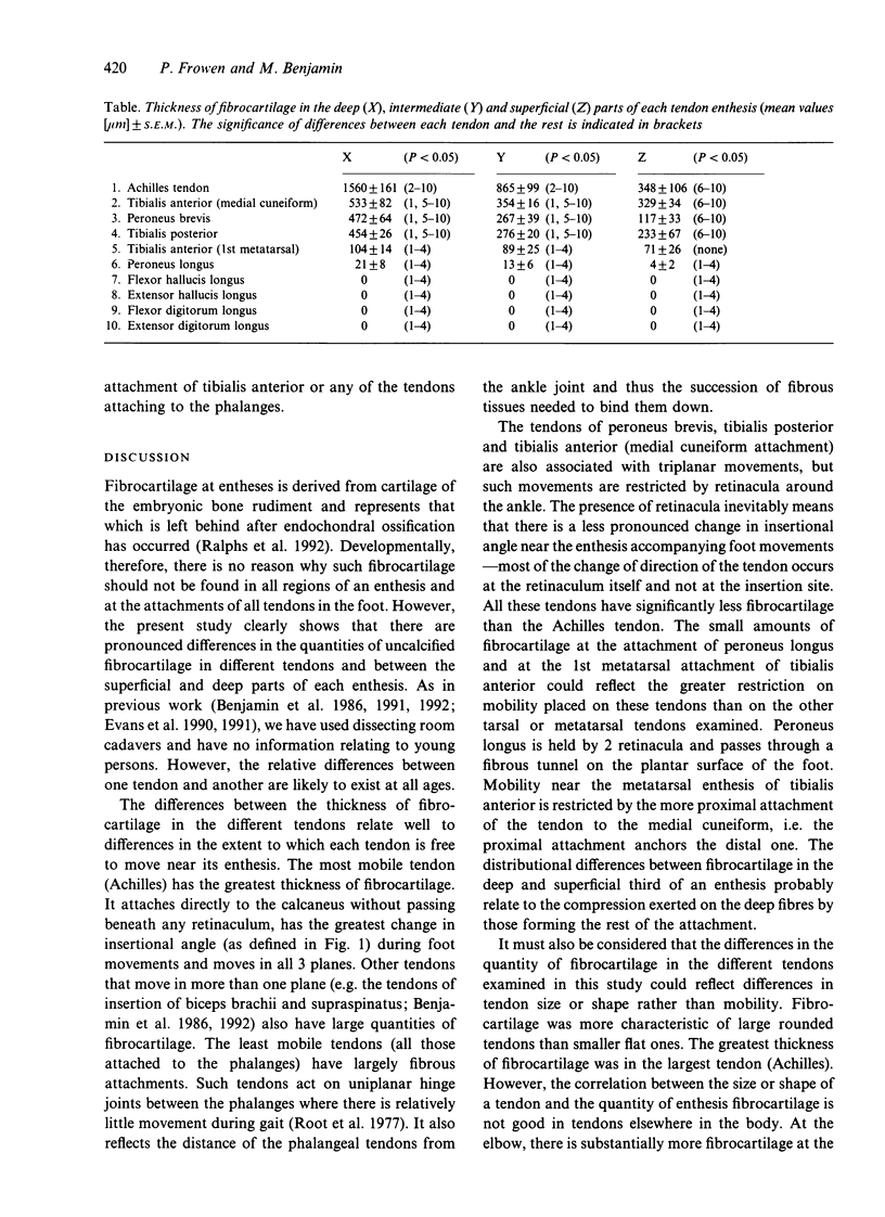
It has been suggested that fibrocartilage at entheses (tendon-bone junctions) prevents collagen fibres bending at the hard tissue interface. We have investigated this function by exploring the relationship between the presence or amount of fibrocartilage at the attachments of the major extrinsic muscles in the foot, and the extent to which these tendons bend near their entheses during movement. The tendons were taken from each of 5 formalin-fixed dissecting room cadavers and prepared for routine histology, and sections were collected systematically throughout the blocks. Tendons that attached to the tarsus and metatarsus had fibrocartilaginous entheses, but those attached to the phalanges had fibrous entheses. In all tarsal and metatarsal tendons, the fibrocartilage was significantly thicker (P < 0.05) in the deepest part of the enthesis. Here the greatest amount of fibrocartilage was in the Achilles tendon (mean thickness +/- S.E.M.: 1560 +/- 161 microns). There were moderate amounts at the medial cuneiform attachment of tibialis anterior (533 +/- 82 microns), peroneus brevis (472 +/- 64 microns) and tibialis posterior (454 +/- 26 microns), small quantities at the first metatarsal attachment of tibialis anterior (104 +/- 14 microns) and peroneus longus (21 +/- 8 microns), but only traces at the attachments of the flexor and extensor tendons of the phalanges. The differences can be related to variations in the freedom of movement of the tendons near their attachments. This depends on the extent to which the tendons are bound by retinacula and the range of movement of the joint nearest the enthesis. The results suggest that more 'mobile' tendons have more fibrocartilage.(ABSTRACT TRUNCATED AT 250 WORDS)
Full text
PDF
Images in this article

Selected References
These references are in PubMed. This may not be the complete list of references from this article.
- Benjamin M., Evans E. J., Copp L. The histology of tendon attachments to bone in man. J Anat. 1986 Dec;149:89–100. [PMC free article] [PubMed] [Google Scholar]
- Benjamin M., Evans E. J., Rao R. D., Findlay J. A., Pemberton D. J. Quantitative differences in the histology of the attachment zones of the meniscal horns in the knee joint of man. J Anat. 1991 Aug;177:127–134. [PMC free article] [PubMed] [Google Scholar]
- Benjamin M., Newell R. L., Evans E. J., Ralphs J. R., Pemberton D. J. The structure of the insertions of the tendons of biceps brachii, triceps and brachialis in elderly dissecting room cadavers. J Anat. 1992 Apr;180(Pt 2):327–332. [PMC free article] [PubMed] [Google Scholar]
- Evans E. J., Benjamin M., Pemberton D. J. Fibrocartilage in the attachment zones of the quadriceps tendon and patellar ligament of man. J Anat. 1990 Aug;171:155–162. [PMC free article] [PubMed] [Google Scholar]
- Evans E. J., Benjamin M., Pemberton D. J. Variations in the amount of calcified tissue at the attachments of the quadriceps tendon and patellar ligament in man. J Anat. 1991 Feb;174:145–151. [PMC free article] [PubMed] [Google Scholar]
- Ralphs J. R., Tyers R. N., Benjamin M. Development of functionally distinct fibrocartilages at two sites in the quadriceps tendon of the rat: the suprapatella and the attachment to the patella. Anat Embryol (Berl) 1992;185(2):181–187. doi: 10.1007/BF00185920. [DOI] [PubMed] [Google Scholar]