Abstract
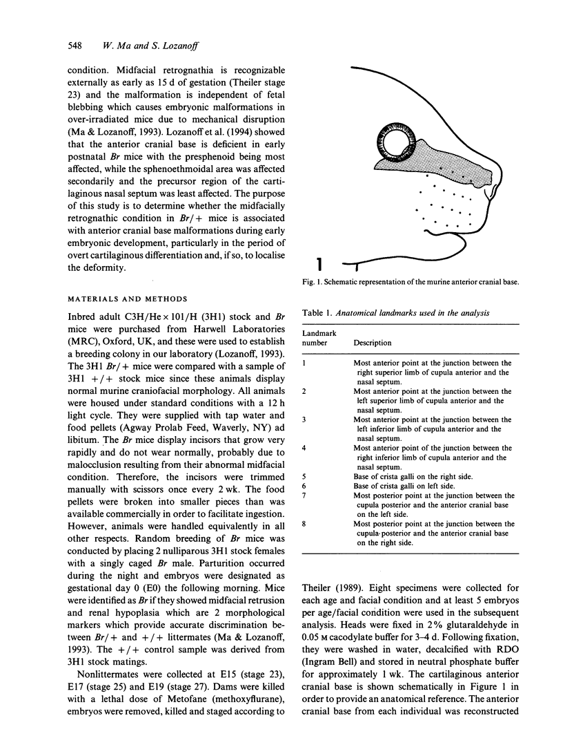
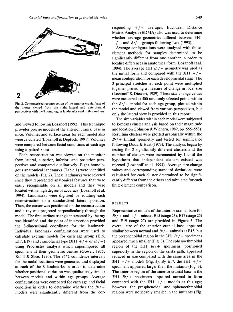
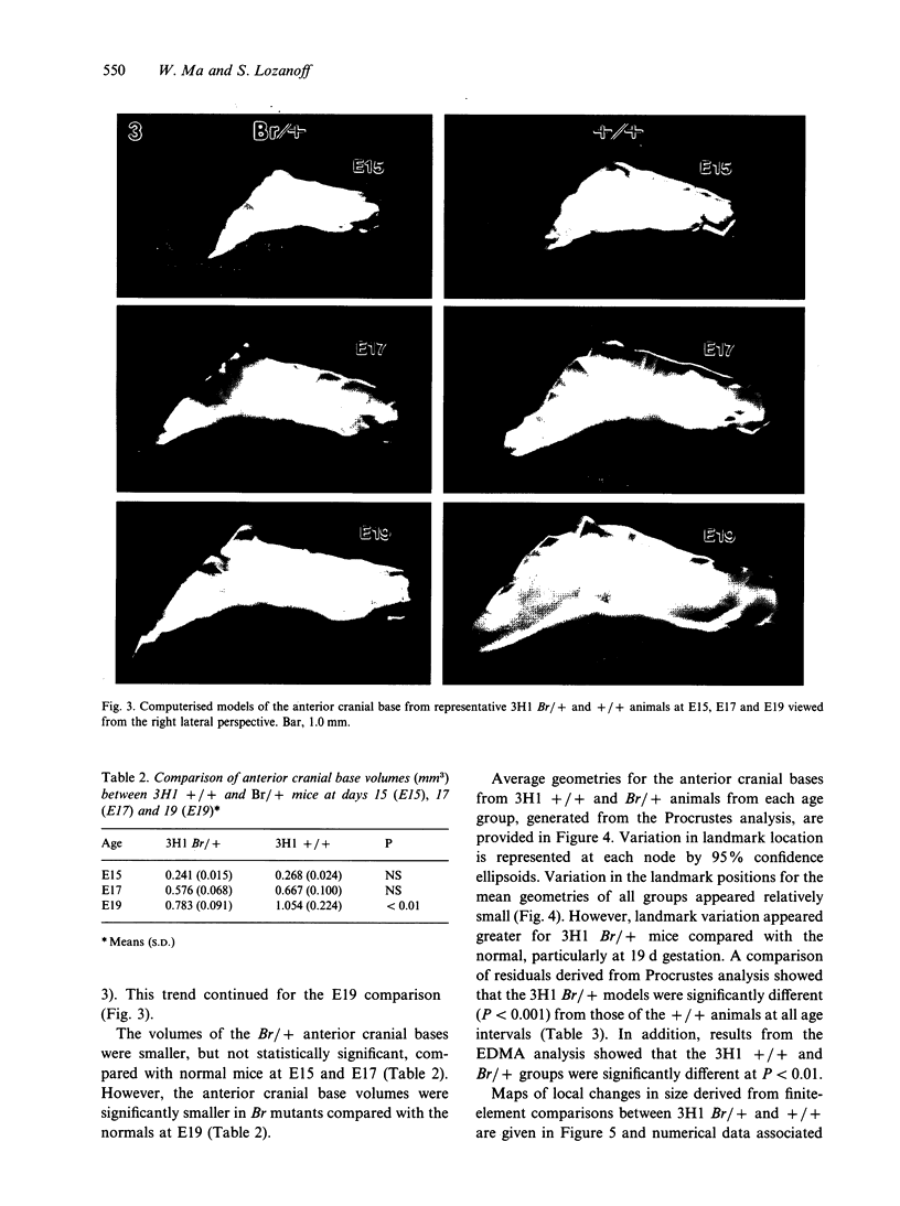
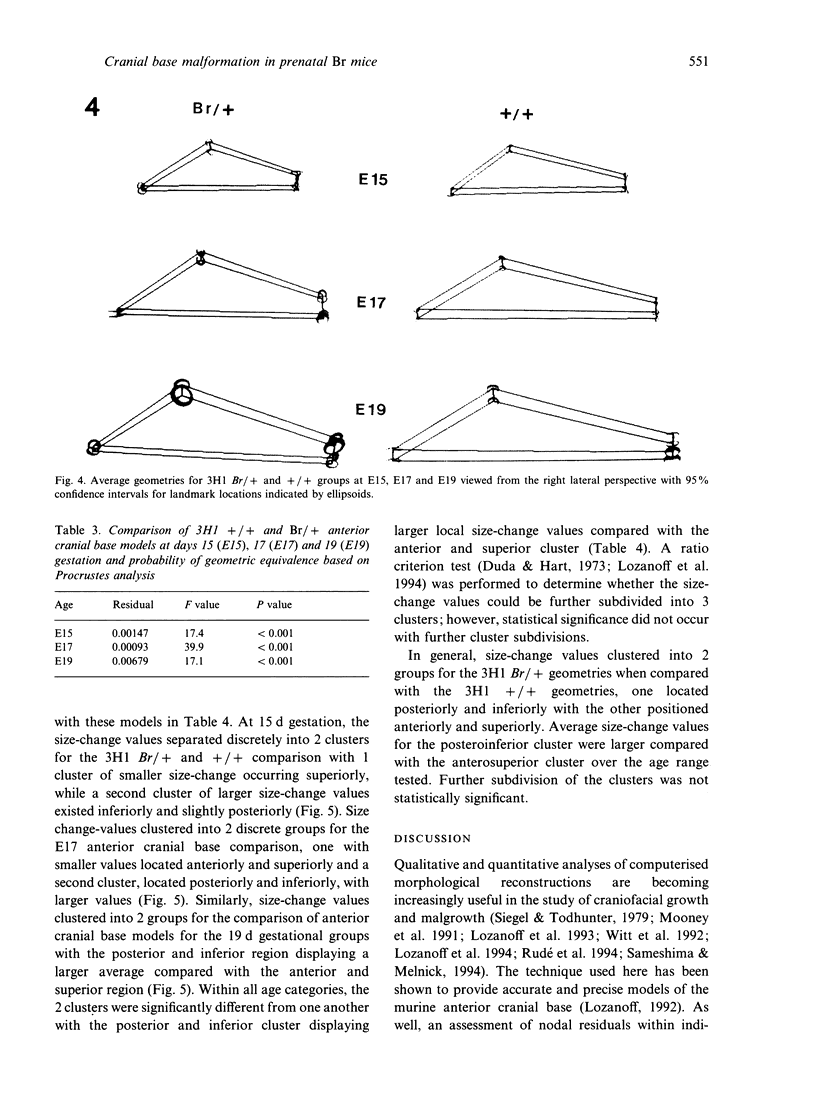
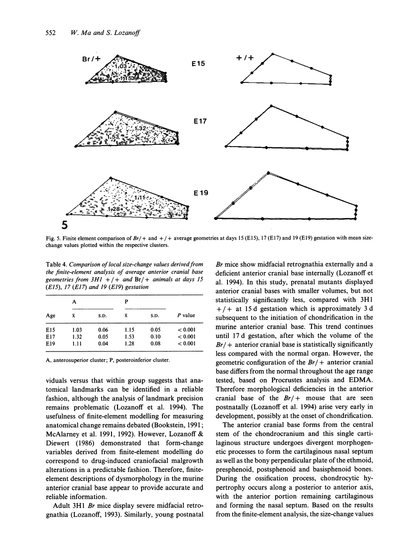
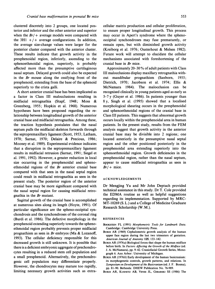
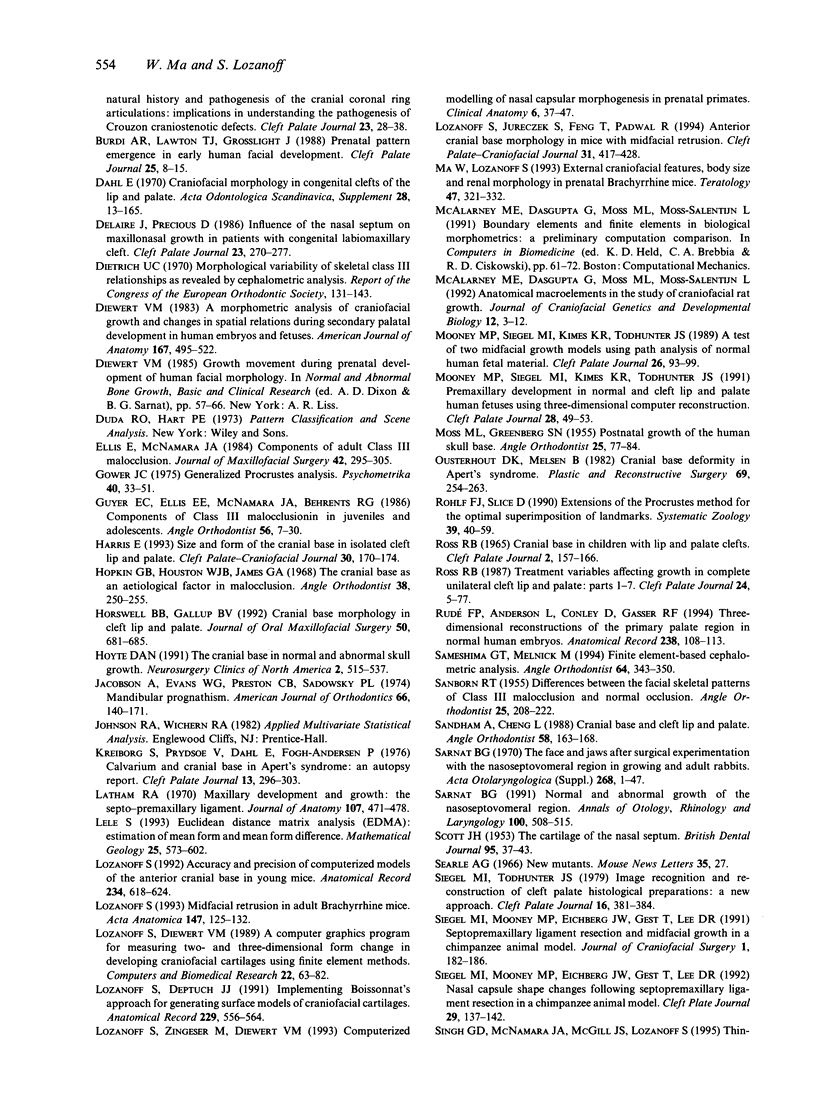
The role of the anterior cranial base in the establishment of midfacial retrognathia remains unclear. The purpose of this study was to determine whether morphological deficiencies occur in the developing anterior cranial base of the retrognathic Brachyrrhine (3H1 Br/+) mouse mutant shortly after overt cartilaginous differentiation and to localise any malformations. Crania from 2 groups of 3H1 Br/+ and +/+ mice, each consisting of 15 animals, were collected at gestational days 15, 17, and 19 (Theiler stages 23, 25, 27). The anterior cranial base from each specimen was subjected to computerised reconstruction and 8 homologous anatomical landmarks were digitised on each model. The landmark configurations were subjected to Procrustes analysis and significant differences between models were determined at each age. In order to localise differences between forms, average landmark configurations derived from Procrustes analysis were subjected to finite-element analysis. Two cluster models were generated based on size-change values. One cluster was located anteriorly and superiorly while the second was located posteriorly and inferiorly within the anterior cranial base. Results indicate that the size-change values for the posterior and inferior cluster increased more rapidly compared with the anterior and superior region over the age range tested. These data indicate that the midfacial retrognathia in Br/+ mice is associated with abnormal growth activity in the presphenoid component of the presumptive anterior cranial base. In addition, the deficiency is present in the presphenoid at the time of overt cartilaginous differentiation.
Full text
PDF

Images in this article
Selected References
These references are in PubMed. This may not be the complete list of references from this article.
- Burdi A. R., Kusnetz A. B., Venes J. L., Gebarski S. S. The natural history and pathogenesis of the cranial coronal ring articulations: implications in understanding the pathogenesis of the Crouzon craniostenotic defects. Cleft Palate J. 1986 Jan;23(1):28–39. [PubMed] [Google Scholar]
- Burdi A. R., Lawton T. J., Grosslight J. Prenatal pattern emergence in early human facial development. Cleft Palate J. 1988 Jan;25(1):8–15. [PubMed] [Google Scholar]
- Delaire J., Precious D. Influence of the nasal septum on maxillonasal growth in patients with congenital labiomaxillary cleft. Cleft Palate J. 1986 Oct;23(4):270–277. [PubMed] [Google Scholar]
- Dietrich U. C. Morphological variability of skeletal Class 3 relationships as revealed by cephalometric analysis. Rep Congr Eur Orthod Soc. 1970:131–143. [PubMed] [Google Scholar]
- Diewert V. M. A morphometric analysis of craniofacial growth and changes in spatial relations during secondary palatal development in human embryos and fetuses. Am J Anat. 1983 Aug;167(4):495–522. doi: 10.1002/aja.1001670407. [DOI] [PubMed] [Google Scholar]
- Diewert V. M. Growth movements during prenatal development of human facial morphology. Prog Clin Biol Res. 1985;187:57–66. [PubMed] [Google Scholar]
- Ellis E., 3rd, McNamara J. A., Jr Components of adult Class III malocclusion. J Oral Maxillofac Surg. 1984 May;42(5):295–305. doi: 10.1016/0278-2391(84)90109-5. [DOI] [PubMed] [Google Scholar]
- Guyer E. C., Ellis E. E., 3rd, McNamara J. A., Jr, Behrents R. G. Components of class III malocclusion in juveniles and adolescents. Angle Orthod. 1986 Jan;56(1):7–30. doi: 10.1043/0003-3219(1986)056<0007:COCIMI>2.0.CO;2. [DOI] [PubMed] [Google Scholar]
- Harris E. F. Size and form of the cranial base in isolated cleft lip and palate. Cleft Palate Craniofac J. 1993 Mar;30(2):170–174. doi: 10.1597/1545-1569_1993_030_0170_safotc_2.3.co_2. [DOI] [PubMed] [Google Scholar]
- Hopkin G. B., Houston W. J., James G. A. The cranial base as an aetiological factor in malocclusion. Angle Orthod. 1968 Jul;38(3):250–255. doi: 10.1043/0003-3219(1968)038<0250:TCBAAA>2.0.CO;2. [DOI] [PubMed] [Google Scholar]
- Horswell B. B., Gallup B. V. Cranial base morphology in cleft lip and palate: a cephalometric study from 7 to 18 years of age. J Oral Maxillofac Surg. 1992 Jul;50(7):681–686. doi: 10.1016/0278-2391(92)90095-h. [DOI] [PubMed] [Google Scholar]
- Hoyte D. A. The cranial base in normal and abnormal skull growth. Neurosurg Clin N Am. 1991 Jul;2(3):515–537. [PubMed] [Google Scholar]
- Jacobson A., Evans W. G., Preston C. B., Sadowsky P. L. Mandibular prognathism. Am J Orthod. 1974 Aug;66(2):140–171. doi: 10.1016/0002-9416(74)90233-4. [DOI] [PubMed] [Google Scholar]
- Kreiborg S., Prydsoe U., Dahl E., Fogh-Anderson P. Clinical conference I. Calvarium and cranial base in Apert's syndrome: an autopsy report. Cleft Palate J. 1976 Jul;13:296–303. [PubMed] [Google Scholar]
- Latham R. A. Maxillary development and growth: the septo-premaxillary ligament. J Anat. 1970 Nov;107(Pt 3):471–478. [PMC free article] [PubMed] [Google Scholar]
- Lozanoff S. Accuracy and precision of computerized models of the anterior cranial base in young mice. Anat Rec. 1992 Dec;234(4):618–624. doi: 10.1002/ar.1092340417. [DOI] [PubMed] [Google Scholar]
- Lozanoff S., Deptuch J. J. Implementing Boissonnat's method for generating surface models of craniofacial cartilages. Anat Rec. 1991 Apr;229(4):556–564. doi: 10.1002/ar.1092290416. [DOI] [PubMed] [Google Scholar]
- Lozanoff S., Diewert V. M. A computer graphics program for measuring two- and three-dimensional form change in developing craniofacial cartilages using finite element methods. Comput Biomed Res. 1989 Feb;22(1):63–82. doi: 10.1016/0010-4809(89)90016-5. [DOI] [PubMed] [Google Scholar]
- Lozanoff S., Jureczek S., Feng T., Padwal R. Anterior cranial base morphology in mice with midfacial retrusion. Cleft Palate Craniofac J. 1994 Nov;31(6):417–428. doi: 10.1597/1545-1569_1994_031_0417_acbmim_2.3.co_2. [DOI] [PubMed] [Google Scholar]
- Lozanoff S. Midfacial retrusion in adult brachyrrhine mice. Acta Anat (Basel) 1993;147(2):125–132. doi: 10.1159/000147492. [DOI] [PubMed] [Google Scholar]
- Ma W., Lozanoff S. External craniofacial features, body size, and renal morphology in prenatal brachyrrhine mice. Teratology. 1993 Apr;47(4):321–332. doi: 10.1002/tera.1420470409. [DOI] [PubMed] [Google Scholar]
- McAlarney M. E., Dasgupta G., Moss M. L., Moss-Salentijn L. Anatomical macroelements in the study of craniofacial rat growth. J Craniofac Genet Dev Biol. 1992 Jan-Mar;12(1):3–12. [PubMed] [Google Scholar]
- Mooney M. P., Siegel M. I., Kimes K. R., Todhunter J. A test of two midfacial growth models using path analysis of normal human fetal material. Cleft Palate J. 1989 Apr;26(2):93–99. [PubMed] [Google Scholar]
- Mooney M. P., Siegel M. I., Kimes K. R., Todhunter J. Premaxillary development in normal and cleft lip and palate human fetuses using three-dimensional computer reconstruction. Cleft Palate Craniofac J. 1991 Jan;28(1):49–54. doi: 10.1597/1545-1569_1991_028_0049_pdinac_2.3.co_2. [DOI] [PubMed] [Google Scholar]
- Ousterhout D. K., Melsen B. Cranial base deformity in Apert's syndrome. Plast Reconstr Surg. 1982 Feb;69(2):254–263. doi: 10.1097/00006534-198202000-00013. [DOI] [PubMed] [Google Scholar]
- ROSS R. B. CRANIAL BASE IN CHILDREN WITH LIP AND PALATE CLEFTS. Cleft Palate J. 1965 Apr;31:157–166. [PubMed] [Google Scholar]
- Ross R. B. Treatment variables affecting facial growth in complete unilateral cleft lip and palate. Cleft Palate J. 1987 Jan;24(1):5–77. [PubMed] [Google Scholar]
- Rudé F. P., Anderson L., Conley D., Gasser R. F. Three-dimensional reconstructions of the primary palate region in normal human embryos. Anat Rec. 1994 Jan;238(1):108–113. doi: 10.1002/ar.1092380112. [DOI] [PubMed] [Google Scholar]
- Sameshima G. T., Melnick M. Finite element-based cephalometric analysis. Angle Orthod. 1994;64(5):343–350. doi: 10.1043/0003-3219(1994)064<0343:FECA>2.0.CO;2. [DOI] [PubMed] [Google Scholar]
- Sandham A., Cheng L. Cranial base and cleft lip and palate. Angle Orthod. 1988 Apr;58(2):163–168. doi: 10.1043/0003-3219(1988)058<0163:CBACLA>2.0.CO;2. [DOI] [PubMed] [Google Scholar]
- Sarnat B. G. Normal and abnormal growth at the nasoseptovomeral region. Ann Otol Rhinol Laryngol. 1991 Jun;100(6):508–515. doi: 10.1177/000348949110000615. [DOI] [PubMed] [Google Scholar]
- Sarnat B. G. The face and jaws after surgical experimentation with the septovomeral region in growing and adult rabbits. Acta Otolaryngol Suppl. 1970;268:1–30. doi: 10.3109/00016487009131762. [DOI] [PubMed] [Google Scholar]
- Siegel M. I., Mooney M. P., Eichberg J. W., Gest T., Lee D. R. Nasal capsule shape changes following septopremaxillary ligament resection in a chimpanzee animal model. Cleft Palate Craniofac J. 1992 Mar;29(2):137–142. doi: 10.1597/1545-1569_1992_029_0137_ncscfs_2.3.co_2. [DOI] [PubMed] [Google Scholar]
- Siegel M. I., Mooney M. P., Eichberg J. W., Gest T., Lee D. R. Septopremaxillary ligament resection and midfacial growth in a chimpanzee animal model. J Craniofac Surg. 1990 Oct;1(4):182–186. doi: 10.1097/00001665-199001040-00006. [DOI] [PubMed] [Google Scholar]
- Siegel M. I., Todhunter J. S. Image recognition and the reconstruction of cleft palate histological preparations: a new approach. Cleft Palate J. 1979 Oct;16(4):381–384. [PubMed] [Google Scholar]
- Trotman C. A., Ross R. B. Craniofacial growth in bilateral cleft lip and palate: ages six years to adulthood. Cleft Palate Craniofac J. 1993 May;30(3):261–273. doi: 10.1597/1545-1569_1993_030_0261_cgibcl_2.3.co_2. [DOI] [PubMed] [Google Scholar]
- Witt P. D., Hardesty R. A., Zuppan C., Rouse G., Hasso A. N., Boyne P. Fetal kleeblattschädel cranium: morphologic, radiographic, and histologic analysis. Cleft Palate Craniofac J. 1992 Jul;29(4):363–368. doi: 10.1597/1545-1569_1992_029_0363_fkdcmr_2.3.co_2. [DOI] [PubMed] [Google Scholar]
- van Limborgh J. A new view of the control of the morphogenesis of the skull. Acta Morphol Neerl Scand. 1970 Nov;8(2):143–160. [PubMed] [Google Scholar]
- van Limborgh J. The role of genetic and local environmental factors in the control of postnatal craniofacial morphogenesis. Acta Morphol Neerl Scand. 1972 Oct;10(1):37–47. [PubMed] [Google Scholar]


