Abstract
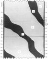
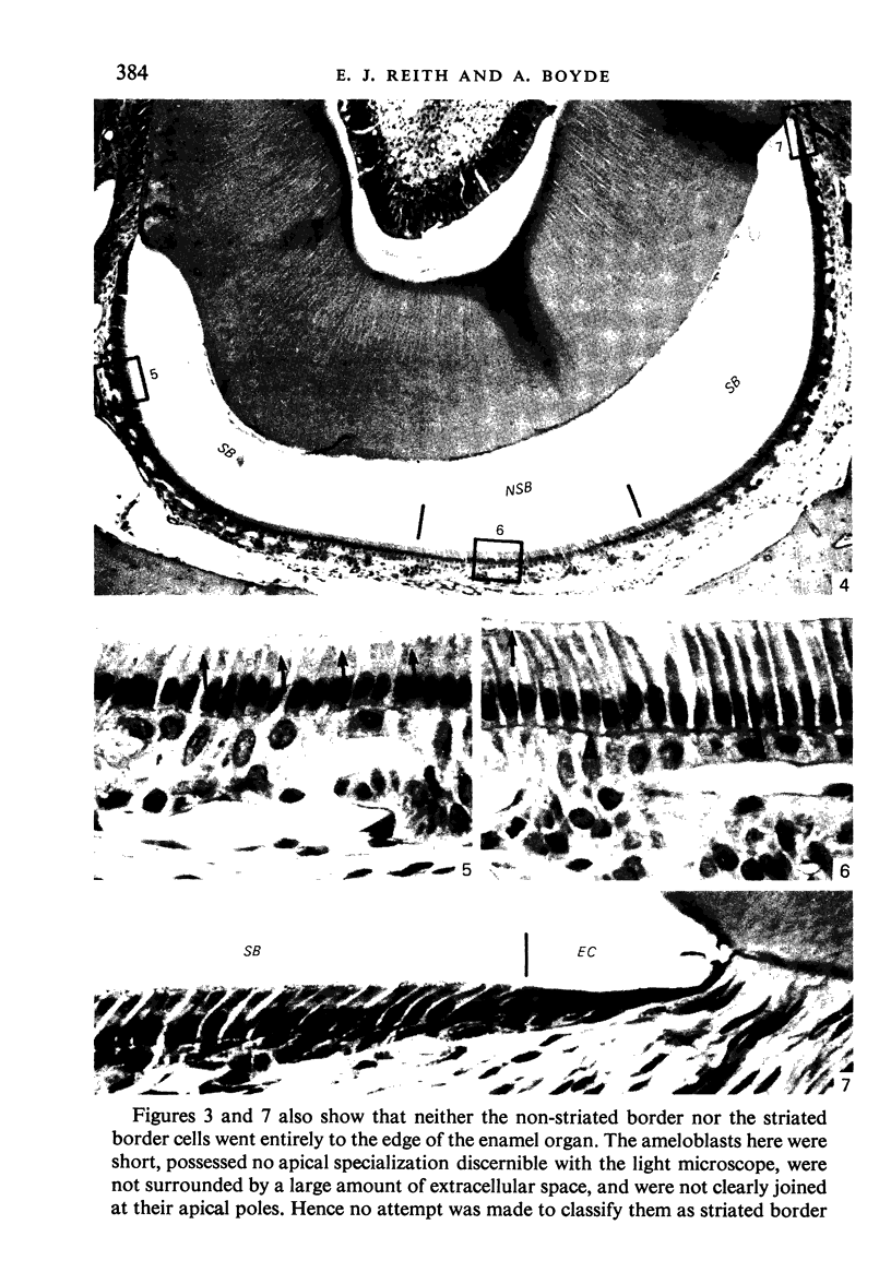
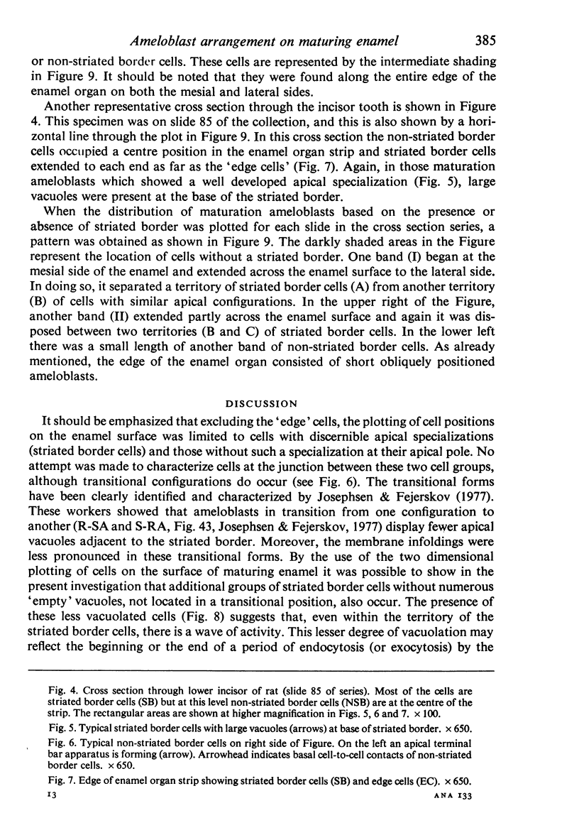
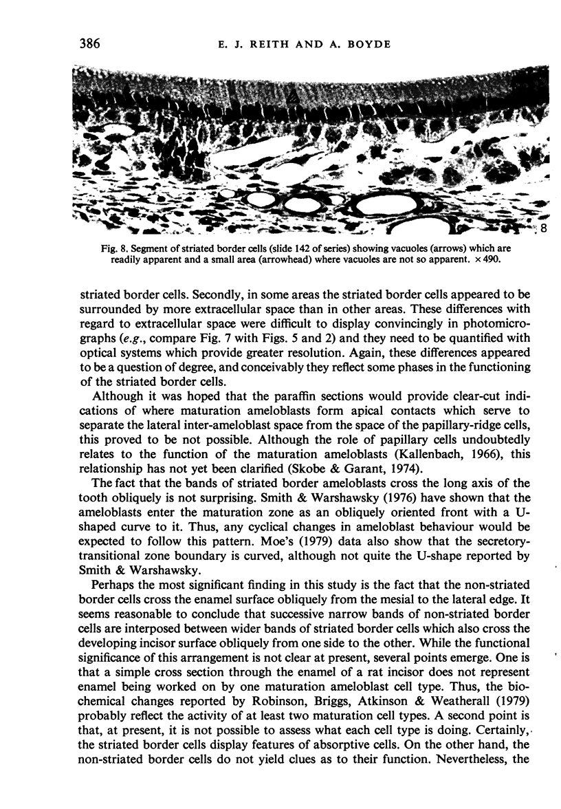
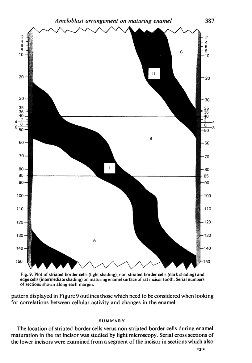
The location of striated border cells versus non-striated border cells during enamel maturation in the rat incisor was studied by light microscopy. Serial cross sections of the lower incisors were examined from a segment of the incisor in sections which also included the first molar tooth. In the series recorded here every cross section showed both striated and non-striated border cells. A map showing the distribution of cells on the enamel surface, as plotted from their position in each of the cross sections, reveals that the non-striated border cells traverse the enamel as oblique bands. Between the narrow bands of non-striated border cells were wide bands of striated border cells. The non-striated border cells were joined at their basal nuclear poles by contacts which appeared to separate the lateral inter-ameloblast space from the space between the papillary-ridge cells. Neither the striated border cells nor the non-striated border cells went entirely to the edge of the enamel organs. The above pattern of striated border cells and non-striated border cells is regarded to be a manifestation of the cyclical activity of maturation ameloblasts.
Full text
PDF
Images in this article
Selected References
These references are in PubMed. This may not be the complete list of references from this article.
- Boyde A., Reith E. J. Scanning electron microscopy of rat maturation ameloblasts. Cell Tissue Res. 1977 Mar 9;178(2):221–228. doi: 10.1007/BF00219049. [DOI] [PubMed] [Google Scholar]
- Boyde A., Reith E. J. Scanning electron microscopy of the lateral cell surfaces of rat incisor ameloblasts. J Anat. 1976 Dec;122(Pt 3):603–610. [PMC free article] [PubMed] [Google Scholar]
- Josephsen K., Fejerskov O. Ameloblast modulation in the maturation zone of the rat incisor enamel organ. A light and electron microscopic study. J Anat. 1977 Sep;124(Pt 1):45–70. [PMC free article] [PubMed] [Google Scholar]
- Kallenbach E. Electron microscopy of the papillary layer of rat incisor enamel organ during enamel maturation. J Ultrastruct Res. 1966 Mar;14(5):518–533. doi: 10.1016/s0022-5320(66)80079-5. [DOI] [PubMed] [Google Scholar]
- Kallenbach E. Fine structure of rat incisor ameloblasts during enamel maturation. J Ultrastruct Res. 1968 Jan;22(1):90–119. doi: 10.1016/s0022-5320(68)90051-8. [DOI] [PubMed] [Google Scholar]
- Kallenbach E. Fine structure of rat incisor enamel organ during late pigmentation and regression stages. J Ultrastruct Res. 1970 Jan;30(1):38–63. doi: 10.1016/s0022-5320(70)90063-8. [DOI] [PubMed] [Google Scholar]
- MARSLAND E. A. A histological investigation of amelogenesis in rats. I. Matrix formation. Br Dent J. 1951 Nov 20;91(10):251–261. [PubMed] [Google Scholar]
- Moe H. Physiological cell death of secretory ameloblasts in the rat incisor. Cell Tissue Res. 1979 Apr 12;197(3):443–451. doi: 10.1007/BF00233569. [DOI] [PubMed] [Google Scholar]
- REITH E. J. The ultrastructure of ameloblasts during matrix formation and the maturation of enamel. J Biophys Biochem Cytol. 1961 Apr;9:825–839. doi: 10.1083/jcb.9.4.825. [DOI] [PMC free article] [PubMed] [Google Scholar]
- Reith E. J., Boyde A. A correlated scanning and transmission electron microscopic study of maturation ameloblasts in developing molar teeth of rats. Cell Tissue Res. 1979 Apr 12;197(3):421–431. doi: 10.1007/BF00233567. [DOI] [PubMed] [Google Scholar]
- Reith E. J. The stages of amelogenesis as observed in molar teeth of young rats. J Ultrastruct Res. 1970 Jan;30(1):111–151. doi: 10.1016/s0022-5320(70)90068-7. [DOI] [PubMed] [Google Scholar]
- Robinson C., Briggs H. D., Atkinson P. J., Weatherell J. A. Matrix and mineral changes in developing enamel. J Dent Res. 1979 Mar;58(SPEC):871–882. doi: 10.1177/00220345790580024101. [DOI] [PubMed] [Google Scholar]
- SYMONS N. B. Alkaline phosphatase activity in the developing teeth of the rat. J Anat. 1955 Apr;89(2):238–245. [PMC free article] [PubMed] [Google Scholar]
- Skobe Z., Garant P. R. Electron microscopy of horseradish peroxidase uptake by papillary cells of the mouse incisor enamel organ. Arch Oral Biol. 1974 May;19(5):387–395. doi: 10.1016/0003-9969(74)90180-0. [DOI] [PubMed] [Google Scholar]
- Smith C. E., Warshawsky H. Movement of entire cell populations during renewal of the rat incisor as shown by radoioautography after labeling with 3H-thymidine. The concept of a continuously differentiating cross-sectional segment. (With an appendix on the development of the periodontal ligament). Am J Anat. 1976 Feb;145(2):225–259. doi: 10.1002/aja.1001450206. [DOI] [PubMed] [Google Scholar]
- Warshawsky H., Smith C. E. Morphological classification of rat incisor ameloblasts. Anat Rec. 1974 Aug;179(4):423–446. doi: 10.1002/ar.1091790403. [DOI] [PubMed] [Google Scholar]


