Abstract
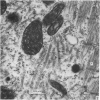
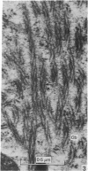
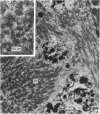
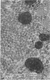
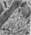
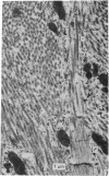
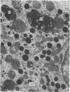
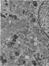
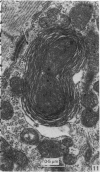
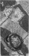
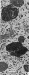
Ageing changes in the fine structure of atrial myocardial cells were studied in rats ranging from 2-32 months in age. The most striking change observed was the increasingly frequent appearance, from about 6 months onwards, of helically aggregated strands containing filaments which in respects other than arrangement bore resemblance to thick myofilaments. Other ageing changes included the accumulation of dense bodies and various types of mitochondrial degradation.
Full text
PDF
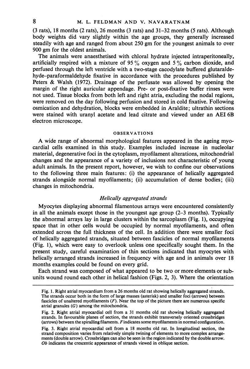
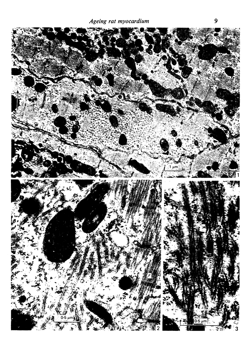
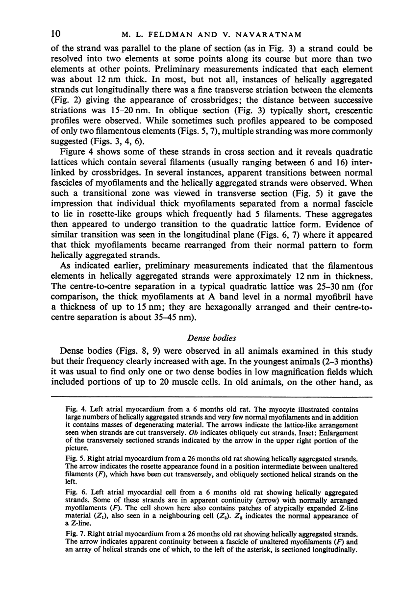
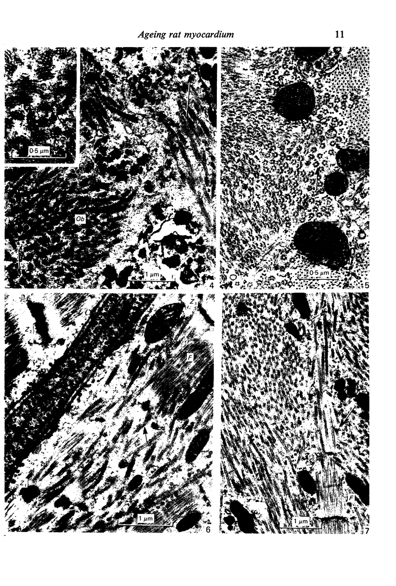
Images in this article
Selected References
These references are in PubMed. This may not be the complete list of references from this article.
- Abu-Erreish G. M., Sanadi D. R. Age-related changes in cytochrome concentration of myocardial mitochondria. Mech Ageing Dev. 1978 Jun;7(6):425–432. doi: 10.1016/0047-6374(78)90083-0. [DOI] [PubMed] [Google Scholar]
- Cavoto F. V., Kelliher G. J., Roberts J. Electrophysiological changes in the rat atrium with age. Am J Physiol. 1974 Jun;226(6):1293–1297. doi: 10.1152/ajplegacy.1974.226.6.1293. [DOI] [PubMed] [Google Scholar]
- Goldberg P. B., Baskin S. I., Roberts J. Effects of aging on ionic movements of atrial muscle. Fed Proc. 1975 Feb;34(2):188–190. [PubMed] [Google Scholar]
- Heller L. J., Whitehorn W. V. Age-associated alterations in myocardial contractile properties. Am J Physiol. 1972 Jun;222(6):1613–1619. doi: 10.1152/ajplegacy.1972.222.6.1613. [DOI] [PubMed] [Google Scholar]
- Herbener G. H. A morphometric study of age-dependent changes in mitochondrial population of mouse liver and heart. J Gerontol. 1976 Jan;31(1):8–12. doi: 10.1093/geronj/31.1.8. [DOI] [PubMed] [Google Scholar]
- Huxley H. E., Brown W. The low-angle x-ray diagram of vertebrate striated muscle and its behaviour during contraction and rigor. J Mol Biol. 1967 Dec 14;30(2):383–434. doi: 10.1016/s0022-2836(67)80046-9. [DOI] [PubMed] [Google Scholar]
- Koobs D. H., Schultz R. L., Jutzy R. V. The origin of lipofuscin and possible consequences to the myocardium. Arch Pathol Lab Med. 1978 Feb;102(2):66–68. [PubMed] [Google Scholar]
- Peters A., Walsh T. M. A study of the organization of apical dendrites in the somatic sensory cortex of the rat. J Comp Neurol. 1972 Mar;144(3):253–268. doi: 10.1002/cne.901440302. [DOI] [PubMed] [Google Scholar]
- Sachs H. G., Colgan J. A., Lazarus M. L. Ultrastructure of the aging myocardium: a morphometric approach. Am J Anat. 1977 Sep;150(1):63–71. doi: 10.1002/aja.1001500105. [DOI] [PubMed] [Google Scholar]
- Tate E. L., Herbener G. H. A morphometric study of the density of mitochondrial cristae in heart and liver of aging mice. J Gerontol. 1976 Mar;31(2):129–134. doi: 10.1093/geronj/31.2.129. [DOI] [PubMed] [Google Scholar]
- Tomanek R. J., Karlsson U. L. Myocardial ultrastructure of young and senescent rats. J Ultrastruct Res. 1973 Feb;42(3):201–220. doi: 10.1016/s0022-5320(73)90050-6. [DOI] [PubMed] [Google Scholar]
- Travis D. F., Travis A. Ultrastructural changes in the left ventricular rat myocardial cells with age. J Ultrastruct Res. 1972 Apr;39(1):124–148. doi: 10.1016/s0022-5320(72)80013-3. [DOI] [PubMed] [Google Scholar]


