Abstract
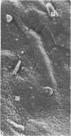
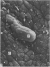
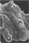
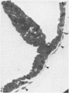
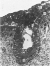
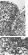
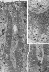
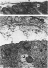
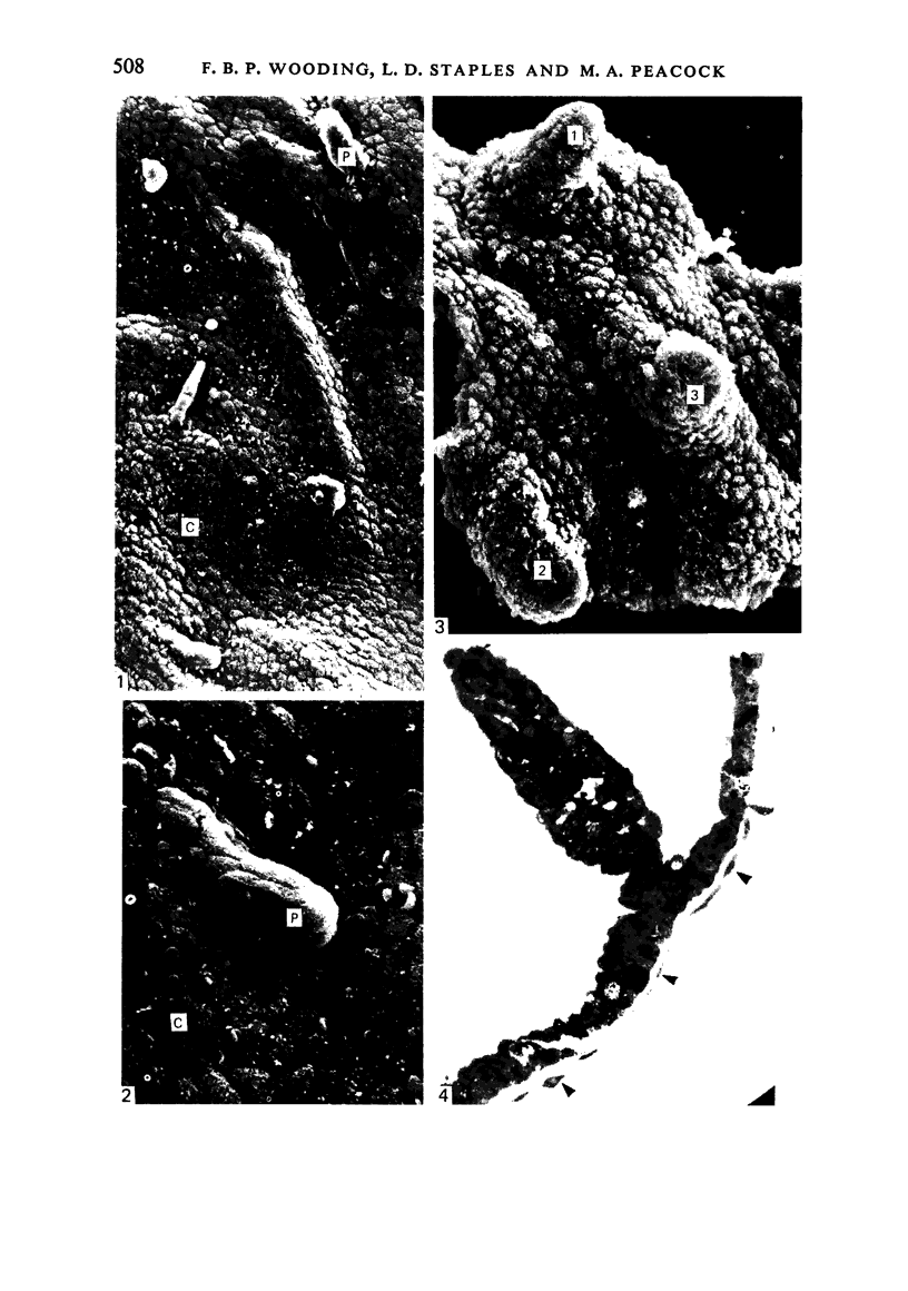
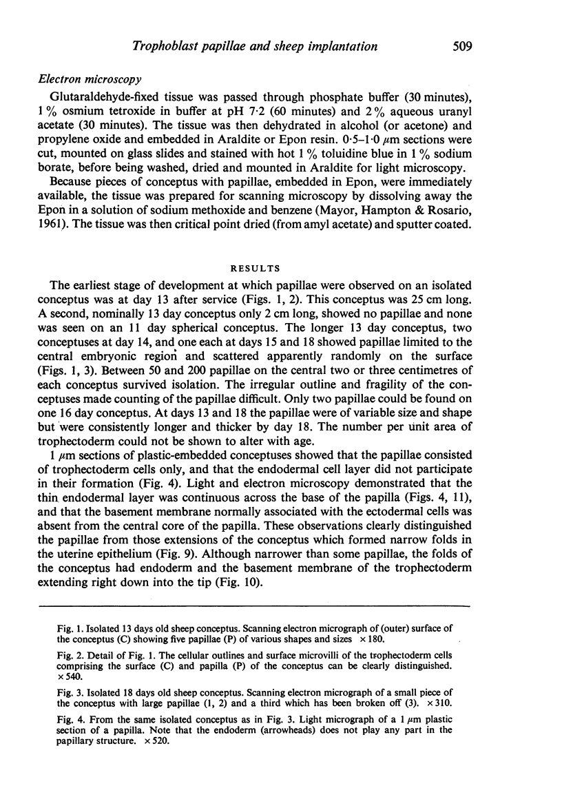
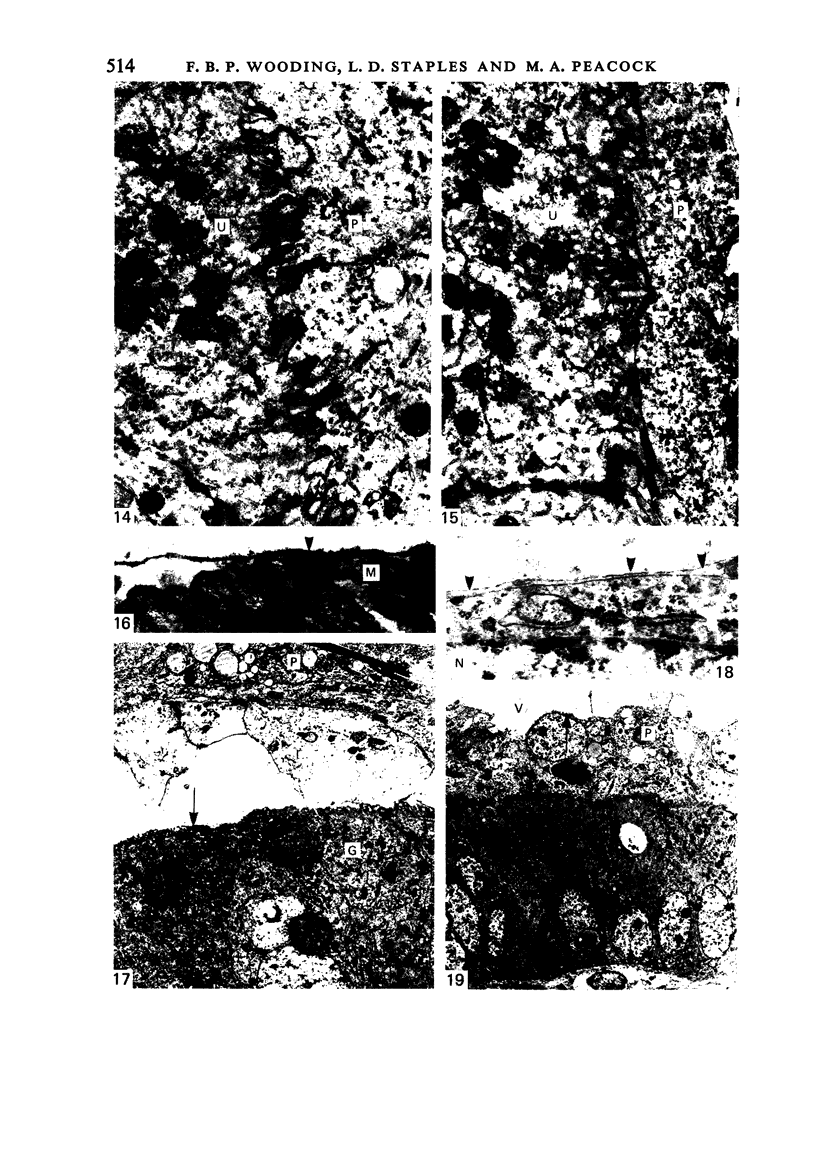
A study of 13-18 days old sheep conceptuses has consistently demonstrated the presence of multicellular protrusions (papillae) from the trophectoderm surface. These papillae were shown to be restricted to the embryonic region of conceptuses flushed out of the uterus. After perfusion fixation of the uterus on day 16 of pregnancy, the papillae can be observed penetrating well down into the lumina of the uterine glands. The papillae have not been observed at or after day 20. It is suggested that the papillae may play an important but transient role in anchoring the embryonic region of the conceptus against the uterine epithelium to allow the initiation of the cellular changes characteristic of implantation.
Full text
PDF
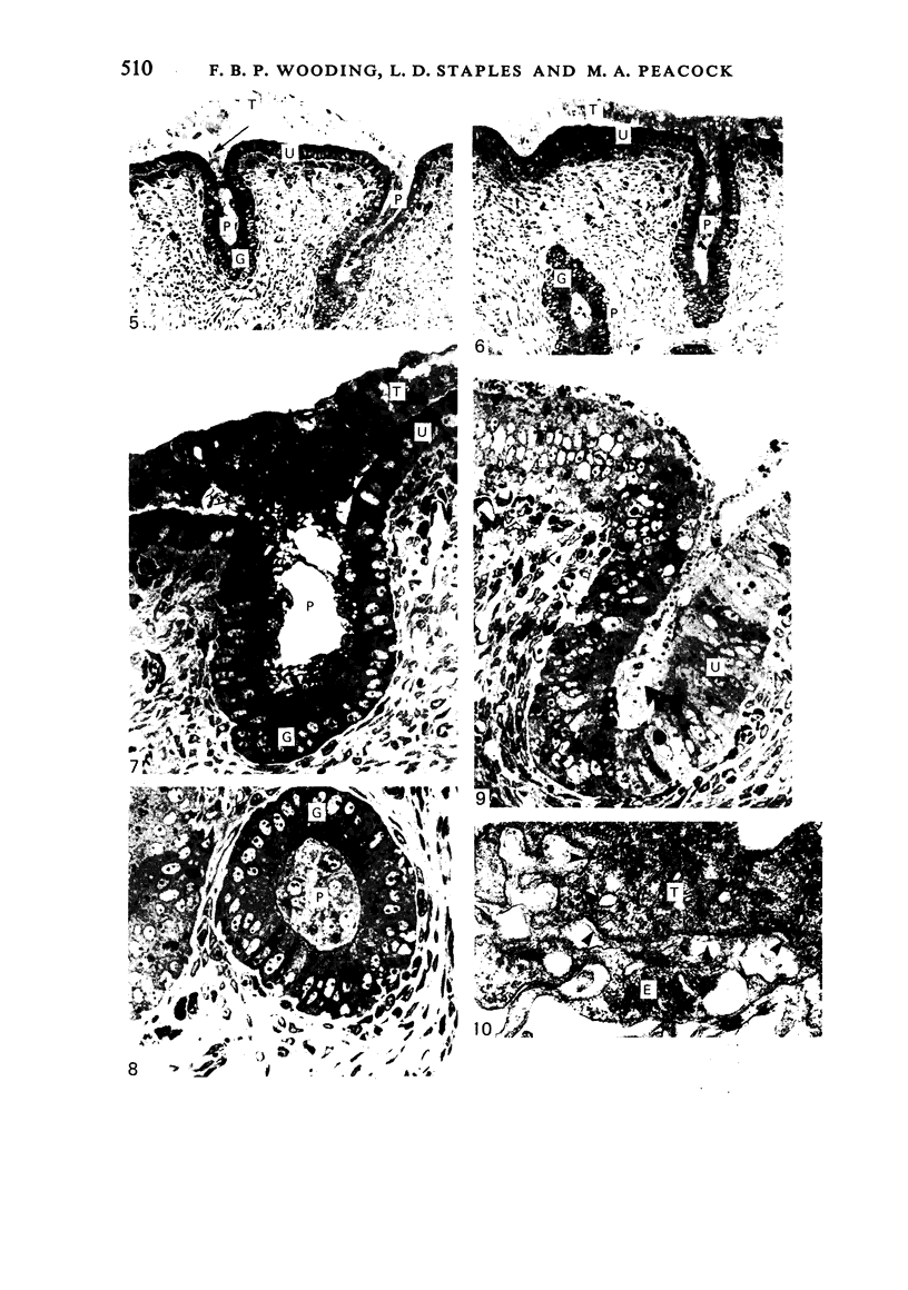
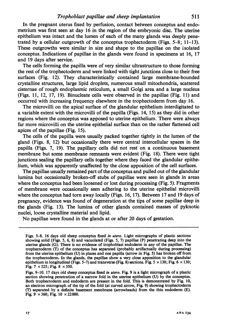
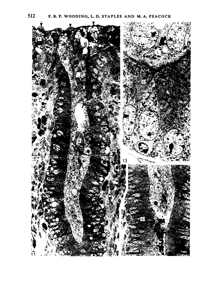
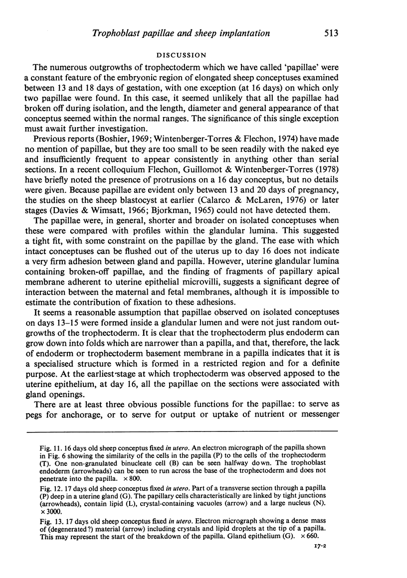
Images in this article
Selected References
These references are in PubMed. This may not be the complete list of references from this article.
- BJOERKMAN N. FINE STRUCTURE OF THE OVINE PLACENTOME. J Anat. 1965 Apr;99:283–297. [PMC free article] [PubMed] [Google Scholar]
- Boshier D. P. A histological and histochemical examination of implantation and early placentome formation in sheep. J Reprod Fertil. 1969 Jun;19(1):51–61. doi: 10.1530/jrf.0.0190051. [DOI] [PubMed] [Google Scholar]
- Calarco P. G., McLaren A. Ultrastructural observations of preimplantation stages of the sheep. J Embryol Exp Morphol. 1976 Dec;36(3):609–622. [PubMed] [Google Scholar]
- Davies J., Wimsatt W. A. Observation on the fine structure of the sheep placenta. Acta Anat (Basel) 1966;65(1):182–223. doi: 10.1159/000142872. [DOI] [PubMed] [Google Scholar]
- Lawn A. M., Chiquoine A. D., Amoroso E. C. The development of the placenta in the sheep and goat: an electron microscope study. J Anat. 1969 Nov;105(Pt 3):557–578. [PMC free article] [PubMed] [Google Scholar]
- MAYOR H. D., HAMPTON J. C., ROSARIO B. A simple method for removing the resin from epoxy-embedded tissue. J Biophys Biochem Cytol. 1961 Apr;9:909–910. doi: 10.1083/jcb.9.4.909. [DOI] [PMC free article] [PubMed] [Google Scholar]
- Wintenberger-Torrés S., Fléchon J. E. Ultrastructural evolution of the trophoblast cells of the pre-implantation sheep blastocyst from day 8 to day 18. J Anat. 1974 Sep;118(Pt 1):143–153. [PMC free article] [PubMed] [Google Scholar]


