

Abstract
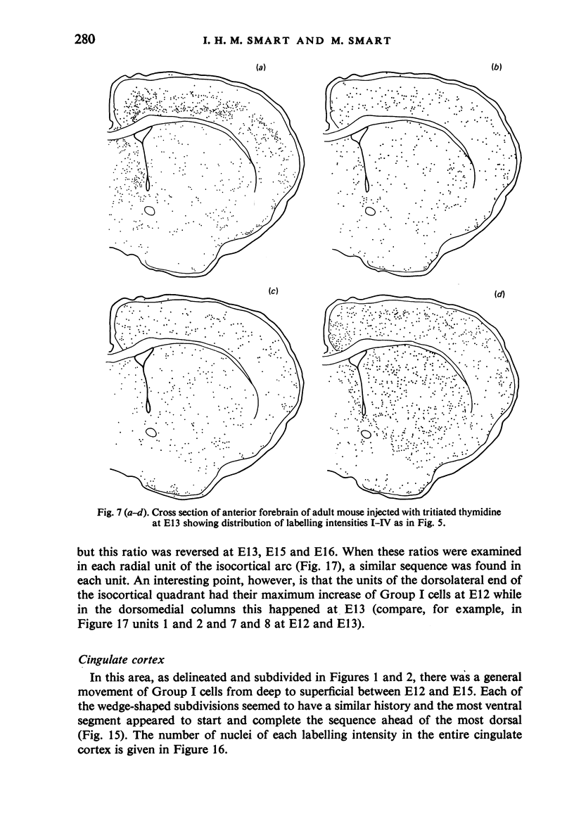
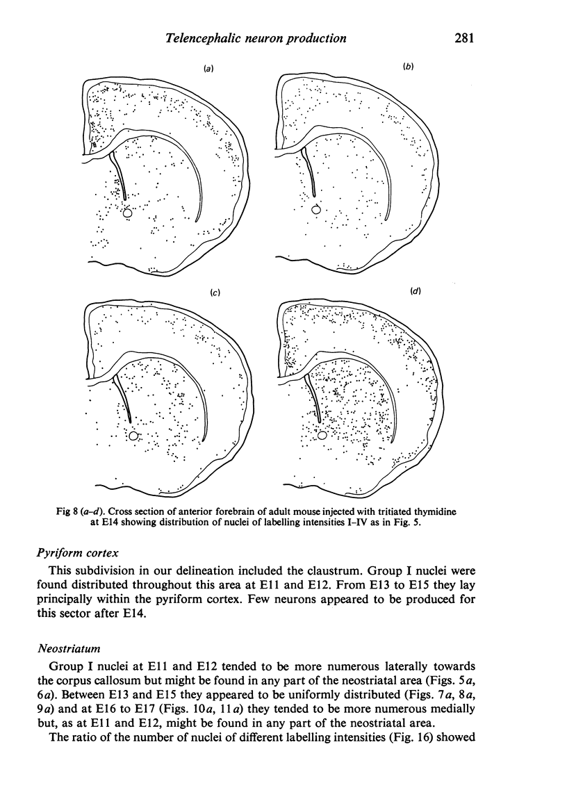
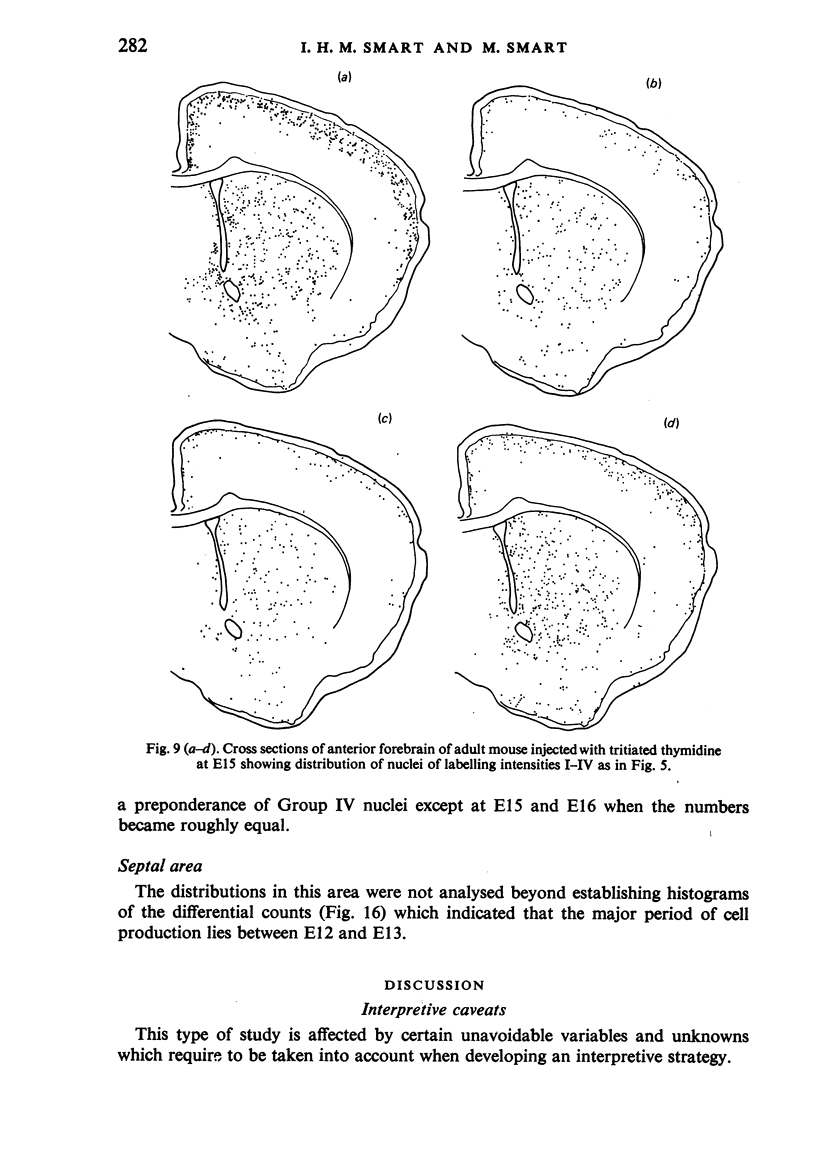
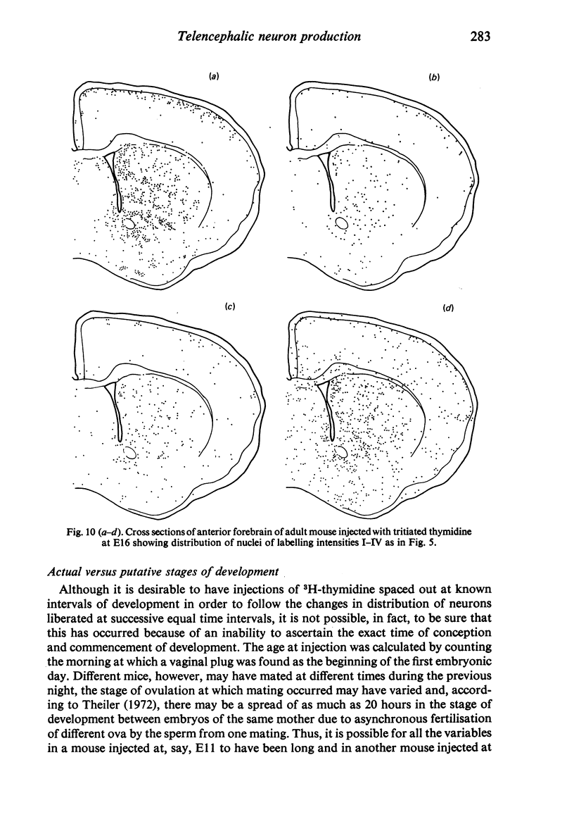
The distribution of cells of different labelling intensities in the anterior forebrain of adult mice injected with tritiated thymidine at daily intervals during prenatal life was determined by mapping the location of labelled cells on enlarged photographs of autoradiographed sections. The isocortical arc was subdivided into an arbitrary number of radially orientated units. Each radial unit was found to have a similar sequence of arrival and distribution of labelled cells; the ventrolateral units, however, entered and completed the sequence ahead of dorsomedial units indicating the presence of a wave of differentiation spreading in this direction across the generative layers giving rise to cortical neurons. An attempt was made to identify (from differential grain counts) comparable samples of first and second generation cells produced after each pulse of labelled thymidine. The changing ratio between the two generations suggested that there may be two peaks in neuron birth during the generative period.
Full text
PDF


Selected References
These references are in PubMed. This may not be the complete list of references from this article.
- Altman J., Das G. D. Autoradiographic and histological studies of postnatal neurogenesis. I. A longitudinal investigation of the kinetics, migration and transformation of cells incorporating tritiated thymidine in neonate rats, with special reference to postnatal neurogenesis in some brain regions. J Comp Neurol. 1966 Mar;126(3):337–389. doi: 10.1002/cne.901260302. [DOI] [PubMed] [Google Scholar]
- Angevine J. B., Jr, Sidman R. L. Autoradiographic study of cell migration during histogenesis of cerebral cortex in the mouse. Nature. 1961 Nov 25;192:766–768. doi: 10.1038/192766b0. [DOI] [PubMed] [Google Scholar]
- Berry M., Rogers A. W. The migration of neuroblasts in the developing cerebral cortex. J Anat. 1965 Oct;99(Pt 4):691–709. [PMC free article] [PubMed] [Google Scholar]
- Bisconte J. C., Marty R. Analyse chronoarchitectonique du cereveau de rat par radioautographie. I. Histogenèse du télencéphale. J Hirnforsch. 1975;16(1):55–74. [PubMed] [Google Scholar]
- Bisconte J. C., Marty R. Etude quantitative du marquage radioautographique dans le système nerveux du rat. II. Caractéristiques finales dans le cerveau de l'animal adulte: lois d'interprétation et concept de chronoarchitectonie corticale. Exp Brain Res. 1975;22(1):37–56. doi: 10.1007/BF00235410. [DOI] [PubMed] [Google Scholar]
- Bisconte J. C., Marty R. Etude quantitative du marquage radioautogrphique dans le système nerveux du rat. I. Caractéristiques primitives dans le tube neural. Exp Brain Res. 1974;21(5):455–461. doi: 10.1007/BF00237164. [DOI] [PubMed] [Google Scholar]
- Caviness V. S., Jr Architectonic map of neocortex of the normal mouse. J Comp Neurol. 1975 Nov 15;164(2):247–263. doi: 10.1002/cne.901640207. [DOI] [PubMed] [Google Scholar]
- Duckett S., Pearse A. G. The cells of Cajal-Retzius in the developing human brain. J Anat. 1968 Jan;102(Pt 2):183–187. [PMC free article] [PubMed] [Google Scholar]
- England J. M., Rogers A. W. The statistical analysis of autoradiographs. I. Grain count distributions over uniformly labelled sources. J Microsc. 1970;92(3):159–165. doi: 10.1111/j.1365-2818.1970.tb02250.x. [DOI] [PubMed] [Google Scholar]
- Fernández V., Bravo H. Autoradiographic study of development of the cerebral cortex in the rabbit. Brain Behav Evol. 1974;9(5):317–332. doi: 10.1159/000123674. [DOI] [PubMed] [Google Scholar]
- HUBEL D. H., WIESEL T. N. Receptive fields, binocular interaction and functional architecture in the cat's visual cortex. J Physiol. 1962 Jan;160:106–154. doi: 10.1113/jphysiol.1962.sp006837. [DOI] [PMC free article] [PubMed] [Google Scholar]
- Hicks S. P., D'Amato C. J. Cell migrations to the isocortex in the rat. Anat Rec. 1968 Mar;160(3):619–634. doi: 10.1002/ar.1091600311. [DOI] [PubMed] [Google Scholar]
- Hoshino K., Matsuzawa T., Murakami U. Charasteristics of the cell cycle of matrix cells in the mouse embryo during histogenesis of telencephalon. Exp Cell Res. 1973 Mar 15;77(1):89–94. doi: 10.1016/0014-4827(73)90556-9. [DOI] [PubMed] [Google Scholar]
- Hubel D. H., Wiesel T. N. Laminar and columnar distribution of geniculo-cortical fibers in the macaque monkey. J Comp Neurol. 1972 Dec;146(4):421–450. doi: 10.1002/cne.901460402. [DOI] [PubMed] [Google Scholar]
- MOUNTCASTLE V. B. Modality and topographic properties of single neurons of cat's somatic sensory cortex. J Neurophysiol. 1957 Jul;20(4):408–434. doi: 10.1152/jn.1957.20.4.408. [DOI] [PubMed] [Google Scholar]
- Marin-Padilla M. Early prenatal ontogenesis of the cerebral cortex (neocortex) of the cat (Felis domestica). A Golgi study. I. The primordial neocortical organization. Z Anat Entwicklungsgesch. 1971;134(2):117–145. doi: 10.1007/BF00519296. [DOI] [PubMed] [Google Scholar]
- Marin-Padilla M. Prenatal ontogenetic history of the principal neurons of the neocortex of the cat (Felis domestica). A Golgi study. II. Developmental differences and their significances. Z Anat Entwicklungsgesch. 1972;136(2):125–142. doi: 10.1007/BF00519174. [DOI] [PubMed] [Google Scholar]
- Raedler E., Raedler A., Feldhaus S. Dynamical aspects of neocortical histogenesis in the rat. Anat Embryol (Berl) 1980;158(3):253–269. doi: 10.1007/BF00301816. [DOI] [PubMed] [Google Scholar]
- Rakic P. Neurons in rhesus monkey visual cortex: systematic relation between time of origin and eventual disposition. Science. 1974 Feb 1;183(4123):425–427. doi: 10.1126/science.183.4123.425. [DOI] [PubMed] [Google Scholar]
- Rickmann M., Chronwall B. M., Wolff J. R. On the development of non-pyramidal neurons and axons outside the cortical plate: the early marginal zone as a pallial anlage. Anat Embryol (Berl) 1977 Dec 2;151(3):285–307. doi: 10.1007/BF00318931. [DOI] [PubMed] [Google Scholar]
- Sas E., Sanides F. A comparative Golgi study of Cajal foetal cells. Z Mikrosk Anat Forsch. 1970;82(3):385–396. [PubMed] [Google Scholar]
- Shimada M., Langman J. Cell proliferation, migration and differentiation in the cerebral cortex of the golden hamster. J Comp Neurol. 1970 Jun;139(2):227–244. doi: 10.1002/cne.901390206. [DOI] [PubMed] [Google Scholar]
- Sidman R. L., Rakic P. Neuronal migration, with special reference to developing human brain: a review. Brain Res. 1973 Nov 9;62(1):1–35. doi: 10.1016/0006-8993(73)90617-3. [DOI] [PubMed] [Google Scholar]
- Smart I. H. Proliferative characteristics of the ependymal layer during the early development of the mouse neocortex: a pilot study based on recording the number, location and plane of cleavage of mitotic figures. J Anat. 1973 Oct;116(Pt 1):67–91. [PMC free article] [PubMed] [Google Scholar]
- Smart I. H., Smart M. The location of nuclei of different labelling intensities in autoradiographs of the anterior forebrain of postnatial mice injected with [3H]thymidine on the eleventh and twelfth days post-conception. J Anat. 1977 Apr;123(Pt 2):515–525. [PMC free article] [PubMed] [Google Scholar]