Abstract
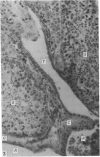
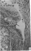
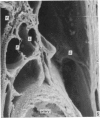
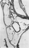
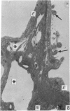
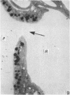
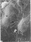
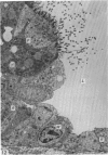
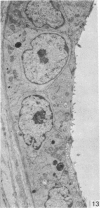
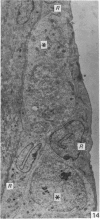
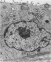
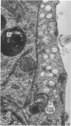
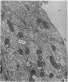
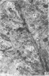
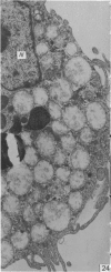
The rete testis in the domestic fowl (Gallus gallus domesticus), Japanese quail (Coturnix coturnix japonica), guinea-fowl (Numida meleagris galeata) and drake (Anas platyrhynchos) was studied histologically and with both the scanning and transmission electron microscopes. All the birds have rete epithelial cells varying between squamous and high cuboidal. A cilium-like structure projects from the luminal portion of most cells into the rete lumen, and the outline of the cells varies from polygonal to elongate. Sparse, stubby microvilli were concentrated on the cell borders. Ultrastructural features suggest only moderate secretory and absorptive activities in the cells. The rete testis of birds is amply supplied with blood as well as lymphatic vessels and nerves. Intraepithelial lymphocytes form part of the rete epithelium, and macrophages are present in large numbers in the rete lumen of the domestic fowl and drake and, to a lesser degree, also in the rete epithelium of the drake.
Full text
PDF
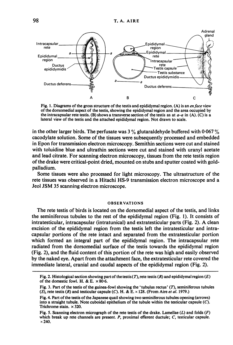
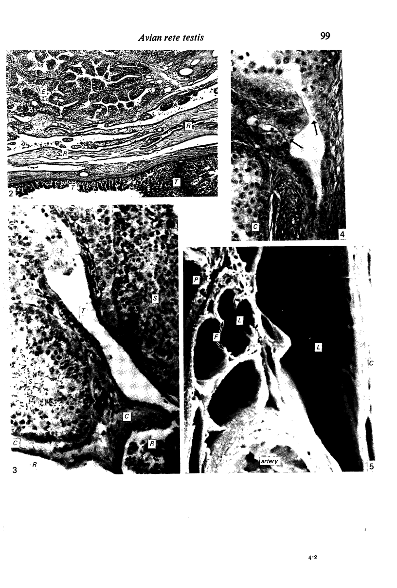
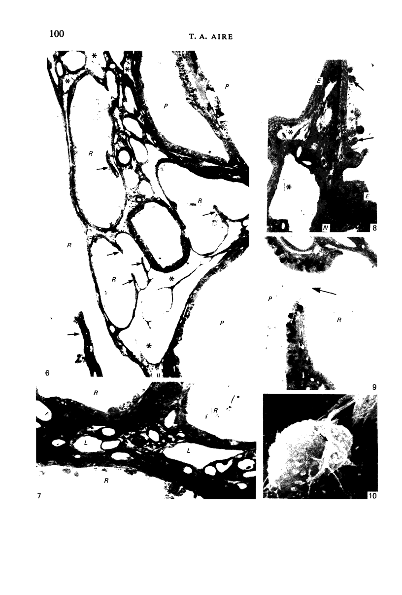
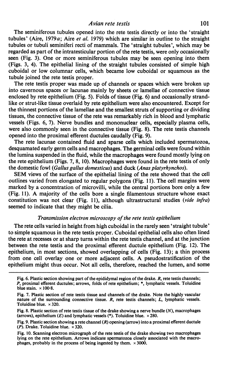
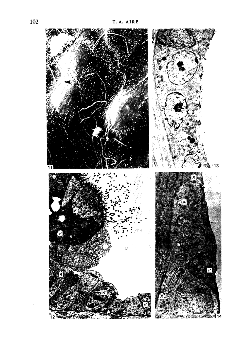
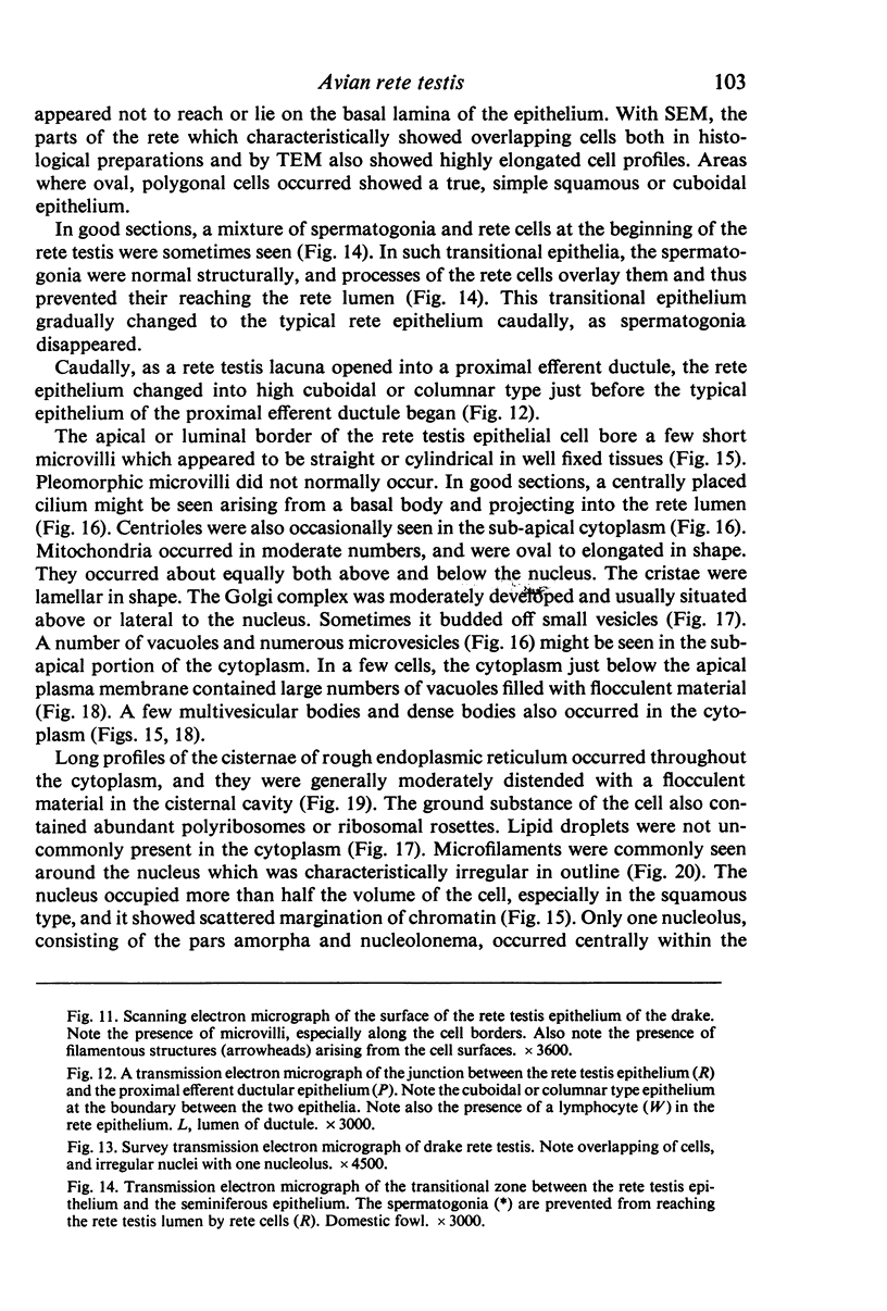
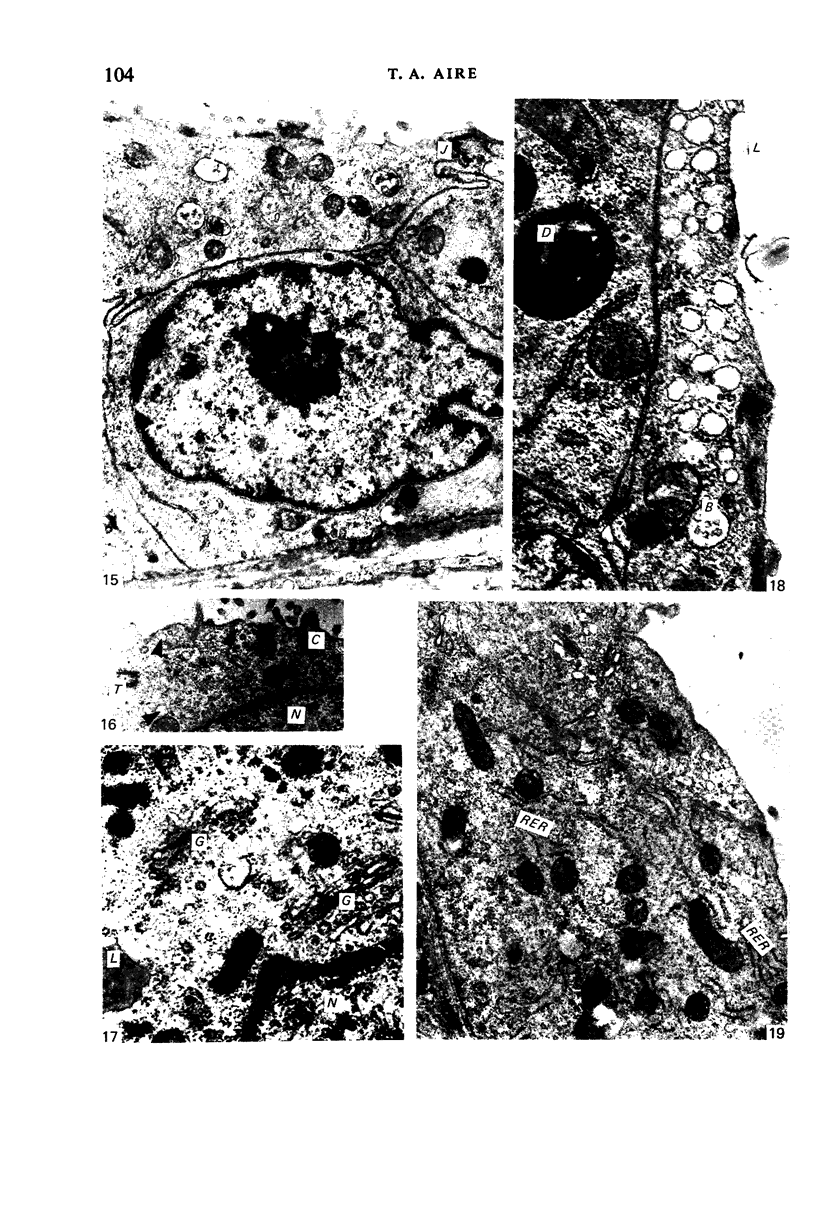
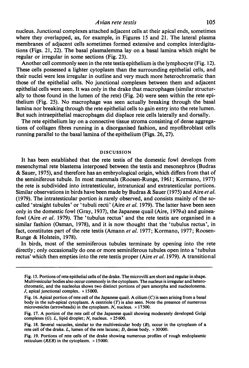
Images in this article
Selected References
These references are in PubMed. This may not be the complete list of references from this article.
- Aire T. A., Ayeni J. S., Olowo-Okorun M. O. The structure of the excurrent ducts of the testis of the guinea-fowl (Numida meleagris). J Anat. 1979 Oct;129(Pt 3):633–643. [PMC free article] [PubMed] [Google Scholar]
- Aire T. A., Malmquist M. Intraepithelial lymphocytes in the excurrent ducts of the testis of the domestic fowl (Gallus domesticus). Acta Anat (Basel) 1979;103(2):142–149. doi: 10.1159/000145005. [DOI] [PubMed] [Google Scholar]
- Aire T. A., Malmqvist M. Macrophages in the excurrent ducts of the testes of normal domestic fowl (Gallus domesticus). Anat Histol Embryol. 1979 Jun;8(2):172–176. doi: 10.1111/j.1439-0264.1979.tb00691.x. [DOI] [PubMed] [Google Scholar]
- Aire T. A. Micro-stereological study of the avian epididymal region. J Anat. 1979 Dec;129(Pt 4):703–706. [PMC free article] [PubMed] [Google Scholar]
- Aire T. A. The epididymal region of the Japanese quail (Coturnix coturnix japonica). Acta Anat (Basel) 1979;103(3):305–312. [PubMed] [Google Scholar]
- Amann R. P., Johnson L., Pickett B. W. Connection between the seminiferous tubules and the efferent ducts in the stallion. Am J Vet Res. 1977 Oct;38(10):1571–1579. [PubMed] [Google Scholar]
- Budras K. D., Sauer T. Morphology of the epididymis of the cock (Gallus domesticus) and its effect upon the steroid sex hormone synthesis. I. Ontogenesis, morphology and distribution of the epididymis. Anat Embryol (Berl) 1975 Dec 23;148(2):175–196. doi: 10.1007/BF00315268. [DOI] [PubMed] [Google Scholar]
- Bustos-Obregón E., Holstein A. F. The rete testis in man: ultrastructural aspects. Cell Tissue Res. 1976 Nov 24;175(1):1–15. doi: 10.1007/BF00220819. [DOI] [PubMed] [Google Scholar]
- Dym M. The mammalian rete testis--a morphological examination. Anat Rec. 1976 Dec;186(4):493–523. doi: 10.1002/ar.1091860404. [DOI] [PubMed] [Google Scholar]
- Hess R. A., Thurston R. J., Biellier H. V. Morphology of the epididymal region and ductus deferens of the turkey (Meleagris gallopavo). J Anat. 1976 Nov;122(Pt 2):241–252. [PMC free article] [PubMed] [Google Scholar]
- Hess R. A., Thurston R. J. Ultrastructure of epithelial cells in the epididymal region of the turkey (Meleagris gallopavo). J Anat. 1977 Dec;124(Pt 3):765–778. [PMC free article] [PubMed] [Google Scholar]
- LADMAN A. J., YOUNG W. C. An electron microscopic study of the ductuli efferentes and rete testis of the guinea pig. J Biophys Biochem Cytol. 1958 Mar 25;4(2):219–226. doi: 10.1083/jcb.4.2.219. [DOI] [PMC free article] [PubMed] [Google Scholar]
- LEESON T. S. Electron microscopy of the rete testis of the rat. Anat Rec. 1962 Sep;144:57–67. doi: 10.1002/ar.1091440108. [DOI] [PubMed] [Google Scholar]
- ROOSEN-RUNGE E. C. The rete testis in the albino rat: its structure, development and morphological significance. Acta Anat (Basel) 1961;45:1–30. doi: 10.1159/000141738. [DOI] [PubMed] [Google Scholar]
- Setchell B. P., Voglmayr J. K., Waites G. M. A blood-testis barrier restricting passage from blood into rete testis fluid but not into lymph. J Physiol. 1969 Jan;200(1):73–85. doi: 10.1113/jphysiol.1969.sp008682. [DOI] [PMC free article] [PubMed] [Google Scholar]
- Tingari M. D. On the structure of the epididymal region and ductus deferens of the domestic fowl (Gallus domesticus). J Anat. 1971 Sep;109(Pt 3):423–435. [PMC free article] [PubMed] [Google Scholar]
- Tingari M. D. The fine structure of the epithelial lining of the ex-current duct system of the testis of the domestic fowl (Gallus domesticus). Q J Exp Physiol Cogn Med Sci. 1972 Jul;57(3):271–295. doi: 10.1113/expphysiol.1972.sp002162. [DOI] [PubMed] [Google Scholar]
- Tuck R. R., Setchell B. P., Waites G. M., Young J. A. The composition of fluid collected by micropuncture and catheterization from the seminiferous tubules and rete testis of rats. Pflugers Arch. 1970;318(3):225–243. doi: 10.1007/BF00593663. [DOI] [PubMed] [Google Scholar]
- Voglmayr J. K., Waites G. M., Setchell B. P. Studies on spermatozoa and fluid collected directly from the testis of the conscious ram. Nature. 1966 May 21;210(5038):861–863. doi: 10.1038/210861b0. [DOI] [PubMed] [Google Scholar]


