

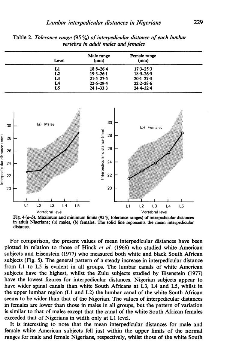
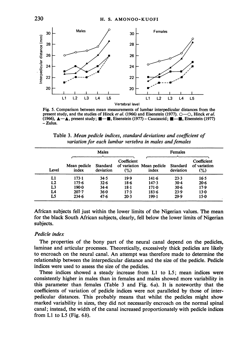
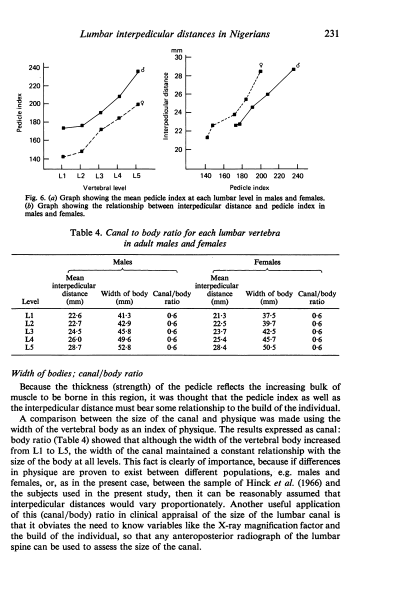
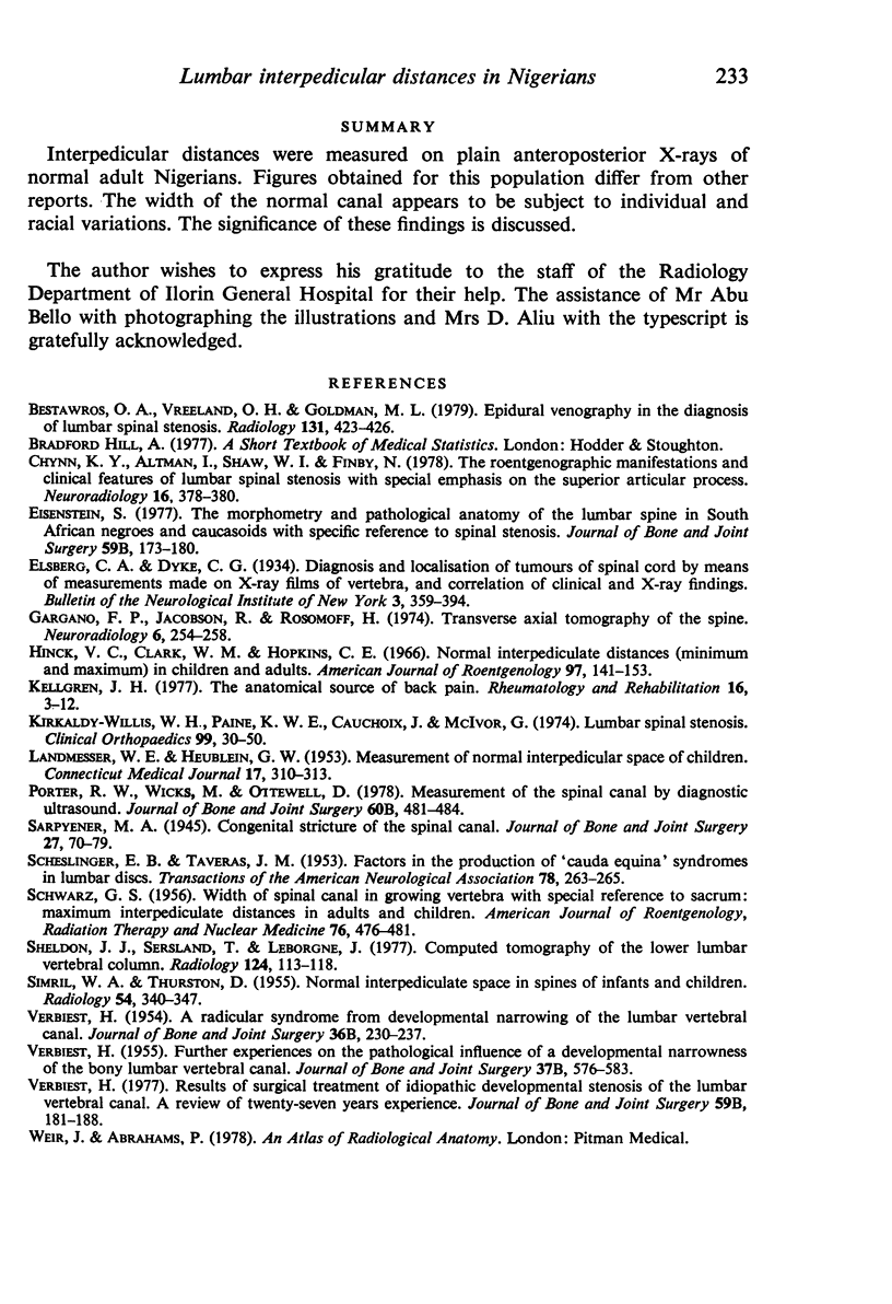
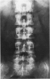
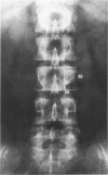
Full text
PDF
Images in this article
Selected References
These references are in PubMed. This may not be the complete list of references from this article.
- Bestawros O. A., Vreeland O. H., Goldman M. L. Epidural venography in the diagnosis of lumbar spinal stenosis. Radiology. 1979 May;131(2):423–426. doi: 10.1148/131.2.423. [DOI] [PubMed] [Google Scholar]
- Chynn K. Y., Altman I., Shaw W. I., Finby N. The roentgenographic manifestations and clinical features of lumbar spinal stenosis with special emphasis on the superior articular process. Neuroradiology. 1978;16:378–380. doi: 10.1007/BF00395310. [DOI] [PubMed] [Google Scholar]
- Eisenstein S. The morphometry and pathological anatomy of the lumbar spine in South African negroes and caucasoids with specific reference to spinal stenosis. J Bone Joint Surg Br. 1977 May;59(2):173–180. doi: 10.1302/0301-620X.59B2.873978. [DOI] [PubMed] [Google Scholar]
- Gargano F. P., Jacobson R., Rosomoff H. Transverse axial tomography of the spine. Neuroradiology. 1974;6(5):254–258. doi: 10.1007/BF00345785. [DOI] [PubMed] [Google Scholar]
- Hinck V. C., Clark W. M., Jr, Hopkins C. E. Normal interpediculate distances (minimum and maximum) in children and adults. Am J Roentgenol Radium Ther Nucl Med. 1966 May;97(1):141–153. doi: 10.2214/ajr.97.1.141. [DOI] [PubMed] [Google Scholar]
- Kellgren J. H. The anatomical source of back pain. Rheumatol Rehabil. 1977 Feb;16(1):3–12. doi: 10.1093/rheumatology/16.1.3. [DOI] [PubMed] [Google Scholar]
- Kirkaldy-Willis W. H., Paine K. W., Cauchoix J., McIvor G. Lumbar spinal stenosis. Clin Orthop Relat Res. 1974 Mar-Apr;(99):30–50. doi: 10.1097/00003086-197403000-00004. [DOI] [PubMed] [Google Scholar]
- LANDMESSER W. E., Jr, HEUBLEIN G. W. Measurement of the normal interpedicular space in the child. Conn State Med J. 1953 Apr;17(4):310–313. [PubMed] [Google Scholar]
- Porter R. W., Wicks M., Ottewell D. Measurement of the spinal canal by diagnostic ultrasound. J Bone Joint Surg Br. 1978 Nov;60-B(4):481–484. doi: 10.1302/0301-620X.60B4.711793. [DOI] [PubMed] [Google Scholar]
- SCHLESINGER E. B., TAVERAS J. M. Factors in the production of cauda equina syndromes in lumbar discs. Trans Am Neurol Assoc. 1953;3(78TH):263–265. [PubMed] [Google Scholar]
- SCHWARZ G. S. The width of the spinal canal in the growing vertebra with special reference to the sacrum; maximum interpediculated distances in adults and children. Am J Roentgenol Radium Ther Nucl Med. 1956 Sep;76(3):476–481. [PubMed] [Google Scholar]
- SIMRIL W. A., THURSTON D. The normal interpediculate space in the spines of infants and children. Radiology. 1955 Mar;64(3):340–347. doi: 10.1148/64.3.340. [DOI] [PubMed] [Google Scholar]
- Sheldon J. J., Sersland T., Leborgne J. Computed tomography of the lower lumbar vertebral column. Normal anatomy and the stenotic canal. Radiology. 1977 Jul;124(1):113–118. doi: 10.1148/124.1.113. [DOI] [PubMed] [Google Scholar]
- VERBIEST H. Further experiences on the pathological influence of a developmental narrowness of the bony lumbar vertebral canal. J Bone Joint Surg Br. 1955 Nov;37-B(4):576–583. doi: 10.1302/0301-620X.37B4.576. [DOI] [PubMed] [Google Scholar]
- Verbiest H. Results of surgical treatment of idiopathic developmental stenosis of the lumbar vertebral canal. A review of twenty-seven years' experience. J Bone Joint Surg Br. 1977 May;59(2):181–188. doi: 10.1302/0301-620X.59B2.141452. [DOI] [PubMed] [Google Scholar]