Abstract
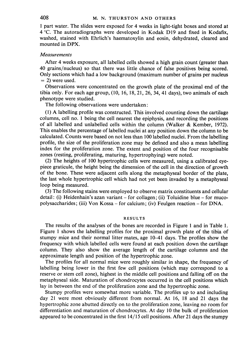
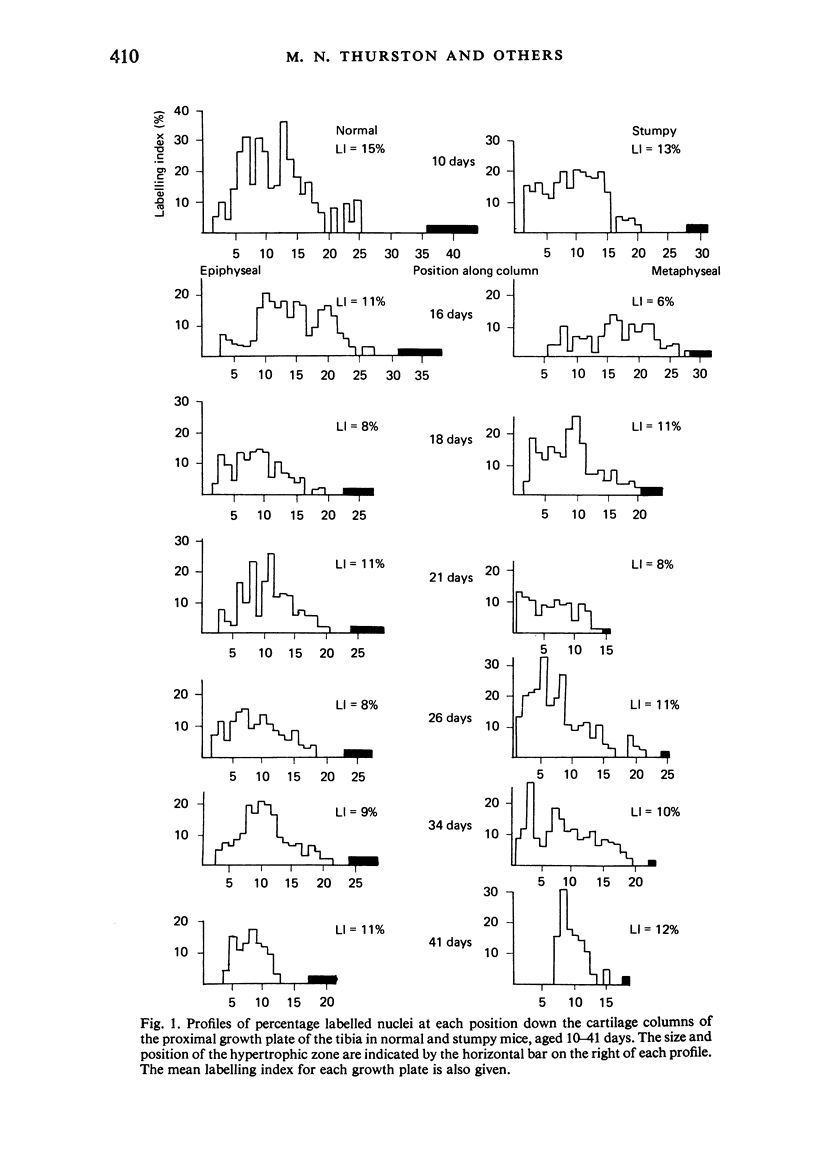
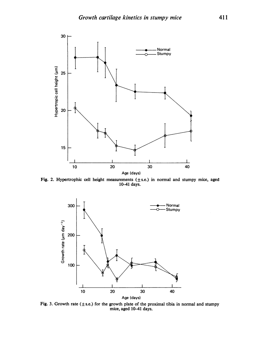
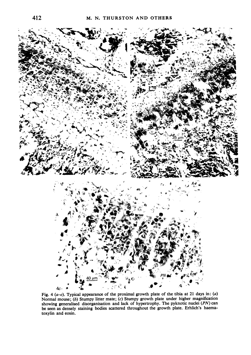
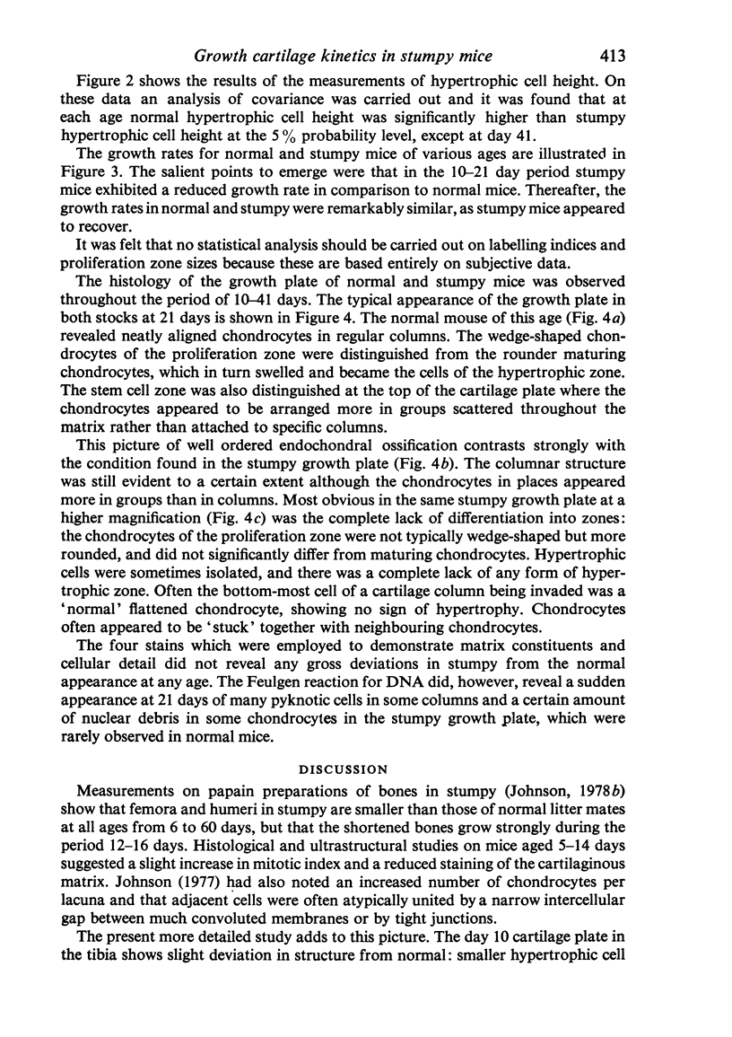
The proximal growth plates of the tibiae in normal and stumpy mice aged 10-41 days were studied. Autoradiographic studies using tritiated thymidine enabled the size of the proliferating cell population and the labelling indices of the growth plates to be determined. Hypertrophic cell heights were also measured. From these data the overall growth rates for the proximal growth plate of the tibia in normal and stumpy mice were calculated. It was found that the major factor responsible for the reduced growth rate in stumpy up to 21 days was the small hypertrophic cell height, while cell proliferation zone size and labelling indices were of minor importance. Histological observations also revealed a lack of organised endochondral ossification, which worsens with age.
Full text
PDF

Images in this article
Selected References
These references are in PubMed. This may not be the complete list of references from this article.
- Ferguson J. M., Wallace M. E., Johnson D. R. A new type of chondrodystrophic mutant in the mouse. J Med Genet. 1978 Apr;15(2):128–131. doi: 10.1136/jmg.15.2.128. [DOI] [PMC free article] [PubMed] [Google Scholar]
- Johnson D. R. The effect of a single injection of papain on growth of the limb bones of the mouse. Growth. 1978 Mar;42(1):27–30. [PubMed] [Google Scholar]
- Johnson D. R. The growth of femur and tibia in three genetically distinct chondrodystrophic mutants of the house mouse. J Anat. 1978 Feb;125(Pt 2):267–275. [PMC free article] [PubMed] [Google Scholar]
- Johnson D. R. Ultrastructural observations on stumpy (stm), a new chondrodystrophic mutant in the mouse. J Embryol Exp Morphol. 1977 Jun;39:279–284. [PubMed] [Google Scholar]
- Kember N. F. Comparative patterns of cell division in epiphyseal cartilage plates in the rat. J Anat. 1972 Jan;111(Pt 1):137–142. [PMC free article] [PubMed] [Google Scholar]
- Rimoin D. L. The chondrodystrophies. Adv Hum Genet. 1975;5:1–118. doi: 10.1007/978-1-4615-9068-2_1. [DOI] [PubMed] [Google Scholar]
- Walker K. V., Kember N. F. Cell kinetics of growth cartilage in the rat tibia. II. Measurements during ageing. Cell Tissue Kinet. 1972 Sep;5(5):409–419. doi: 10.1111/j.1365-2184.1972.tb00379.x. [DOI] [PubMed] [Google Scholar]


