Abstract
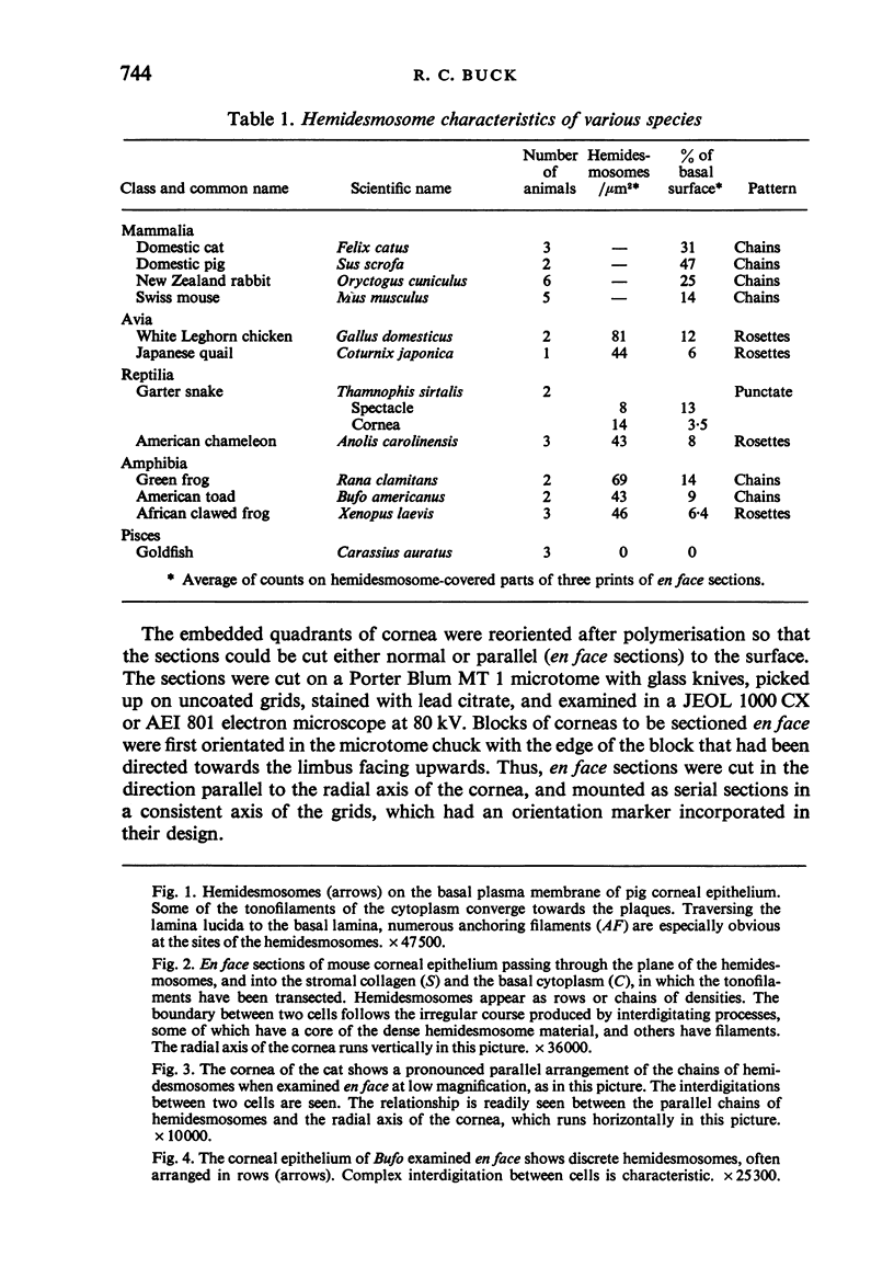
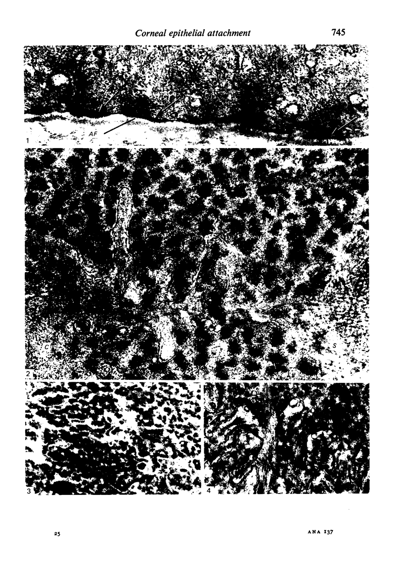
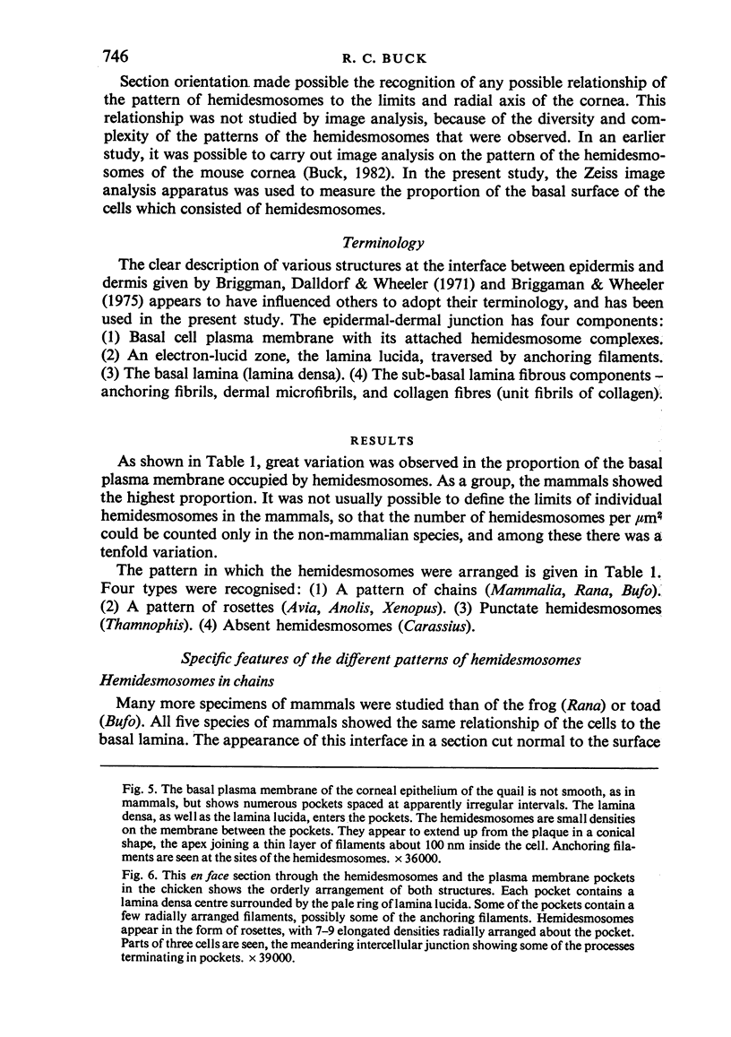
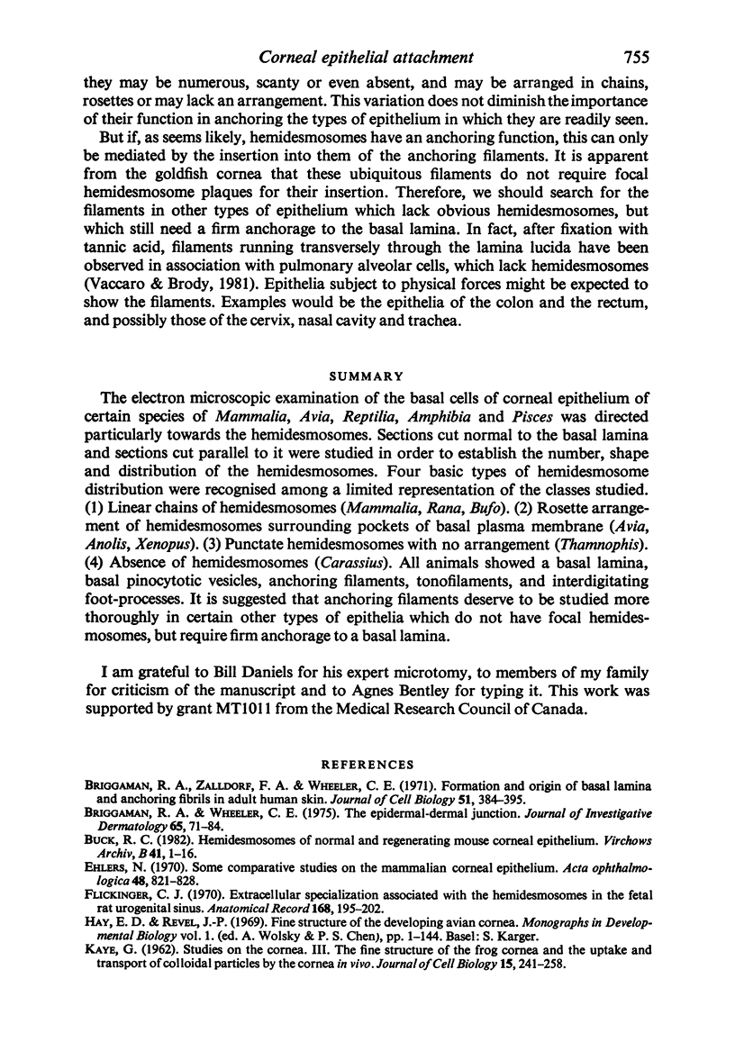
The electron microscopic examination of the basal cells of corneal epithelium certain species of Mammalia, Avia, Reptilia, Amphibia and Pisces was directed particularly towards the hemidesmosomes. Sections cut normal to the basal lamina and sections cut parallel to it were studied in order to establish the number, shape and distribution of the hemidesmosomes. Four basic types of hemidesmosome distribution were recognised among a limited representation of the classes studied. (1) Linear chains of hemidesmosomes (Mammalia, Rana, Bufo). (2) Rosette arrangement of hemidesmosomes surrounding pockets of basal plasma membrane (Avia, Anolis, Xenopus). (3) Punctate hemidesmosomes with no arrangement (Thamnophis). (4) Absence of hemidesmosomes (Carassius). All animals showed a basal lamina, basal pinocytotic vesicles, anchoring filaments, tonofilaments, and interdigitating foot-processes. It is suggested that anchoring filaments deserve to be studied more thoroughly in certain other types of epithelia which do not have focal hemidesmosomes, but require firm anchorage to a basal lamina.
Full text
PDF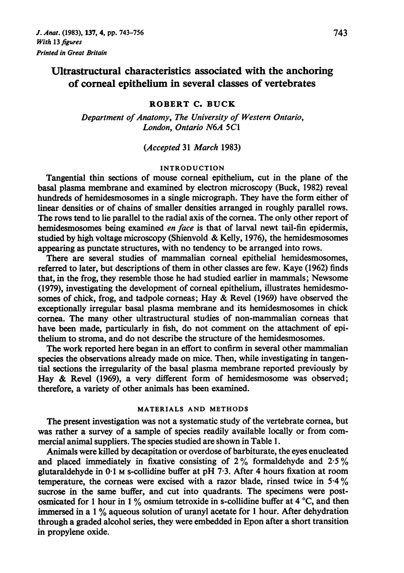
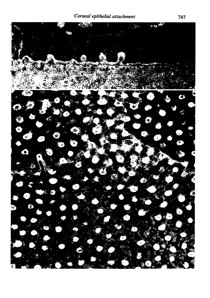
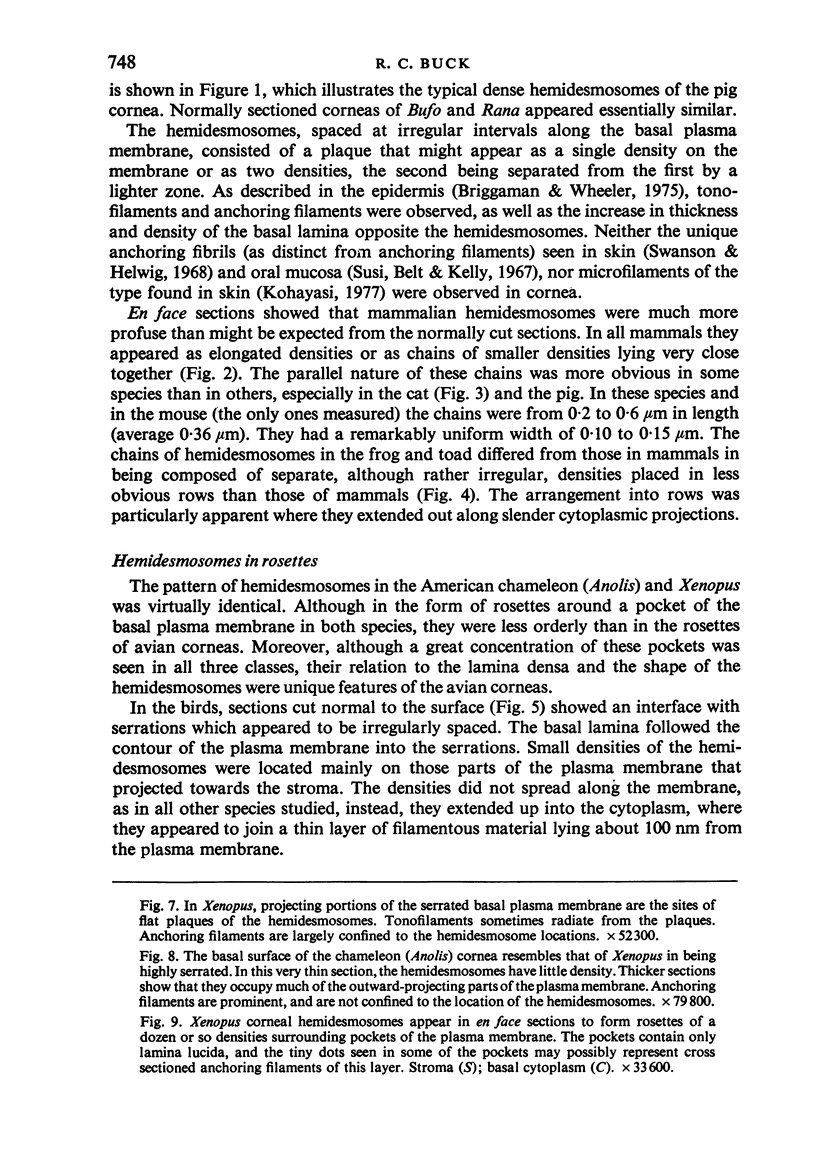
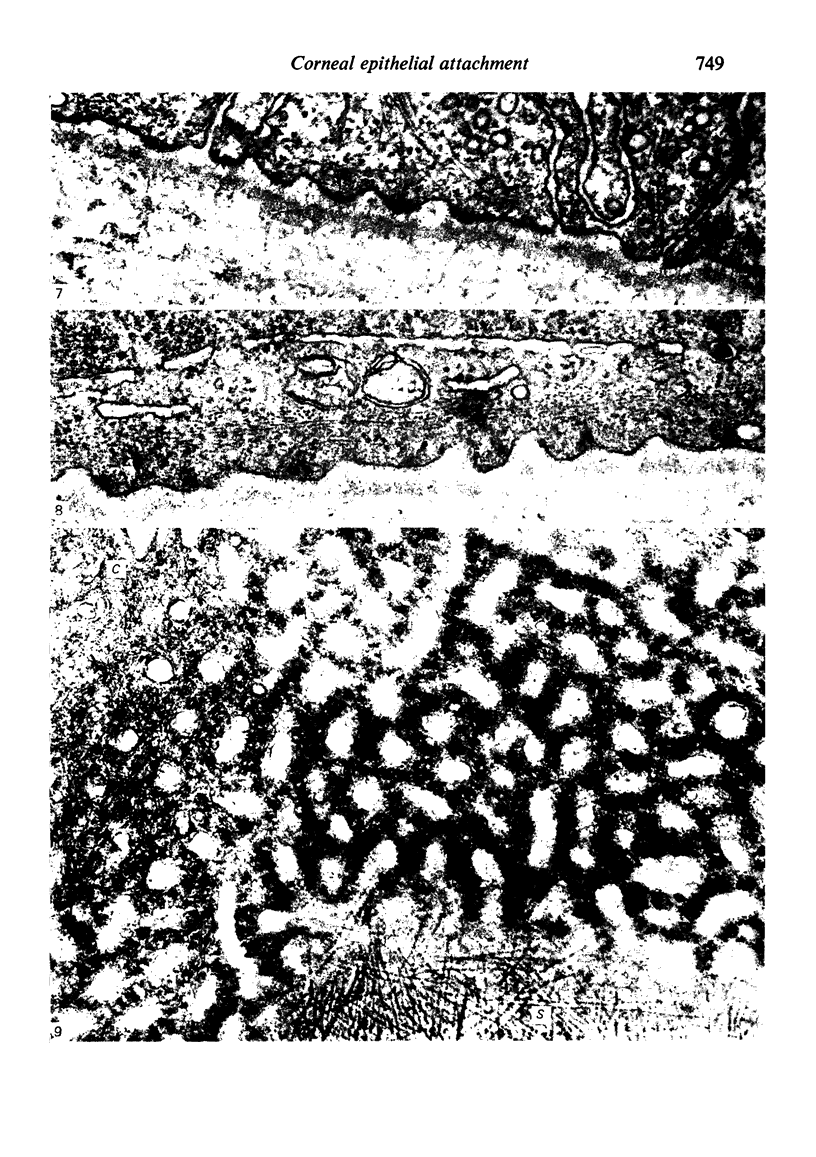
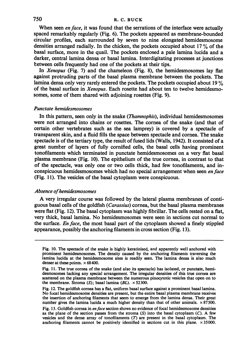
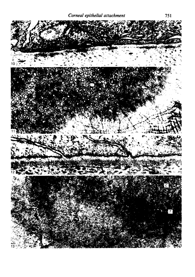
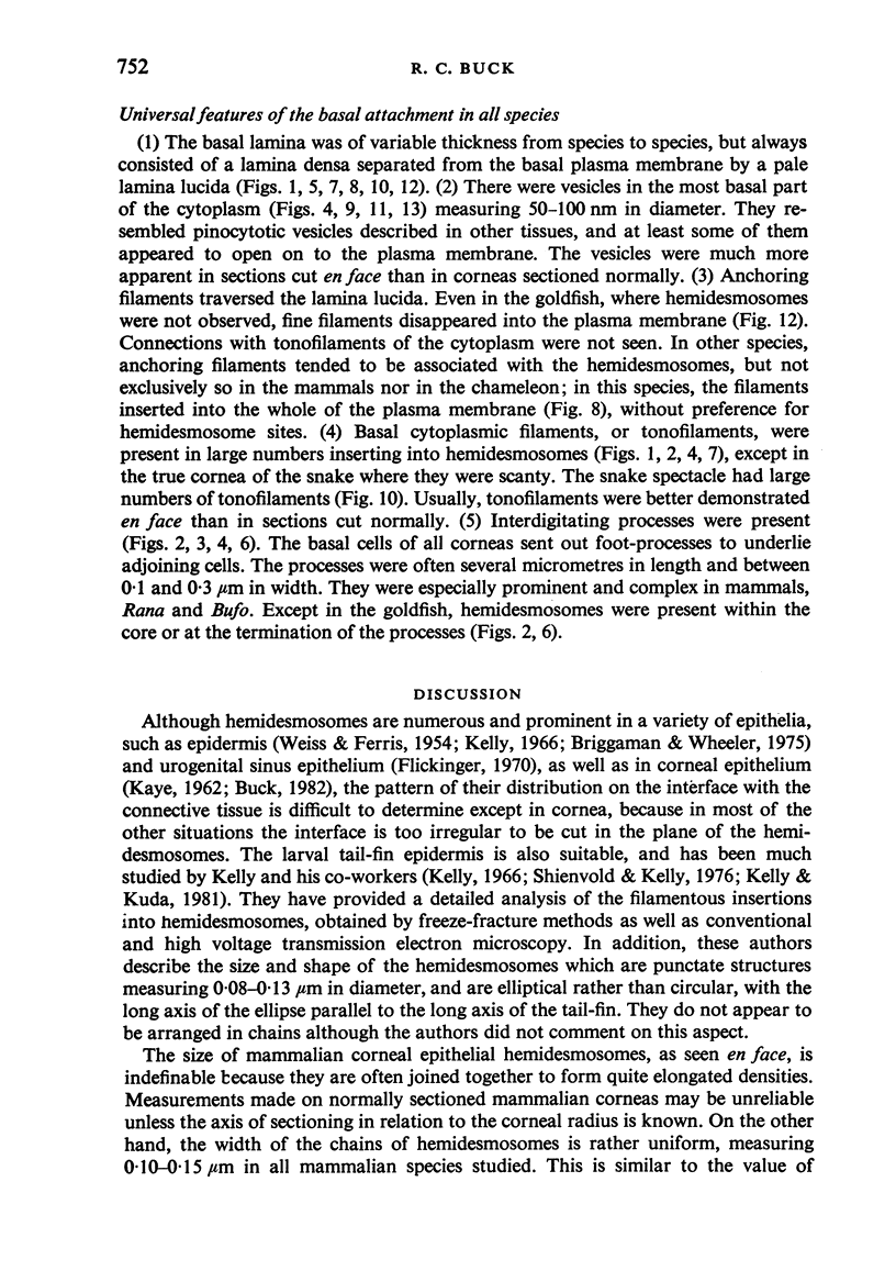
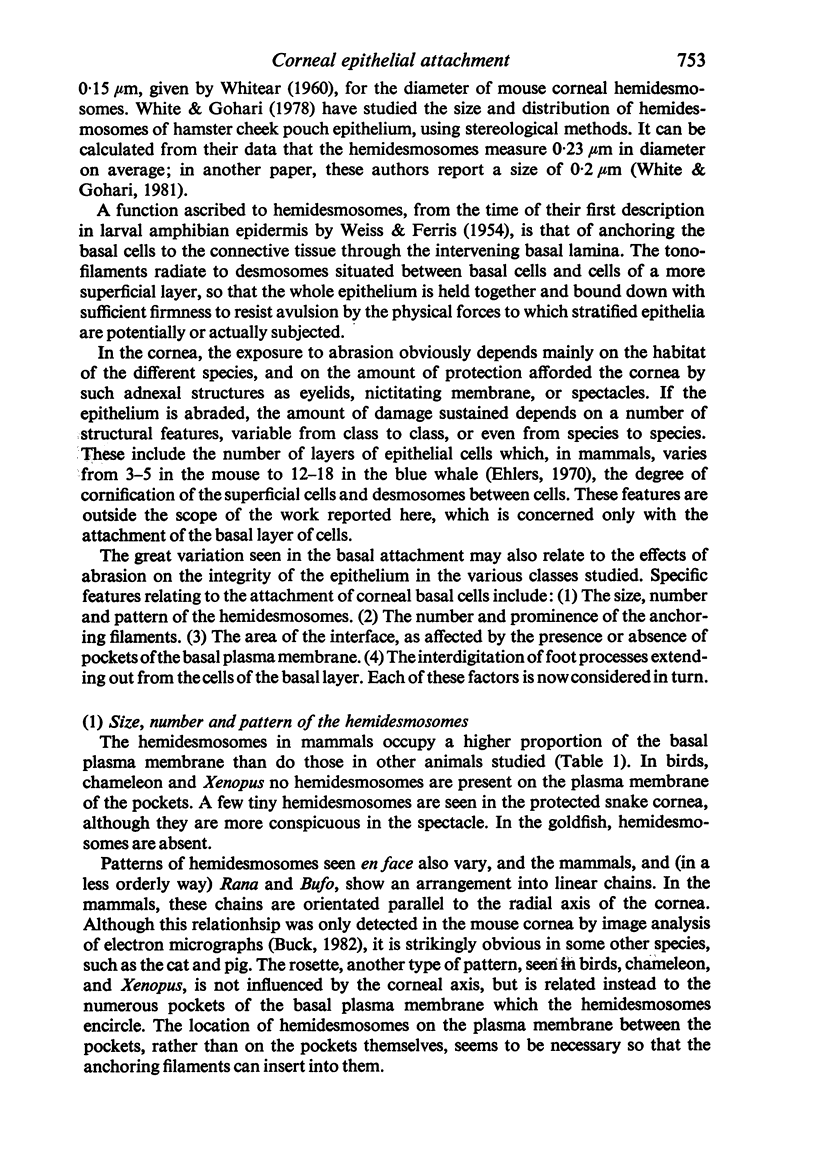
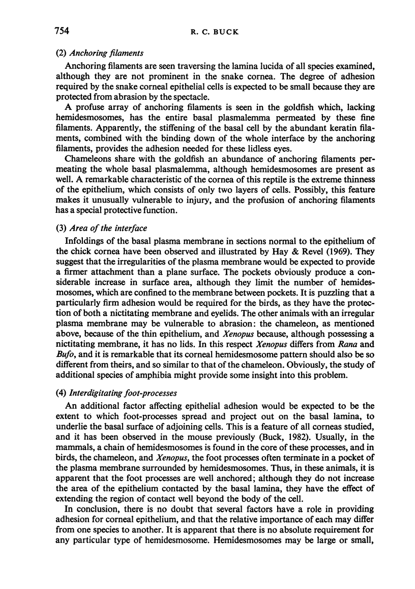
Images in this article
Selected References
These references are in PubMed. This may not be the complete list of references from this article.
- Briggaman R. A., Dalldorf F. G., Wheeler C. E., Jr Formation and origin of basal lamina and anchoring fibrils in adult human skin. J Cell Biol. 1971 Nov;51(21):384–395. doi: 10.1083/jcb.51.2.384. [DOI] [PMC free article] [PubMed] [Google Scholar]
- Briggaman R. A., Wheeler C. E., Jr The epidermal-dermal junction. J Invest Dermatol. 1975 Jul;65(1):71–84. doi: 10.1111/1523-1747.ep12598050. [DOI] [PubMed] [Google Scholar]
- Buck R. C. Hemidesmosomes of normal and regenerating mouse corneal epithelium. Virchows Arch B Cell Pathol Incl Mol Pathol. 1982;41(1-2):1–16. doi: 10.1007/BF02890267. [DOI] [PubMed] [Google Scholar]
- Flickinger C. J. Extracellular specializations associated with hemidesmosomes in the fetal rat urogenital sinus. Anat Rec. 1970 Oct;168(2):195–202. doi: 10.1002/ar.1091680206. [DOI] [PubMed] [Google Scholar]
- Hay E. D., Revel J. P. Fine structure of the developing avian cornea. Monogr Dev Biol. 1969;1:1–144. [PubMed] [Google Scholar]
- KAYE G. I. Studies on the cornea. III. The fine structure of the frog cornea and the uptake and transport of colloidal particles by the cornea in vivo. J Cell Biol. 1962 Nov;15:241–258. doi: 10.1083/jcb.15.2.241. [DOI] [PMC free article] [PubMed] [Google Scholar]
- Kelly D. E. Fine structure of desmosomes. , hemidesmosomes, and an adepidermal globular layer in developing newt epidermis. J Cell Biol. 1966 Jan;28(1):51–72. doi: 10.1083/jcb.28.1.51. [DOI] [PMC free article] [PubMed] [Google Scholar]
- Kelly D. E., Kuda A. M. Traversing filaments in desmosomal and hemidesmosomal attachments: freeze-fracture approaches toward their characterization. Anat Rec. 1981 Jan;199(1):1–14. doi: 10.1002/ar.1091990102. [DOI] [PubMed] [Google Scholar]
- Kobayasi T. Anchoring of basal lamina to elastic fibers by elastic fibrils. J Invest Dermatol. 1977 Jun;68(6):389–390. doi: 10.1111/1523-1747.ep12496956. [DOI] [PubMed] [Google Scholar]
- Newsome D. A. Embryology and biology of the ocular surface. Int Ophthalmol Clin. 1979 Summer;19(2):53–72. [PubMed] [Google Scholar]
- Shienvold F. L., Kelly D. E. The hemidesmosome: new fine structural features revealed by freeze-fracture techniques. Cell Tissue Res. 1976 Sep 20;172(3):289–307. doi: 10.1007/BF00399513. [DOI] [PubMed] [Google Scholar]
- Susi F. R., Belt W. D., Kelly J. W. Fine structure of fibrillar complexes associated with the basement membrane in human oral mucosa. J Cell Biol. 1967 Aug;34(2):686–690. doi: 10.1083/jcb.34.2.686. [DOI] [PMC free article] [PubMed] [Google Scholar]
- Swanson J. L., Helwig E. B. Special fibrils of human dermis. J Invest Dermatol. 1968 Feb;50(2):195–199. [PubMed] [Google Scholar]
- Vaccaro C. A., Brody J. S. Structural features of alveolar wall basement membrane in the adult rat lung. J Cell Biol. 1981 Nov;91(2 Pt 1):427–437. doi: 10.1083/jcb.91.2.427. [DOI] [PMC free article] [PubMed] [Google Scholar]
- WEISS P., FERRIS W. Electronmicrograms of larval amphibian epidermis. Exp Cell Res. 1954 May;6(2):546–549. doi: 10.1016/0014-4827(54)90210-4. [DOI] [PubMed] [Google Scholar]
- WHITEAR M. An electron microscope study of the cornea in mice, with special reference to the innervation. J Anat. 1960 Jul;94:387–409. [PMC free article] [PubMed] [Google Scholar]
- White F. H., Gohari K. A quantitative study of lamina densa alterations in hamster cheek pouch carcinogenesis. J Pathol. 1981 Dec;135(4):277–294. doi: 10.1002/path.1711350405. [DOI] [PubMed] [Google Scholar]


