

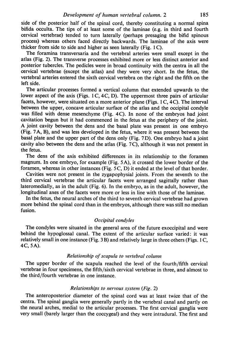
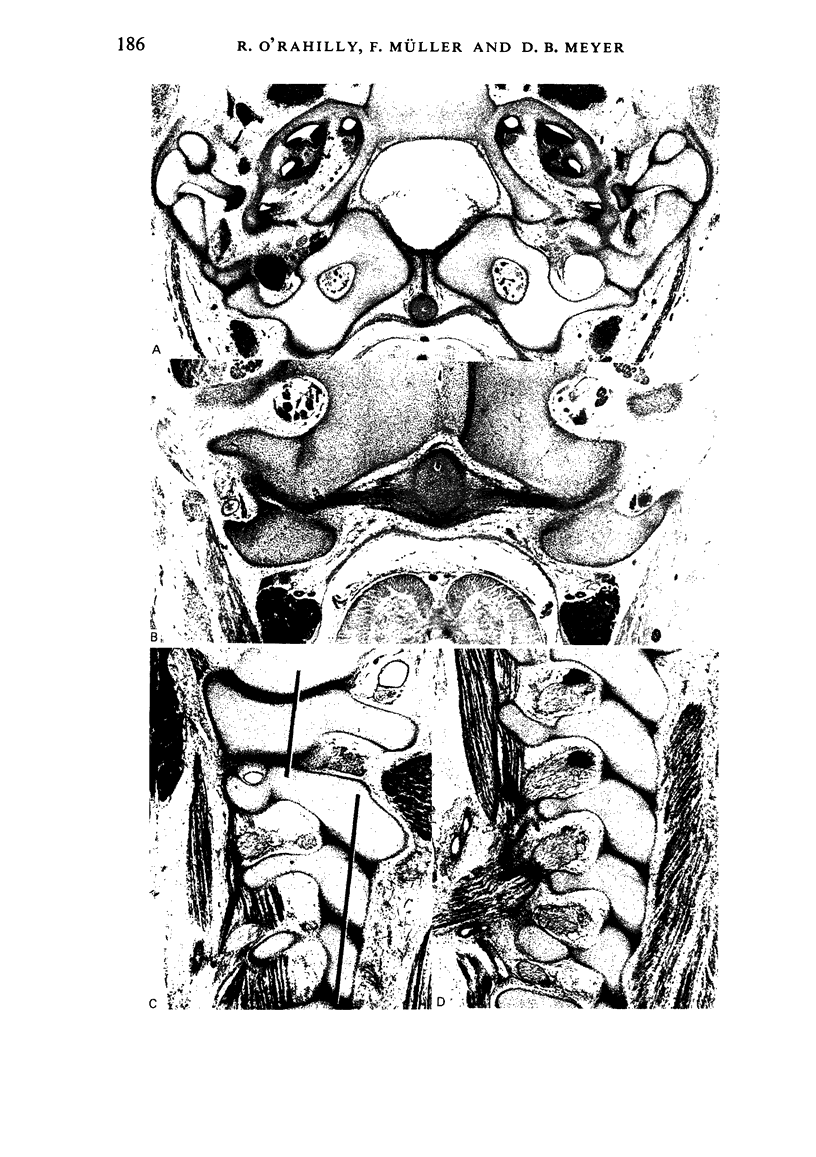
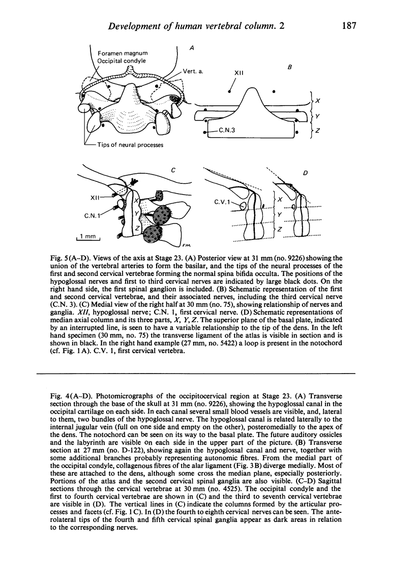
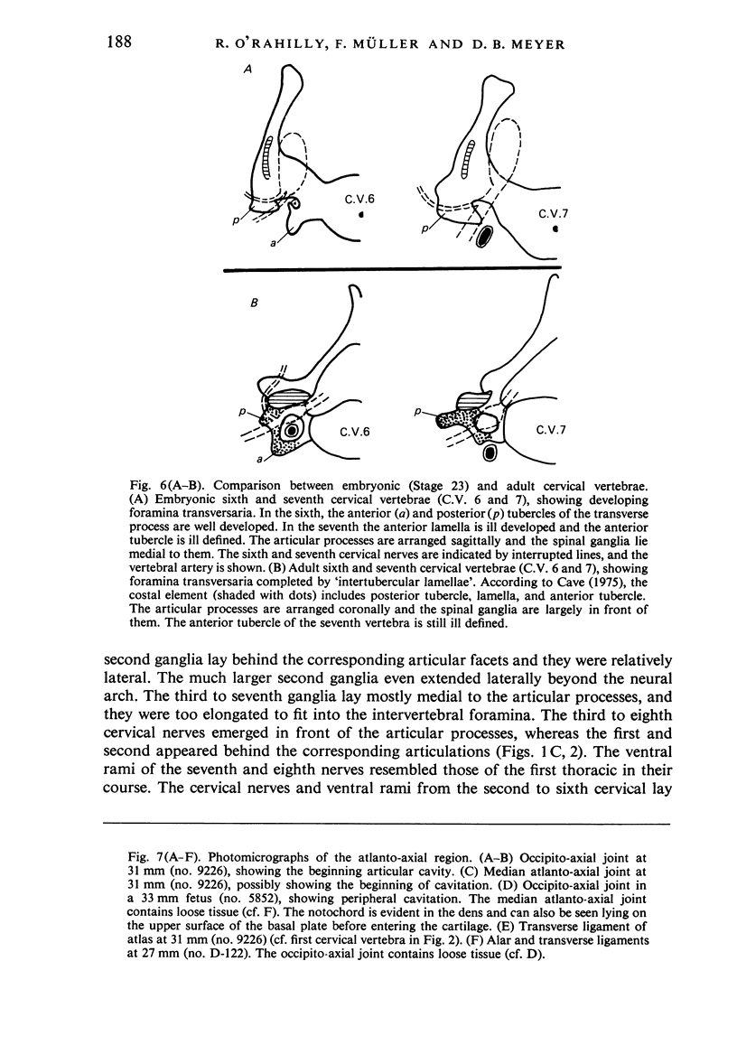
Abstract
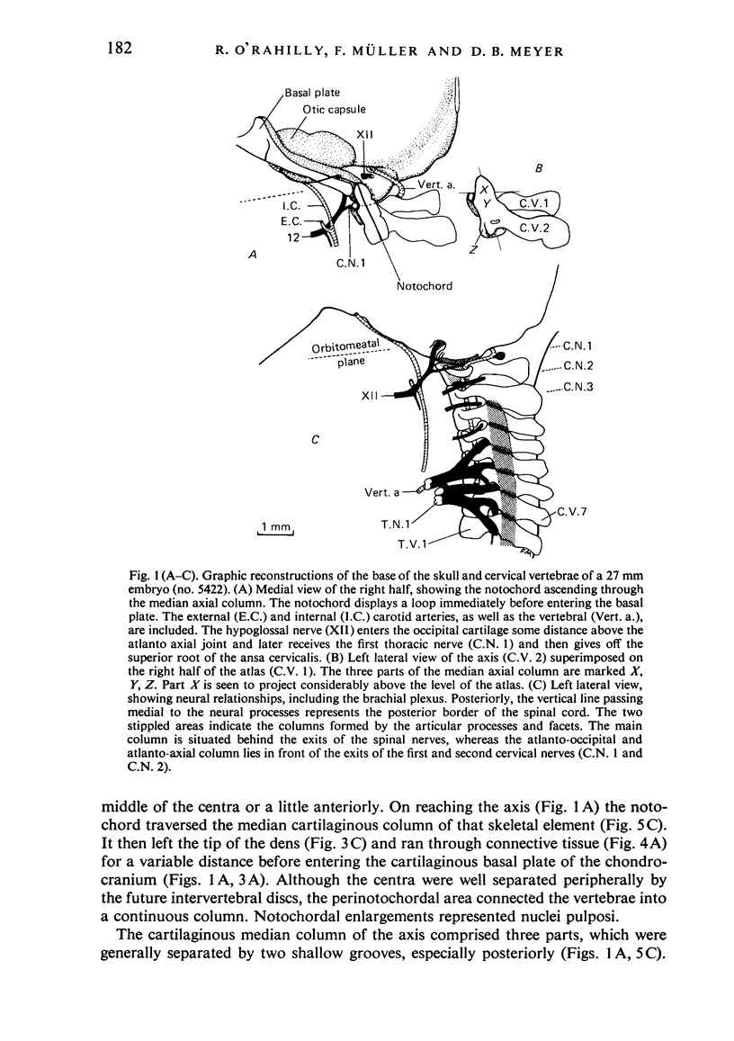
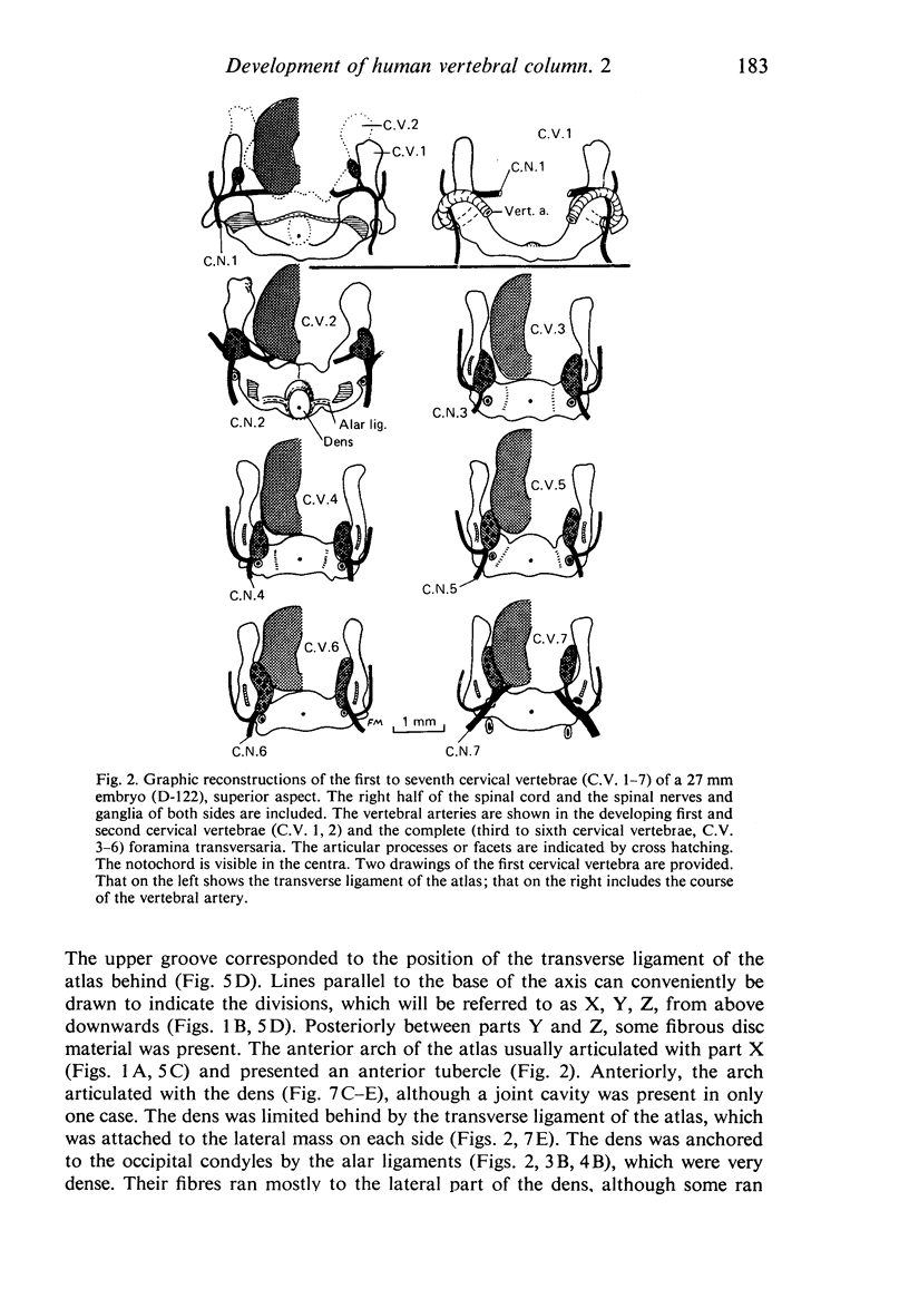
The present investigation of the cervical region of the vertebral column at eight post-ovulatory weeks is the first such study based on precise reconstructions of staged embryos. At the end of the embryonic period proper, a typical vertebra is a U-shaped piece of cartilage characterized by spina bifida occulta. The notochord ascends through the centra and leaves the dens to enter the basal plate of the skull. The median column of the axis comprises three parts (designated X, Y, Z) which persist well into the fetal period. They are related to the first, second and third cervical nerves, respectively. Part X may project into the foramen magnum and form an occipito-axial joint. Part Z appears to be the centrum of the axis. The articular columns of the cervical vertebrae are twofold, as in the adult: an anterior (atlanto-occipital and atlanto-axial) and a posterior (from the lower aspect of the axis downwards). Alar and transverse ligaments are present. Cavitation is not found in the embryonic period in either the atlanto-occipital or zygapophysial joints, and is generally not present in the median atlanto-axial joint either. Most of the transverse processes exhibit anterior and posterior tubercles. An 'intertubercular lamella' may or may not be present, i.e. the foramina transversaria are being formed around the vertebral artery. The spinal ganglia are generally partly in the vertebral canal and partly on the neural arches, medial to the articular processes. During the fetal period, the articular processes shift to a coronal position and this alteration appears to be associated with a corresponding change in the location of the spinal ganglia.
Full text
PDF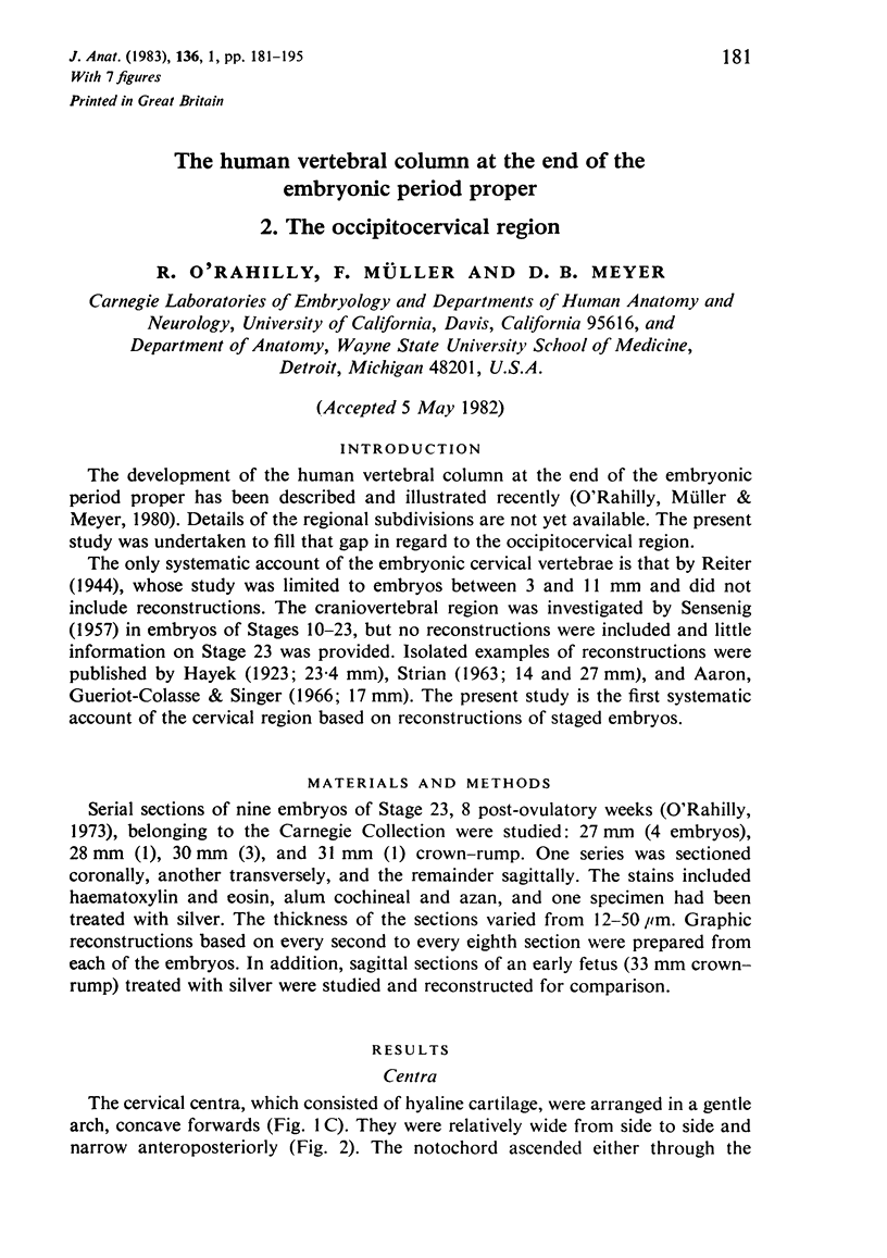
Images in this article

Selected References
These references are in PubMed. This may not be the complete list of references from this article.
- Fischer L., Neidhardt J. H., Gerentes R., Spay G., Giraud M. Structure macroscopique de l'apophyse odontoide d'après l'étude anatomo-radiologique. Lyon Med. 1969 Sep 28;222(34):433–passim. [PubMed] [Google Scholar]
- GANGULY D. N., ROY K. K. A STUDY ON THE CRANIO-VERTEBRAL JOINT IN THE MAN. Anat Anz. 1964 Jun 20;114:433–452. [PubMed] [Google Scholar]
- Gladstone R J, Erichsen-Powell W. Manifestation of Occipital Vertebrae, and Fusion of the Atlas with the Occipital Bone. J Anat Physiol. 1915 Jan;49(Pt 2):190–209. [PMC free article] [PubMed] [Google Scholar]
- Gladstone R. J., Wakeley C. P. Variations of the Occipito-Atlantal Joint in Relation to the Metameric Structure of the Cranio-Vertebral Region. J Anat. 1925 Jan;59(Pt 2):195–216. [PMC free article] [PubMed] [Google Scholar]
- Jenkins F. A., Jr The evolution and development of the dens of the mammalian axis. Anat Rec. 1969 Jun;164(2):173–184. doi: 10.1002/ar.1091640205. [DOI] [PubMed] [Google Scholar]
- LUDWIG K. S. Die Frühentwicklung des Atlas und der Occipitalwirbel beim Menschen. Acta Anat (Basel) 1957;30(1-4):444–461. [PubMed] [Google Scholar]
- Müller F., O'Rahilly R. The human chondrocranium at the end of the embryonic period, proper, with particular reference to the nervous system. Am J Anat. 1980 Sep;159(1):33–58. doi: 10.1002/aja.1001590105. [DOI] [PubMed] [Google Scholar]
- NOBACK C. R., ROBERTSON G. G. Sequences of appearance of ossification centers in the human skeleton during the first five prenatal months. Am J Anat. 1951 Jul;89(1):1–28. doi: 10.1002/aja.1000890102. [DOI] [PubMed] [Google Scholar]
- O'Rahilly R., Meyer D. B. The timing and sequence of events in the development of the human vertebral column during the embryonic period proper. Anat Embryol (Berl) 1979 Oct;157(2):167–176. doi: 10.1007/BF00305157. [DOI] [PubMed] [Google Scholar]
- O'Rahilly R., Muller F., Meyer D. B. The human vertebral column at the end of the embryonic period proper. 1. The column as a whole. J Anat. 1980 Oct;131(Pt 3):565–575. [PMC free article] [PubMed] [Google Scholar]
- STRIAN F. [On growth tendencies in the fetal spine, demonstrated on 2 model series]. Anat Anz. 1963 Jun 29;112:389–408. [PubMed] [Google Scholar]
- Singh-Roy K. K. On Goethe's vertebral theory of the origin of the skull--a recent approach. Anat Anz. 1967;120(3):250–259. [PubMed] [Google Scholar]
- Tilmann B., Lorenz R. The stress at the human atlanto-occipital joint. I. the development of the occipital condyle. Anat Embryol (Berl) 1978 Jun 12;153(3):269–277. doi: 10.1007/BF00315929. [DOI] [PubMed] [Google Scholar]
- ZAWISCH C. Der Ossifikationsprozess des Occipitale und die Rolle des Tectum posterius beim Menschen. Acta Anat (Basel) 1957;30(1-4):988–1007. [PubMed] [Google Scholar]