

Abstract
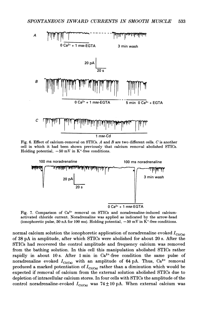
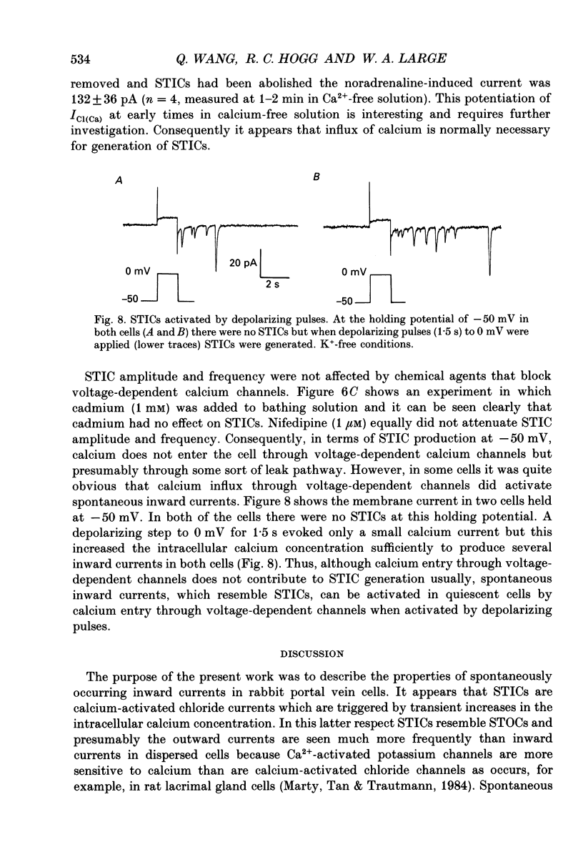
1. Characteristics of spontaneous transient inward currents (STICs) which produced membrane depolarization were analysed with the perforated patch technique in single smooth muscle cells isolated from the rabbit portal vein. 2. In K(+)-free solutions the amplitude of STICs was linearly related to membrane potential and the reversal potential (Er) was -3.0 +/- 0.9 mV. Replacement of external NaCl with NaI shifted Er to -40.0 +/- 1.0 mV. Substitution of external NaCl by NaSCN also moved Er to negative values but replacement of sodium with Tris and choline did not change Er. It is concluded that STICs are generated by an increase in chloride conductance. 3. STICs were abolished or reduced by the chloride channel antagonists anthracene-9-carboxylic acid (1 mM) and 4-acetamido-4'-isothiocyanato-2,2'- stilbene-disulphonic acid (2 mM). 4. STICs were blocked by noradrenaline and caffeine which deplete intracellular calcium stores. This effect was reversible and this result indicates that the primary trigger for STICs is calcium released from intracellular stores and therefore STICs are calcium-activated chloride currents (ICl(Ca)). 5. Removal of calcium from the bathing solution abolished STICs in six out of seven cells but STICs persisted in Ca(2+)-free solution in one cell. When STICs were abolished in Ca(2+)-free external solution the size of the internal calcium store, as estimated from the noradrenaline-induced ICl(Ca), was not altered. It appears that an influx of calcium is usually necessary for STICs to be observed. 6. The frequency and amplitude of STICs were not altered by the voltage-dependent calcium channel antagonist cadmium (1 mM). However, in some quiescent cells influx of calcium through voltage-dependent channels did activate STICs. 7. It was concluded that in isolated portal vein cells STICs represent a Ca(2+)-activated chloride current which leads to spontaneous depolarization of the membrane and may play an important physiological or pathophysiological role to produce smooth muscle contraction.
Full text
PDF


Selected References
These references are in PubMed. This may not be the complete list of references from this article.
- Aickin C. C., Brading A. F. Measurement of intracellular chloride in guinea-pig vas deferens by ion analysis, 36chloride efflux and micro-electrodes. J Physiol. 1982 May;326:139–154. doi: 10.1113/jphysiol.1982.sp014182. [DOI] [PMC free article] [PubMed] [Google Scholar]
- Amédée T., Large W. A., Wang Q. Characteristics of chloride currents activated by noradrenaline in rabbit ear artery cells. J Physiol. 1990 Sep;428:501–516. doi: 10.1113/jphysiol.1990.sp018224. [DOI] [PMC free article] [PubMed] [Google Scholar]
- Beech D. J., Bolton T. B. Two components of potassium current activated by depolarization of single smooth muscle cells from the rabbit portal vein. J Physiol. 1989 Nov;418:293–309. doi: 10.1113/jphysiol.1989.sp017841. [DOI] [PMC free article] [PubMed] [Google Scholar]
- Benham C. D., Bolton T. B., Byrne N. G., Large W. A. Action of externally applied adenosine triphosphate on single smooth muscle cells dispersed from rabbit ear artery. J Physiol. 1987 Jun;387:473–488. doi: 10.1113/jphysiol.1987.sp016585. [DOI] [PMC free article] [PubMed] [Google Scholar]
- Benham C. D., Bolton T. B. Spontaneous transient outward currents in single visceral and vascular smooth muscle cells of the rabbit. J Physiol. 1986 Dec;381:385–406. doi: 10.1113/jphysiol.1986.sp016333. [DOI] [PMC free article] [PubMed] [Google Scholar]
- Byrne N. G., Large W. A. Membrane ionic mechanisms activated by noradrenaline in cells isolated from the rabbit portal vein. J Physiol. 1988 Oct;404:557–573. doi: 10.1113/jphysiol.1988.sp017306. [DOI] [PMC free article] [PubMed] [Google Scholar]
- Clapp L. H., Gurney A. M. Outward currents in rabbit pulmonary artery cells dissociated with a new technique. Exp Physiol. 1991 Sep;76(5):677–693. doi: 10.1113/expphysiol.1991.sp003535. [DOI] [PubMed] [Google Scholar]
- Holman M. E., Kasby C. B., Suthers M. B., Wilson J. A. Some properties of the smooth muscle of rabbit portal vein. J Physiol. 1968 May;196(1):111–132. doi: 10.1113/jphysiol.1968.sp008498. [DOI] [PMC free article] [PubMed] [Google Scholar]
- Hume J. R., Leblanc N. Macroscopic K+ currents in single smooth muscle cells of the rabbit portal vein. J Physiol. 1989 Jun;413:49–73. doi: 10.1113/jphysiol.1989.sp017641. [DOI] [PMC free article] [PubMed] [Google Scholar]
- Marty A., Tan Y. P., Trautmann A. Three types of calcium-dependent channel in rat lacrimal glands. J Physiol. 1984 Dec;357:293–325. doi: 10.1113/jphysiol.1984.sp015501. [DOI] [PMC free article] [PubMed] [Google Scholar]
- Ohya Y., Terada K., Yamaguchi K., Inoue R., Okabe K., Kitamura K., Hirata M., Kuriyama H. Effects of inositol phosphates on the membrane activity of smooth muscle cells of the rabbit portal vein. Pflugers Arch. 1988 Sep;412(4):382–389. doi: 10.1007/BF01907556. [DOI] [PubMed] [Google Scholar]
- Van Helden D. F. Spontaneous and noradrenaline-induced transient depolarizations in the smooth muscle of guinea-pig mesenteric vein. J Physiol. 1991 Jun;437:511–541. doi: 10.1113/jphysiol.1991.sp018609. [DOI] [PMC free article] [PubMed] [Google Scholar]
- Wang Q., Large W. A. Modulation of noradrenaline-induced membrane currents by papaverine in rabbit vascular smooth muscle cells. J Physiol. 1991 Aug;439:501–512. doi: 10.1113/jphysiol.1991.sp018678. [DOI] [PMC free article] [PubMed] [Google Scholar]
- Wang Q., Large W. A. Noradrenaline-evoked cation conductance recorded with the nystatin whole-cell method in rabbit portal vein cells. J Physiol. 1991 Apr;435:21–39. doi: 10.1113/jphysiol.1991.sp018496. [DOI] [PMC free article] [PubMed] [Google Scholar]