Abstract
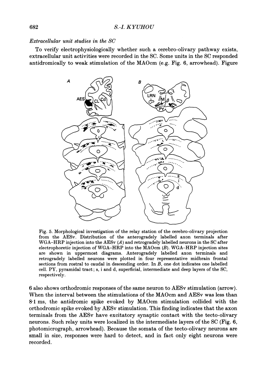
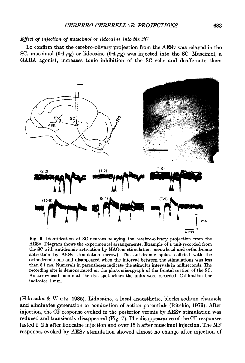
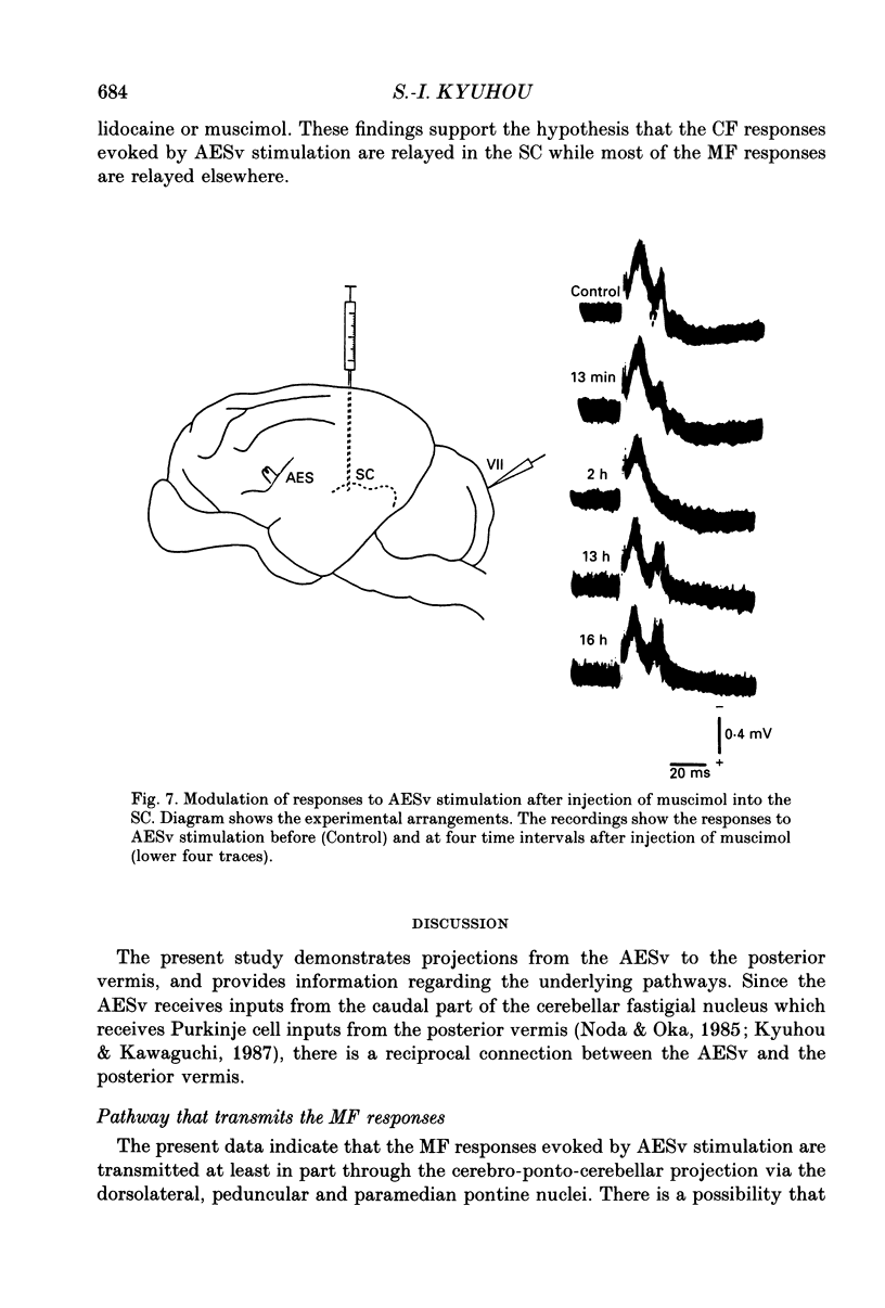
1. Stimulation of the ventral bank of the anterior ectosylvian sulcus (AESv) induced marked mossy fibre (MF) and climbing fibre (CF) responses in the cerebellar posterior vermis (lobules VI-VII) and moderate sized ones in the paraflocculus, paramedian lobules and crus I and II of the cat. The relay stations for these responses to the posterior vermis were investigated morphologically and electrophysiologically. 2. It can be considered that the MF responses were relayed at least in part via the dorsolateral, peduncular and paramedian pontine nuclei, since in these nuclei there were units orthodromically responsive to AESv stimulation and antidromically responsive to stimulation of the posterior vermis. The MF responses are thought to be relayed monosynaptically, since the distribution of axon terminals labelled after injection of wheatgerm agglutinin-conjugated peroxidase (WGA-HRP) into the AESv overlapped in these pontine nuclei with that of neurons labelled after injection of WGA-HRP into the posterior vermis. 3. It is thought that the CF responses are relayed in the caudomedial part of the medial accessory olive (MAOcm), because neurons in the MAOcm were orthodromically responsive to AESv stimulation and antidromically responsive to stimulation of the posterior vermis. 4. It is suggested that the cerebro-olivary projection which transmits the orthodromic responses in the MAOcm is indirect, via the superior colliculus (SC), because injection of WGA-HRP into the AESv labelled axon terminals not in the MAOcm but in the SC, and injection of WGA-HRP into the MAOcm gave rise to retrograde labelling of cells in the SC. Synaptic connections between the axon terminals of the cerebrotectal projection and the tecto-olivary neurons were demonstrated by extracellular unit studies in the SC. 5. The hypothesis that the CF responses were transmitted via the SC was supported by the finding that the CF responses disappeared transiently after muscimol or lidocaine was injected into the SC. 6. These findings provide evidence that the MF responses are transmitted at least in part via the cerebro-ponto-cerebellar projection, while the CF responses are relayed via the cerebro-tecto-olivo-cerebellar projection. These cerebro-cerebellar pathways from the AESv are suggested to participate in conducting visual information to the posterior vermis.
Full text
PDF
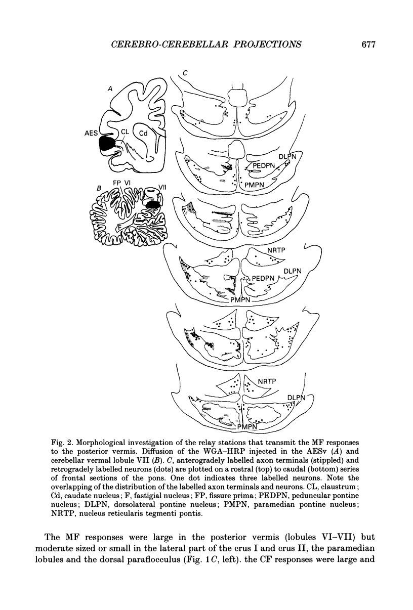
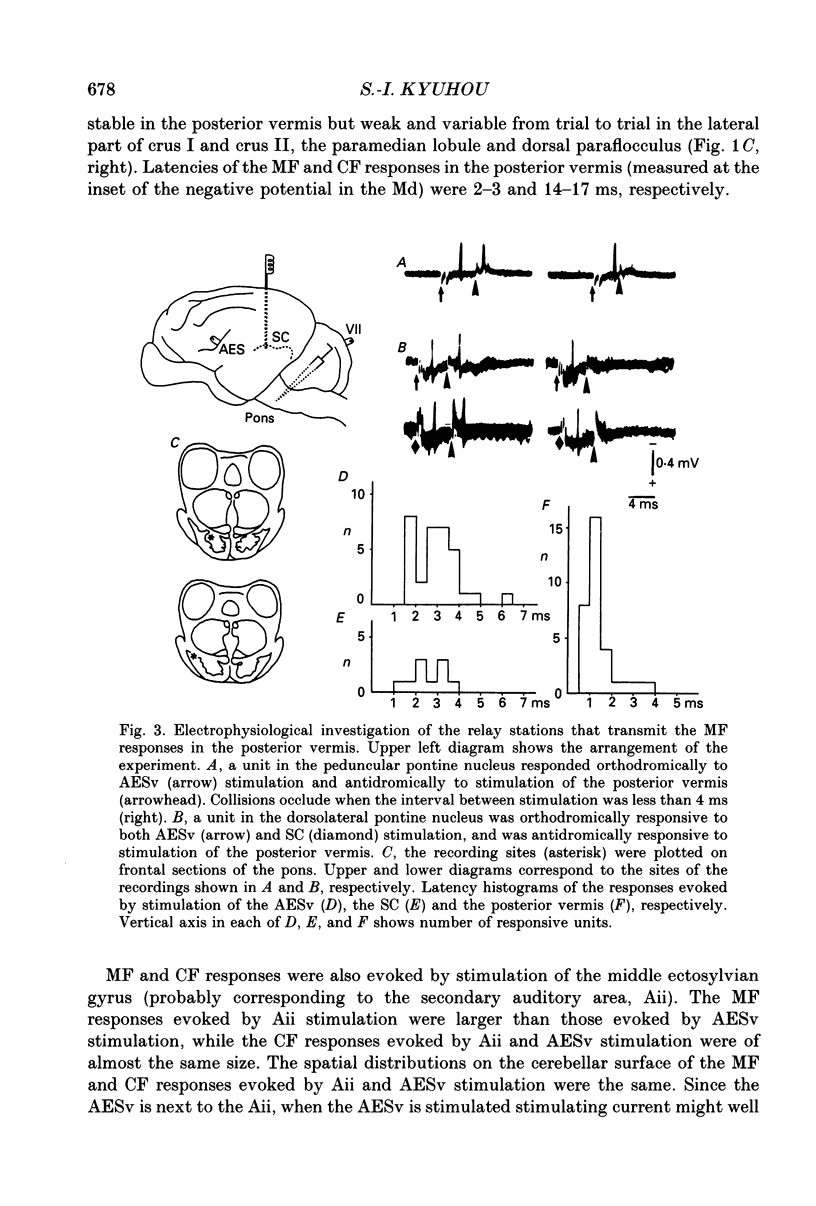
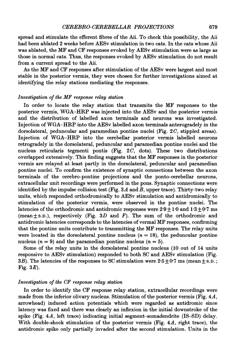
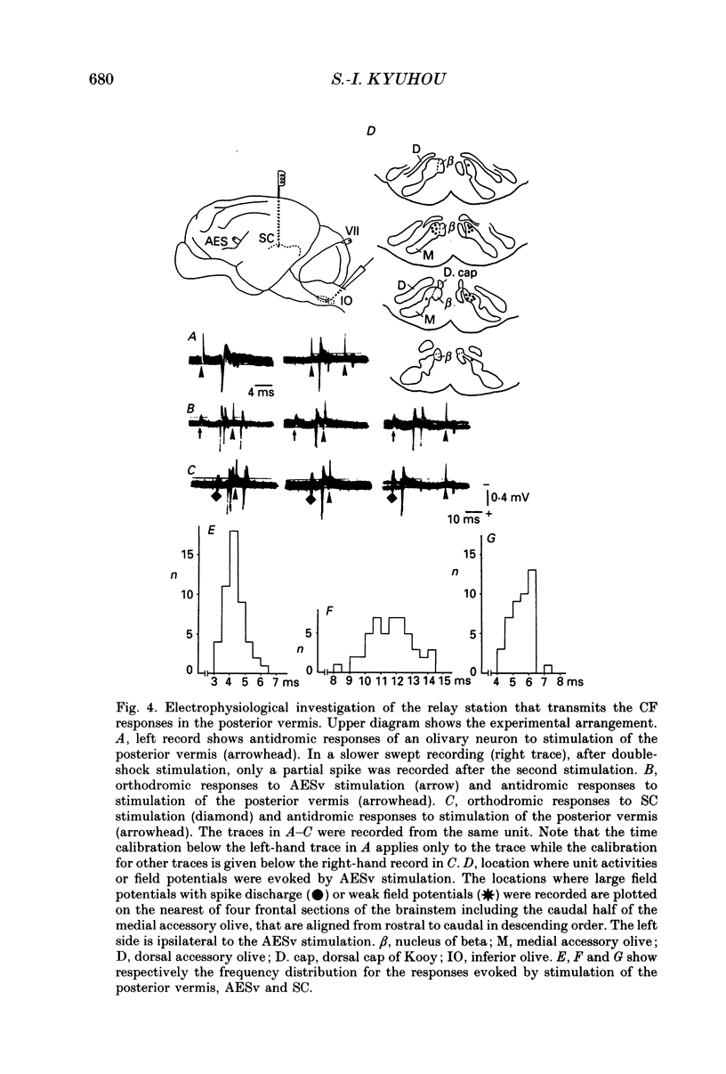
Images in this article
Selected References
These references are in PubMed. This may not be the complete list of references from this article.
- Armstrong B. D., Harvey R. J. Responses in the inferior olive to stimulation of the cerebellar and cerebral cortices in the cat. J Physiol. 1966 Dec;187(3):553–574. doi: 10.1113/jphysiol.1966.sp008108. [DOI] [PMC free article] [PubMed] [Google Scholar]
- Bishop G. A., McCrea R. A., Kitai S. T. A horseradish peroxidase study of the cortico-olivary projection in the cat. Brain Res. 1976 Nov 5;116(2):306–311. doi: 10.1016/0006-8993(76)90908-2. [DOI] [PubMed] [Google Scholar]
- Courville J., Diakiw N. Cerebellar corticonuclear projection in the cat. The vermis of the anterior and posterior lobes. Brain Res. 1976 Jun 25;110(1):1–20. doi: 10.1016/0006-8993(76)90205-5. [DOI] [PubMed] [Google Scholar]
- Eccles J. C., Llinás R., Sasaki K. The mossy fibre-granule cell relay of the cerebellum and its inhibitory control by Golgi cells. Exp Brain Res. 1966;1(1):82–101. doi: 10.1007/BF00235211. [DOI] [PubMed] [Google Scholar]
- Eccles J. C., Provini L., Strata P., Táboríková H. Analysis of electrical potentials evoked in the cerebellar anterior lobe by stimulation of hindlimb and forelimb nerves. Exp Brain Res. 1968;6(3):171–194. doi: 10.1007/BF00235123. [DOI] [PubMed] [Google Scholar]
- Hicks T. P., Benedek G., Thurlow G. A. Organization and properties of neurons in a visual area within the insular cortex of the cat. J Neurophysiol. 1988 Aug;60(2):397–421. doi: 10.1152/jn.1988.60.2.397. [DOI] [PubMed] [Google Scholar]
- Kato N., Kawaguchi S., Miyata H. Cerebro-cerebellar projections from the lateral suprasylvian visual area in the cat. J Physiol. 1988 Jan;395:473–485. doi: 10.1113/jphysiol.1988.sp016930. [DOI] [PMC free article] [PubMed] [Google Scholar]
- Kato N., Kawaguchi S., Miyata H. Post-natal development of the retinal and cerebellar projections onto the lateral suprasylvian area in the cat. J Physiol. 1987 Feb;383:729–743. doi: 10.1113/jphysiol.1987.sp016438. [DOI] [PMC free article] [PubMed] [Google Scholar]
- Kawamura K., Brodal A. The tectopontine projection in the cat: an experimental anatomical study with comments on pathweays for teleceptive impulses to the cerebellum. J Comp Neurol. 1973 Jun 1;149(3):371–390. doi: 10.1002/cne.901490306. [DOI] [PubMed] [Google Scholar]
- Kyuhou S., Kawaguchi S. Cerebellocerebral projection from the fastigial nucleus onto the frontal eye field and anterior ectosylvian visual area in the cat. J Comp Neurol. 1987 May 22;259(4):571–590. doi: 10.1002/cne.902590407. [DOI] [PubMed] [Google Scholar]
- Llinas R., Baker R., Sotelo C. Electrotonic coupling between neurons in cat inferior olive. J Neurophysiol. 1974 May;37(3):560–571. doi: 10.1152/jn.1974.37.3.560. [DOI] [PubMed] [Google Scholar]
- Mizuno N., Mochizuki K., Akimoto C., Matsushima R., Sasaki K. Projections from the parietal cortex to the brain stem nuclei in the cat, with special reference to the parietal cerebro-cerebellar system. J Comp Neurol. 1973 Feb 15;147(4):511–522. doi: 10.1002/cne.901470406. [DOI] [PubMed] [Google Scholar]
- Mower G., Gibson A., Glickstein M. Tectopontine pathway in the cat: laminar distribution of cells of origin and visual properties of target cells in dorsolateral pontine nucleus. J Neurophysiol. 1979 Jan;42(1 Pt 1):1–15. doi: 10.1152/jn.1979.42.1.1. [DOI] [PubMed] [Google Scholar]
- Mucke L., Norita M., Benedek G., Creutzfeldt O. Physiologic and anatomic investigation of a visual cortical area situated in the ventral bank of the anterior ectosylvian sulcus of the cat. Exp Brain Res. 1982;46(1):1–11. doi: 10.1007/BF00238092. [DOI] [PubMed] [Google Scholar]
- Nakamura Y., Kitao Y., Okoyama S. Cortico-Darkschewitsch-olivary projection in the cat: an electron microscope study with the aid of horseradish peroxidase tracing technique. Brain Res. 1983 Sep 5;274(1):140–143. doi: 10.1016/0006-8993(83)90529-2. [DOI] [PubMed] [Google Scholar]
- Noda H., Fujikado T. Involvement of Purkinje cells in evoking saccadic eye movements by microstimulation of the posterior cerebellar vermis of monkeys. J Neurophysiol. 1987 May;57(5):1247–1261. doi: 10.1152/jn.1987.57.5.1247. [DOI] [PubMed] [Google Scholar]
- Noda T., Oka H. Fastigial inputs to the insular cortex in the cat: field potential analysis. Neurosci Lett. 1985 Feb 4;53(3):331–336. doi: 10.1016/0304-3940(85)90560-9. [DOI] [PubMed] [Google Scholar]
- Oka H., Jinnai K. Electrophysiological study of parvocellular red nucleus neurons. Brain Res. 1978 Jun 23;149(1):239–246. doi: 10.1016/0006-8993(78)90605-4. [DOI] [PubMed] [Google Scholar]
- Oka H., Jinnai K., Yamamoto T. The parieto-rubro-olivary pathway in the cat. Exp Brain Res. 1979 Sep;37(1):115–125. doi: 10.1007/BF01474258. [DOI] [PubMed] [Google Scholar]
- Ritchie J. M. A pharmacological approach to the structure of sodium channels in myelinated axons. Annu Rev Neurosci. 1979;2:341–362. doi: 10.1146/annurev.ne.02.030179.002013. [DOI] [PubMed] [Google Scholar]
- Ron S., Robinson D. A. Eye movements evoked by cerebellar stimulation in the alert monkey. J Neurophysiol. 1973 Nov;36(6):1004–1022. doi: 10.1152/jn.1973.36.6.1004. [DOI] [PubMed] [Google Scholar]
- Sasaki K., Matsuda Y., Kawaguchi S., Mizuno N. On the cerebello-thalamo-cerebral pathway for the parietal cortex. Exp Brain Res. 1972;16(1):89–103. doi: 10.1007/BF00233376. [DOI] [PubMed] [Google Scholar]
- Sasaki K., Oka H., Matsuda Y., Shimono T., Mizuno N. Electrophysiological studies of the projections from the parietal association area to the cerebellar cortex. Exp Brain Res. 1975 Jul 11;23(1):91–102. doi: 10.1007/BF00238732. [DOI] [PubMed] [Google Scholar]
- Tamai Y., Miyashita E., Nakai M. Eye movements following cortical stimulation in the ventral bank of the anterior ectosylvian sulcus of the cat. Neurosci Res. 1989 Nov;7(2):159–163. doi: 10.1016/0168-0102(89)90056-4. [DOI] [PubMed] [Google Scholar]


