Abstract
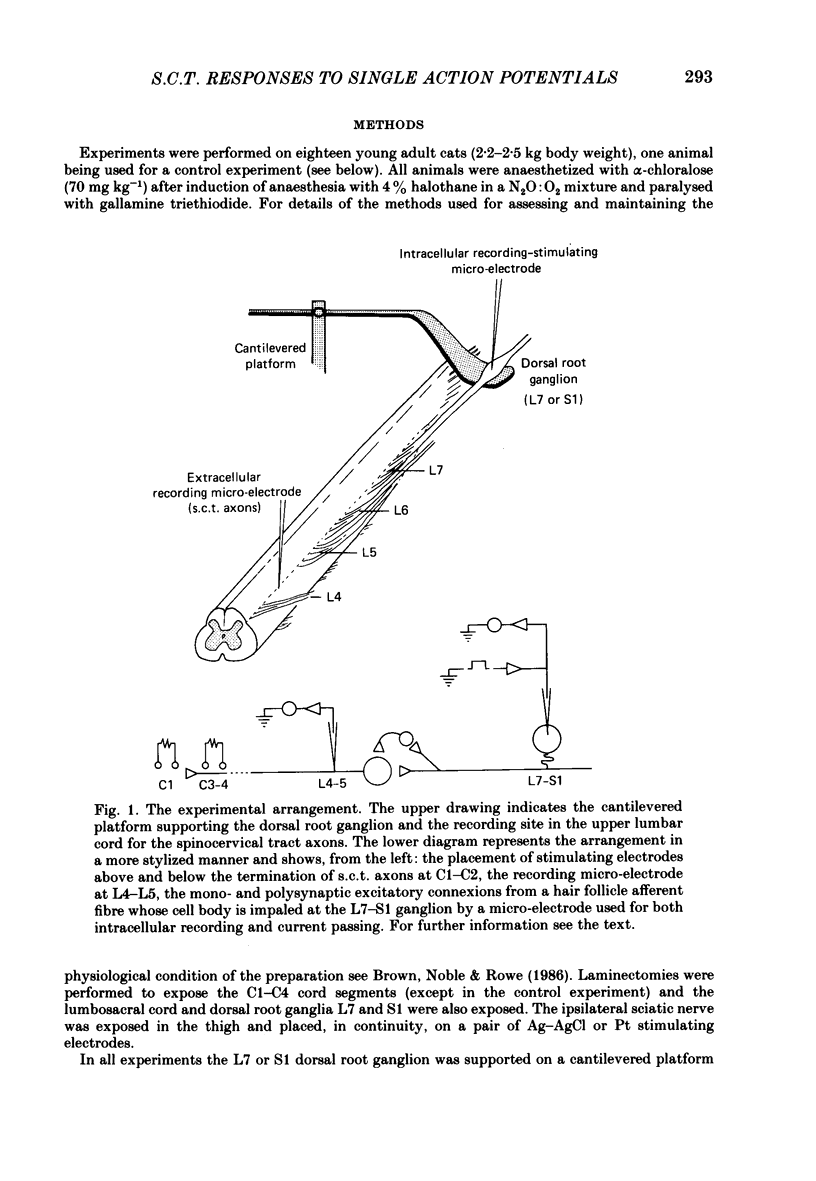
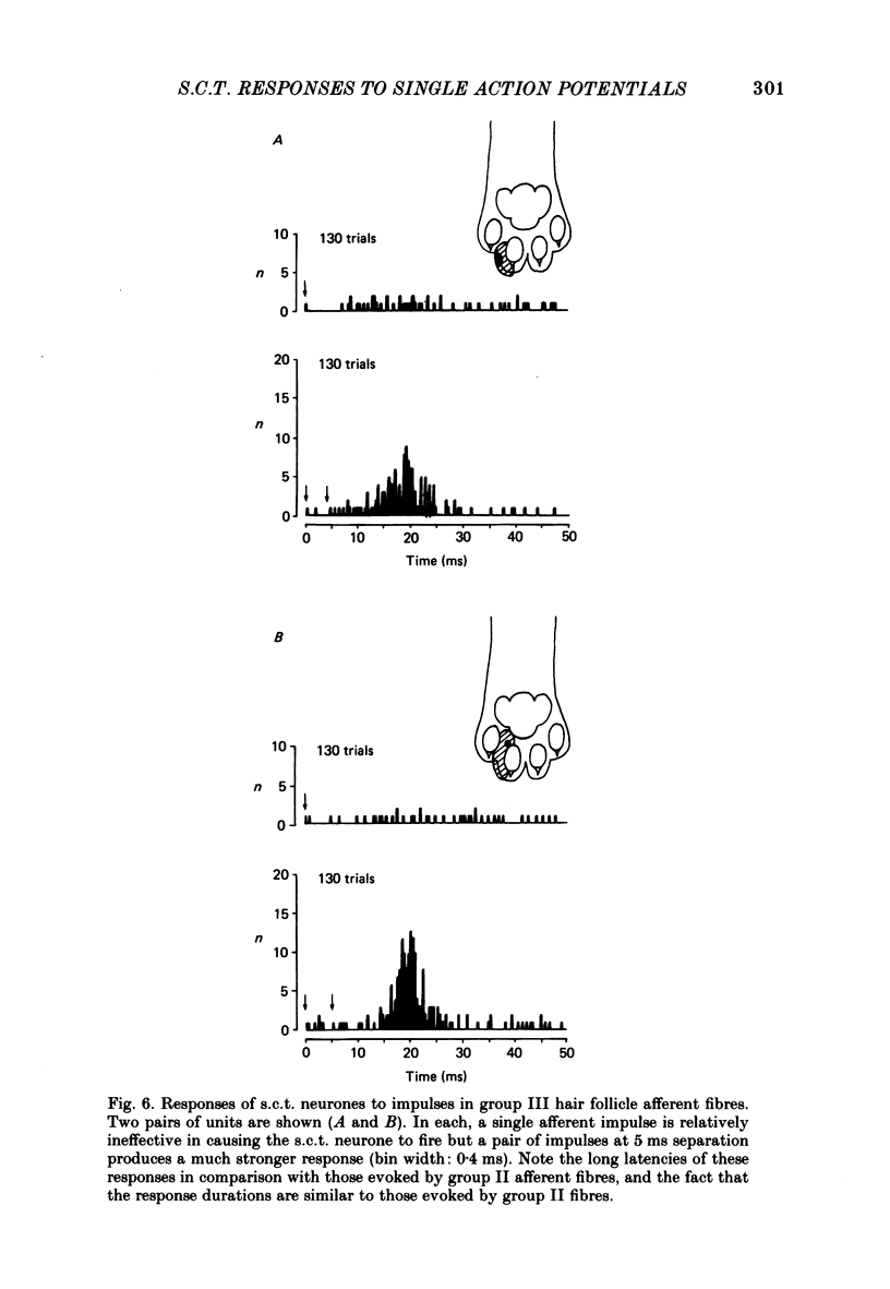
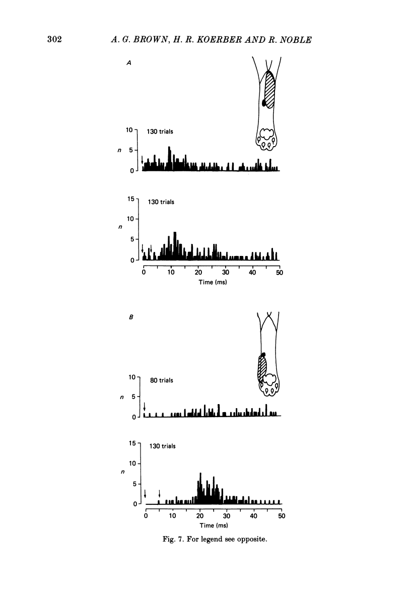
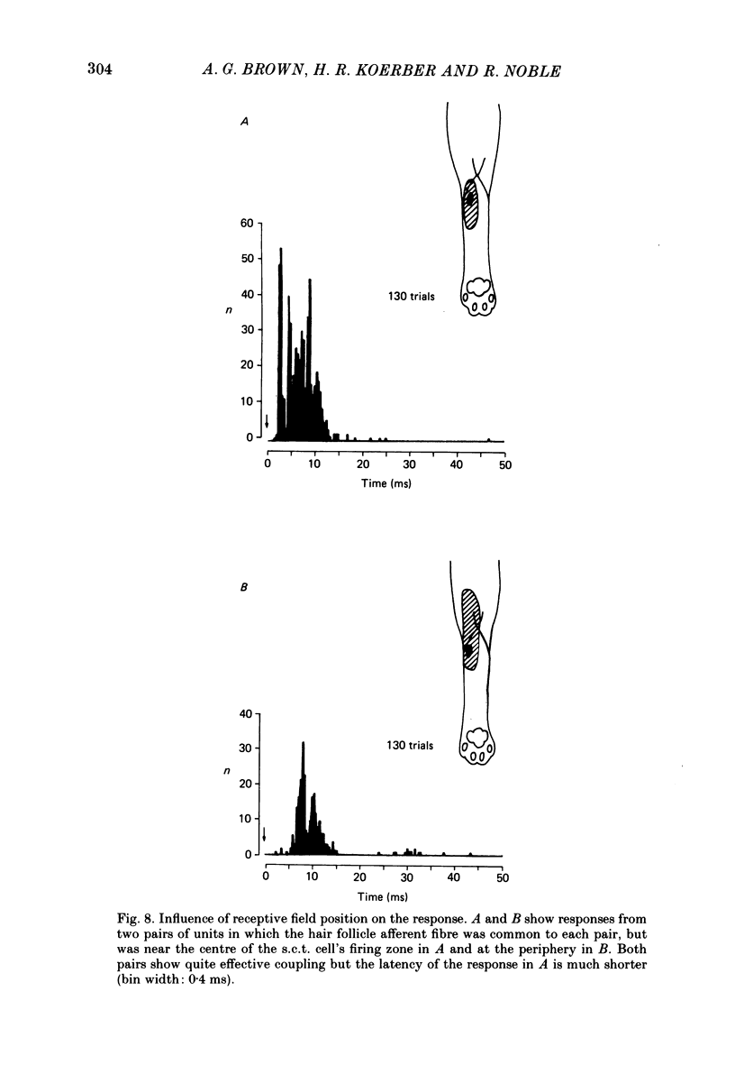
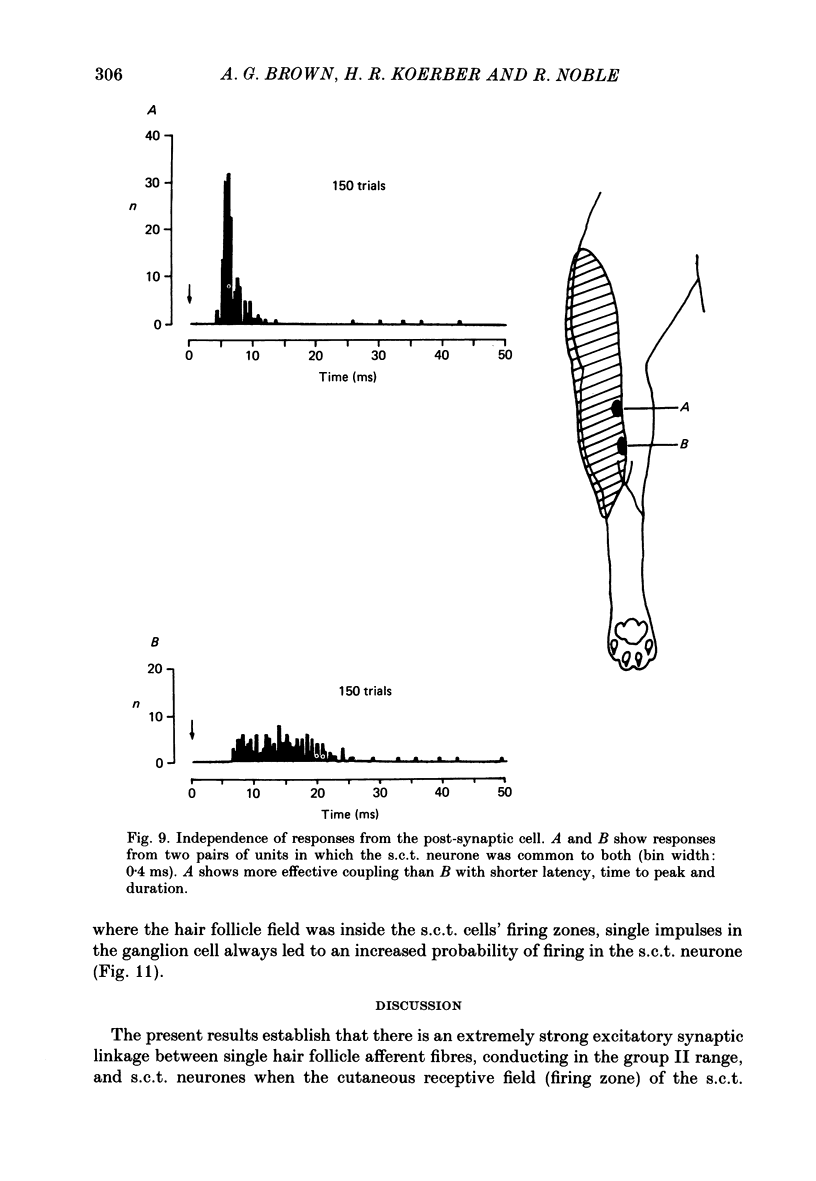
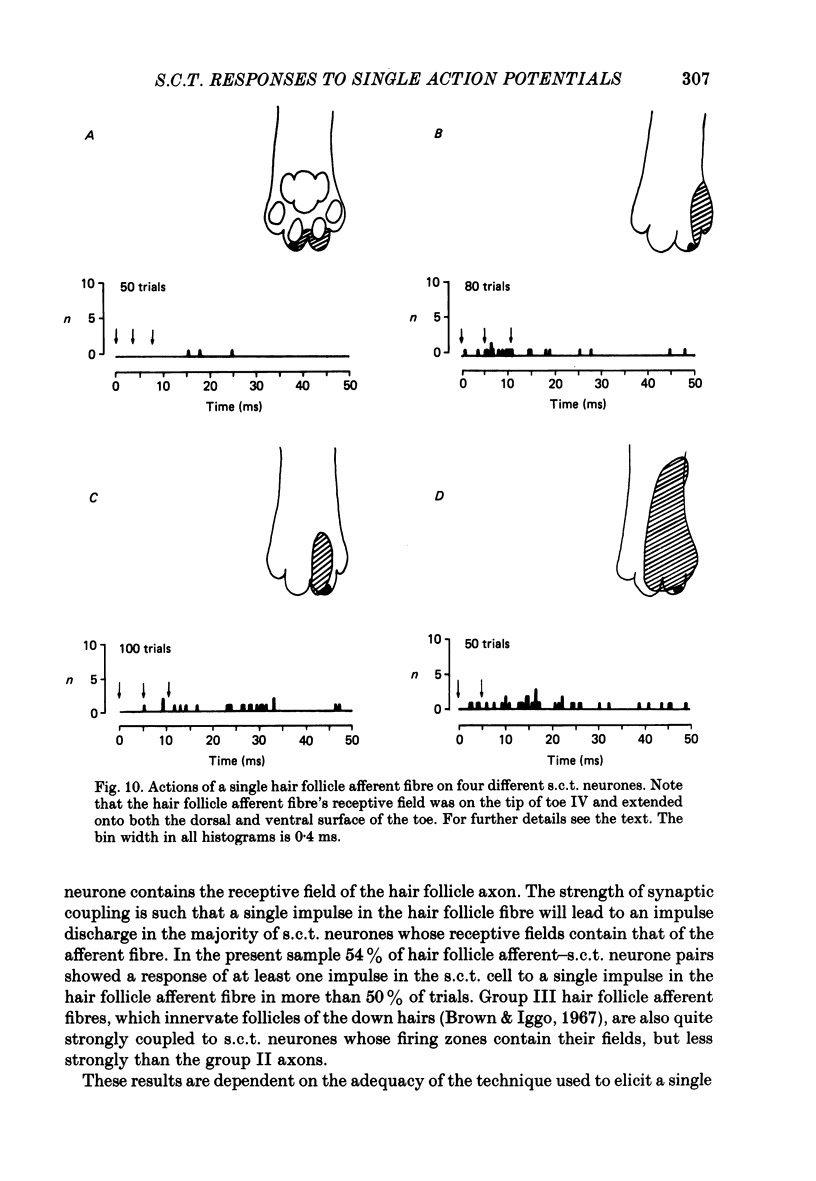
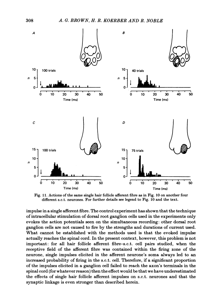
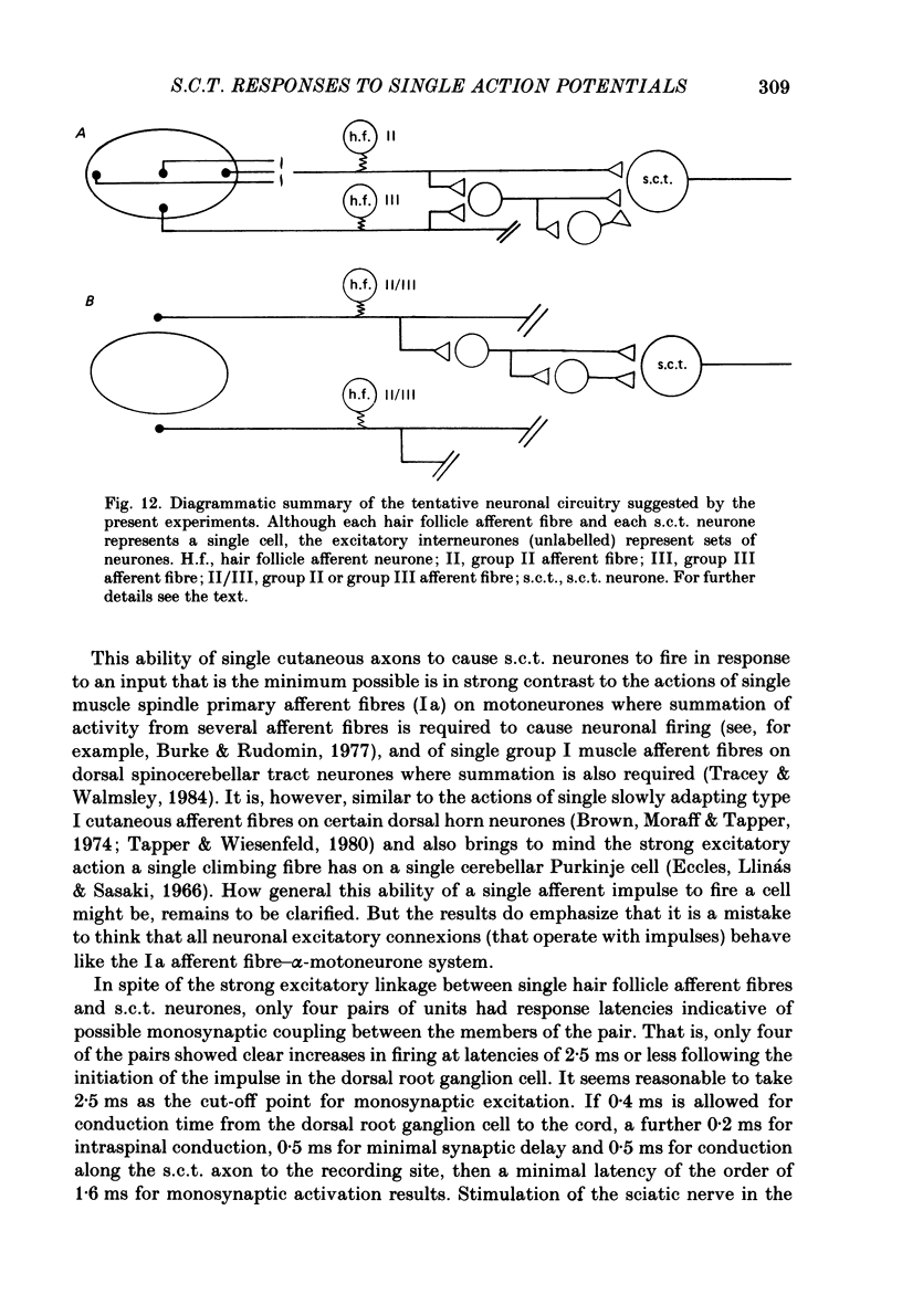
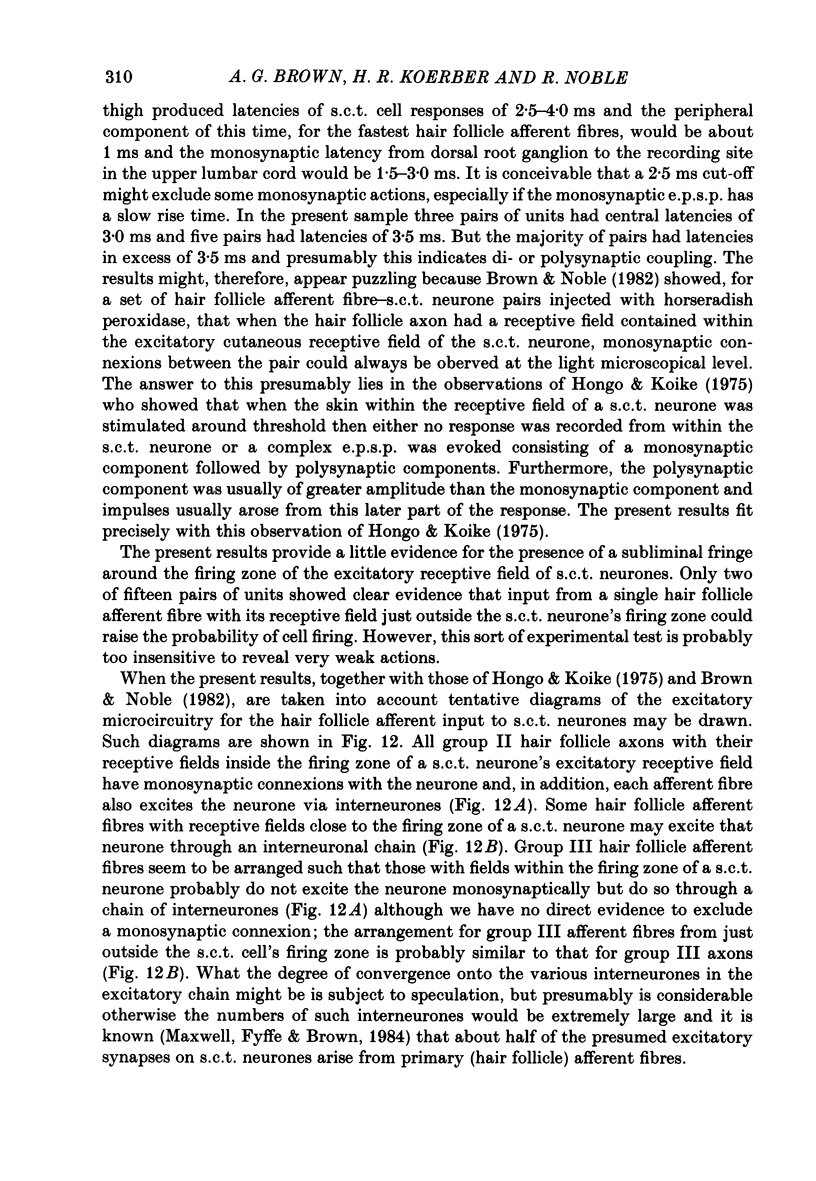
1. In cats under chloralose anaesthesia single dorsal root ganglion cells with axons innervating hair follicles were stimulated intracellularly to produce single impulses. At the same time single spinocervical tract (s.c.t.) neurones were recorded extracellularly, from their axons in the upper lumbar cord. 2. When the receptive field of the afferent fibre was contained within the impulse firing zone of the s.c.t. cell, a single afferent impulse increased the probability of firing of the neurone. In thirty-nine pairs of units, where the afferent fibre had a group II conduction velocity, coupling was very efficient and for seventeen pairs the single afferent impulse produced one or more impulses in the s.c.t. cell in at least 90% of trials. The mean number of impulses evoked in s.c.t. cells by a single group II afferent impulse was 1.47. The latencies of the impulses ranged from 1.5 to 14.0 ms, with times to peak and total durations of 2.5-17.5 ms and 4.5-28.0 ms respectively. For two pairs of units where the afferent fibre had a group III conduction velocity the effectiveness of single afferent impulses was much less and the latencies, but not the durations, of the impulses were longer (12 and 17 ms). 3. When the receptive field of the hair follicle afferent fibre was outside, but close to, the firing zone of the s.c.t. neurone there was no indication that single afferent impulses affected the probability of neuronal discharge for thirteen of fifteen pairs of units. Weak excitation was observed in two pairs and this was clear only when two or more afferent impulses were employed. 4. There was a tendency for hair follicle afferent fibres with their receptive fields at or near the centre of the s.c.t. cell's firing zone to be most effective, producing shorter latency responses with more impulses at higher frequencies. When the afferent's field was peripherally located in the s.c.t. neurone's firing zone there was a wide range of responses but these included those with the longest latencies and very few impulses. 5. The results are discussed with reference to previous work on the spinocervical tract and to the known actions of single impulses on other neuronal types. Suggestions are made for the possible excitatory neuronal circuits linking hair follicle afferent fibres to the s.c.t. neurones.
Full text
PDF
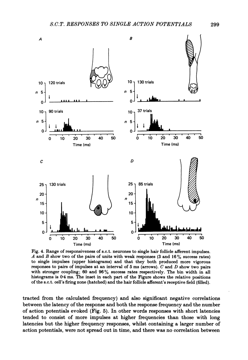
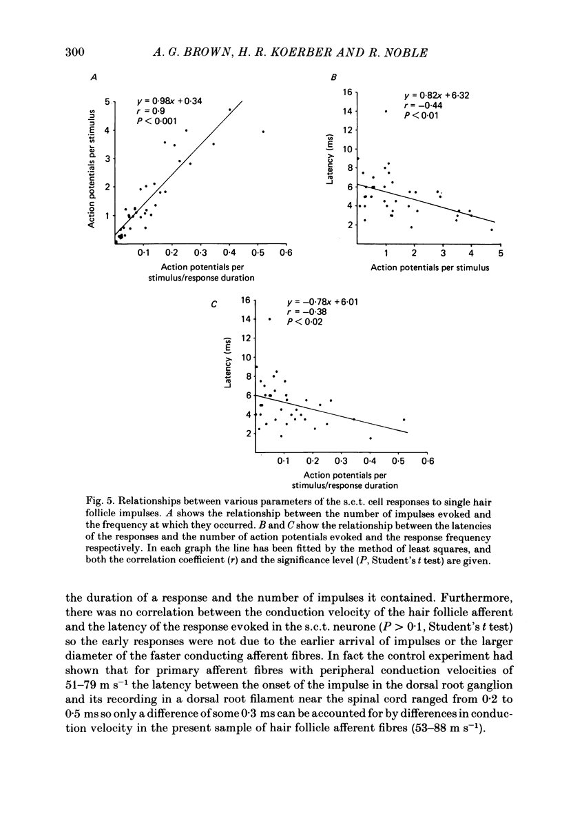

Selected References
These references are in PubMed. This may not be the complete list of references from this article.
- Brown A. G., Franz D. N. Responses of spinocervical tract neurones to natural stimulation of identified cutaneous receptors. Exp Brain Res. 1969;7(3):231–249. doi: 10.1007/BF00239031. [DOI] [PubMed] [Google Scholar]
- Brown A. G., Fyffe R. E., Noble R., Rose P. K., Snow P. J. The density, distribution and topographical organization of spinocervical tract neurones in the cat. J Physiol. 1980 Mar;300:409–428. doi: 10.1113/jphysiol.1980.sp013169. [DOI] [PMC free article] [PubMed] [Google Scholar]
- Brown A. G., Iggo A. A quantitative study of cutaneous receptors and afferent fibres in the cat and rabbit. J Physiol. 1967 Dec;193(3):707–733. doi: 10.1113/jphysiol.1967.sp008390. [DOI] [PMC free article] [PubMed] [Google Scholar]
- Brown A. G., Koerber H. R., Noble R. Actions of trains and pairs of impulses from single primary afferent fibres on single spinocervical tract cells in cat. J Physiol. 1987 Jan;382:313–329. doi: 10.1113/jphysiol.1987.sp016369. [DOI] [PMC free article] [PubMed] [Google Scholar]
- Brown A. G., Koerber H. R., Noble R. An intracellular study of spinocervical tract cell responses to natural stimuli and single hair afferent fibres in cats. J Physiol. 1987 Jan;382:331–354. doi: 10.1113/jphysiol.1987.sp016370. [DOI] [PMC free article] [PubMed] [Google Scholar]
- Brown A. G., Noble R. Connexions between hair follicle afferent fibres and spinocervical tract neurones in the cat: the synthesis of receptive fields. J Physiol. 1982 Feb;323:77–91. doi: 10.1113/jphysiol.1982.sp014062. [DOI] [PMC free article] [PubMed] [Google Scholar]
- Brown A. G., Noble R., Rowe M. J. Receptive field profiles and integrative properties of spinocervical tract cells in the cat. J Physiol. 1986 May;374:335–348. doi: 10.1113/jphysiol.1986.sp016082. [DOI] [PMC free article] [PubMed] [Google Scholar]
- Brown A. G., Rose P. K., Snow P. J. Dendritic trees and cutaneous receptive fields of adjacent spinocervical tract neurones in the cat. J Physiol. 1980 Mar;300:429–440. doi: 10.1113/jphysiol.1980.sp013170. [DOI] [PMC free article] [PubMed] [Google Scholar]
- Brown P. B., Moraff H., Tapper D. N. Functional organization of the cat's dorsal horn: spontaneous activity and central cell response to single impulses in single type I fibers. J Neurophysiol. 1973 Sep;36(5):827–839. doi: 10.1152/jn.1973.36.5.827. [DOI] [PubMed] [Google Scholar]
- Eccles J. C., Llinás R., Sasaki K. The excitatory synaptic action of climbing fibres on the Purkinje cells of the cerebellum. J Physiol. 1966 Jan;182(2):268–296. doi: 10.1113/jphysiol.1966.sp007824. [DOI] [PMC free article] [PubMed] [Google Scholar]
- Maxwell D. J., Fyffe R. E., Brown A. G. Fine structure of normal and degenerating primary afferent boutons associated with characterized spinocervical tract neurons in the cat. Neuroscience. 1984 May;12(1):151–163. doi: 10.1016/0306-4522(84)90144-1. [DOI] [PubMed] [Google Scholar]
- Tapper D. N., Wiesenfeld Z. A dorsal spinal neural network in cat. I. Responses to single impulses in single type I cutaneous input fibers. J Neurophysiol. 1980 Dec;44(6):1190–1213. doi: 10.1152/jn.1980.44.6.1190. [DOI] [PubMed] [Google Scholar]
- Tracey D. J., Walmsley B. Synaptic input from identified muscle afferents to neurones of the dorsal spinocerebellar tract in the cat. J Physiol. 1984 May;350:599–614. doi: 10.1113/jphysiol.1984.sp015220. [DOI] [PMC free article] [PubMed] [Google Scholar]


