Abstract
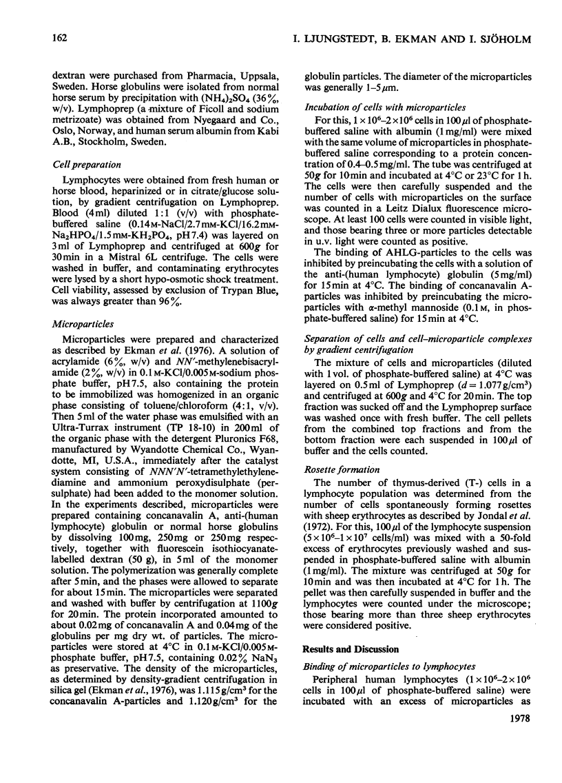
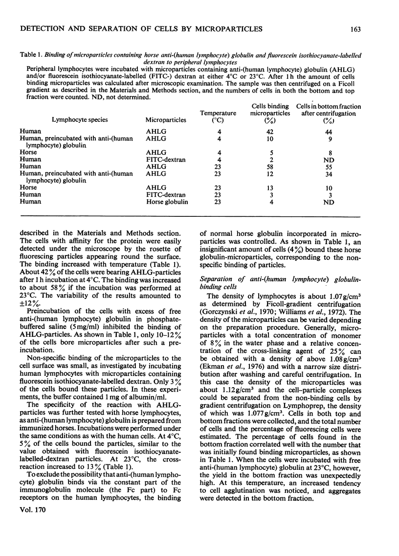
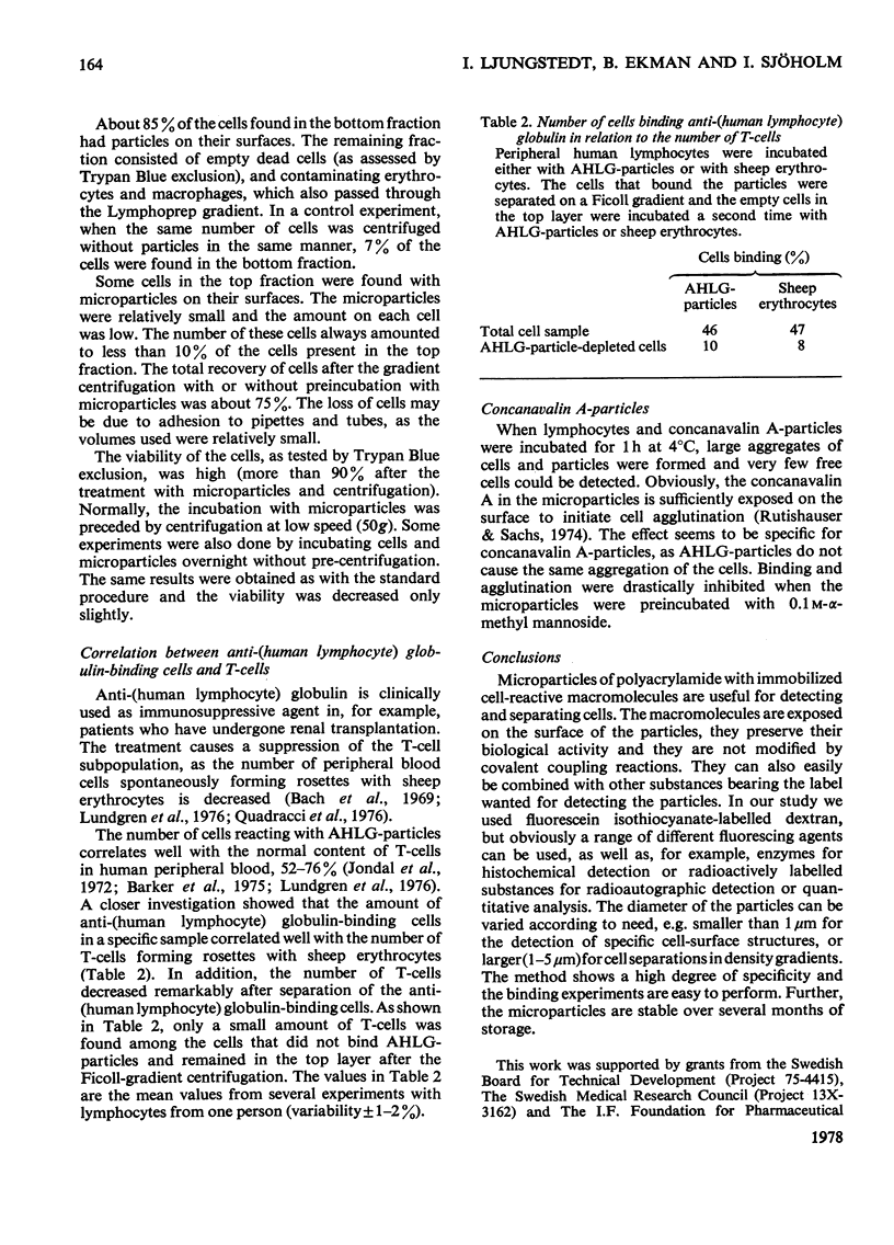
Horse anti-(human lymphocyte) globulin was immobilized together with fluorescein labelled dextran in spherical microparticles of polyacrylamide (AHLG-particles). The particles had a diameter of 1-5 micrometer and a density of 1.12g/cm3, with globulin exposed on the surface. Human lymphocytes bearing the antigen (thymus-derived lymphocytes) bound the particles, which were easily detected by fluorescence microscopy. In this way, about 58% of circulating human lymphocytes were able to bind AHLG-particles at 23 degrees C. Non-specific binding was low, only 3% when human serum albumin was present in the buffer, and only 4% when non-specific horse globulins were incorporated in the microparticles. The cell-particle complexes could be separated from cells that had not reacted by density-gradient centrifugation in Ficoll/metrizoate. The viability was not changed after the separation procedure. The number of cells binding AHLG-particles corresponded well the the relative amount of T-cells. When the cells binding AHLG-particles were separated from the lymphocytes, the number of T-cells decreased remarkably, indicating that the antibodies bind preferably to the T-cell population. Concanavalin A immobilized in microparticles was sufficiently exposed to initiate the agglutination of the lymphocytes. The agglutination was completely inhibited by preincubating the microparticles with alpha-methyl mannoside.
Full text
PDF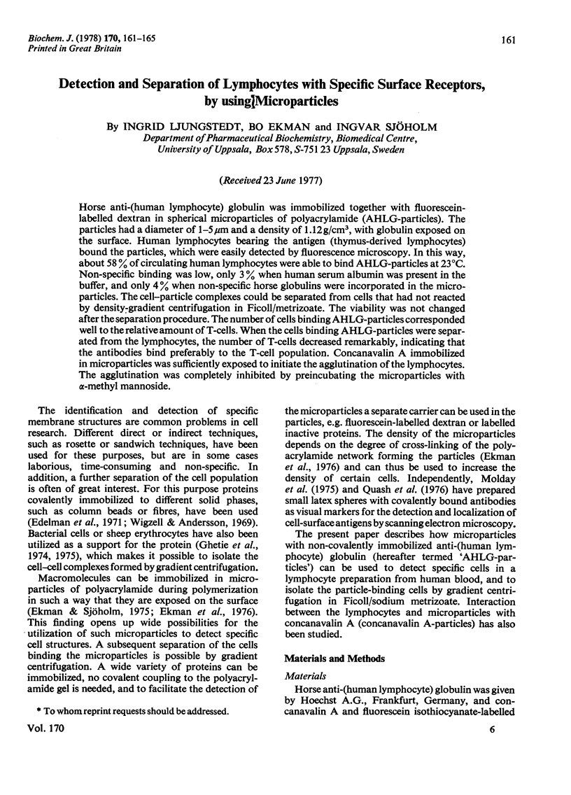
Selected References
These references are in PubMed. This may not be the complete list of references from this article.
- Bach J. F., Dormont J., Dardenne M., Balner H. In vitro rosette inhibition by antihuman antilymphocyte serum. Correlation with skin graft prolongation in subhuman primates. Transplantation. 1969 Sep;8(3):265–280. doi: 10.1097/00007890-196909000-00008. [DOI] [PubMed] [Google Scholar]
- Barker C. R., Worman C. P., Smith J. L. Purification and quantification of T and B lymphocytes by an affinity method. Immunology. 1975 Oct;29(4):765–777. [PMC free article] [PubMed] [Google Scholar]
- Edelman G. M., Rutishauser U., Millette C. F. Cell fractionation and arrangement on fibers, beads, and surfaces. Proc Natl Acad Sci U S A. 1971 Sep;68(9):2153–2157. doi: 10.1073/pnas.68.9.2153. [DOI] [PMC free article] [PubMed] [Google Scholar]
- Ekman B., Lofter C., Sjöholm I. Incorporation of macromolecules in microparticles: preparation and characteristics. Biochemistry. 1976 Nov 16;15(23):5115–5120. doi: 10.1021/bi00668a026. [DOI] [PubMed] [Google Scholar]
- Ekman B., Sjöholm I. Use of macromolecules in microparticles. Nature. 1975 Oct 30;257(5529):825–826. doi: 10.1038/257825a0. [DOI] [PubMed] [Google Scholar]
- Ghetie V., Nilsson K., Sjöquist J. Density gradient separation of lymphoid cells adhering to protein-A-containing staphylococci. Proc Natl Acad Sci U S A. 1974 Dec;71(12):4831–4835. doi: 10.1073/pnas.71.12.4831. [DOI] [PMC free article] [PubMed] [Google Scholar]
- Ghetie V., Stålenheim G., Sjöquist J. Cell separation by staphylococcal protein A-coated erythrocytes. Scand J Immunol. 1975 Sep;4(5-6):471–477. doi: 10.1111/j.1365-3083.1975.tb02652.x. [DOI] [PubMed] [Google Scholar]
- Gorczynski R. M., Miller R. G., Phillips R. A. Homogeneity of antibody-producing cells as analysed by their buoyant density in gradients of Ficoll. Immunology. 1970 Nov;19(5):817–829. [PMC free article] [PubMed] [Google Scholar]
- Jondal M., Holm G., Wigzell H. Surface markers on human T and B lymphocytes. I. A large population of lymphocytes forming nonimmune rosettes with sheep red blood cells. J Exp Med. 1972 Aug 1;136(2):207–215. doi: 10.1084/jem.136.2.207. [DOI] [PMC free article] [PubMed] [Google Scholar]
- Molday R. S., Dreyer W. J., Rembaum A., Yen S. P. New immunolatex spheres: visual markers of antigens on lymphocytes for scanning electron microscopy. J Cell Biol. 1975 Jan;64(1):75–88. doi: 10.1083/jcb.64.1.75. [DOI] [PMC free article] [PubMed] [Google Scholar]
- Quadracci L. J., Ringdén O., Krzymanski M. The effect of uremia and transplantation on lymphocyte subpopulations. Kidney Int. 1976 Aug;10(2):179–184. doi: 10.1038/ki.1976.93. [DOI] [PubMed] [Google Scholar]
- Quash G. A., Niveleau A., Aupoix M., Greenland T. Immunolatex visualisation of cell surface Forssman and polyamine antigens. Exp Cell Res. 1976 Mar 15;98(2):253–261. doi: 10.1016/0014-4827(76)90435-3. [DOI] [PubMed] [Google Scholar]
- Rutishauser U., Sachs L. Receptor mobility and the mechanism of cell-cell binding induced by concanavalin A. Proc Natl Acad Sci U S A. 1974 Jun;71(6):2456–2460. doi: 10.1073/pnas.71.6.2456. [DOI] [PMC free article] [PubMed] [Google Scholar]
- Wigzell H., Andersson B. Cell separation on antigen-coated columns. Elimination of high rate antibody-forming cells and immunological memory cells. J Exp Med. 1969 Jan 1;129(1):23–36. doi: 10.1084/jem.129.1.23. [DOI] [PMC free article] [PubMed] [Google Scholar]
- Williams N., Kraft N., Shortman K. The separation of different cell classes from lymphoid organs. VI. The effect of osmolarity of gradient media on the density distribution of cells. Immunology. 1972 May;22(5):885–899. [PMC free article] [PubMed] [Google Scholar]
- de Grouchy J. Human chromosomes and their anomalies. Postgrad Med J. 1976;52 (Suppl 2):5–16. [PubMed] [Google Scholar]


