

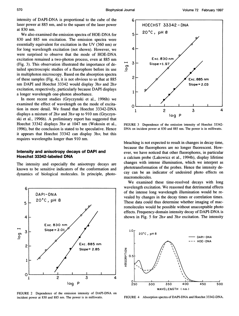
Abstract
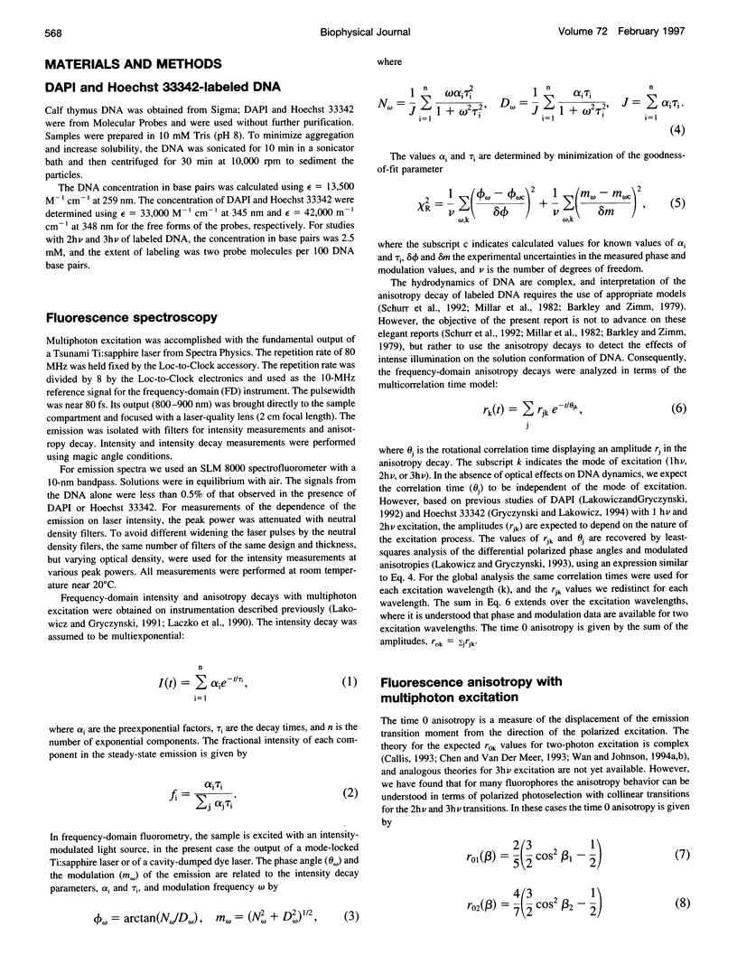
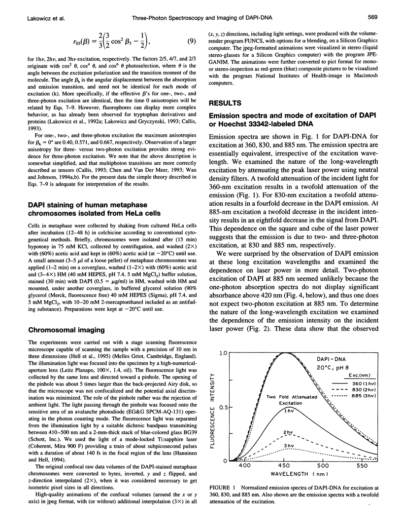
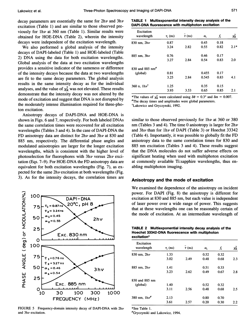
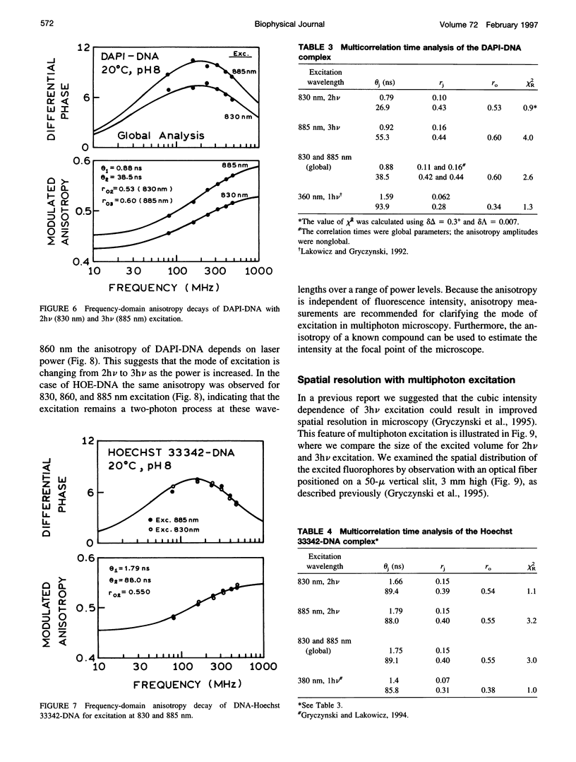
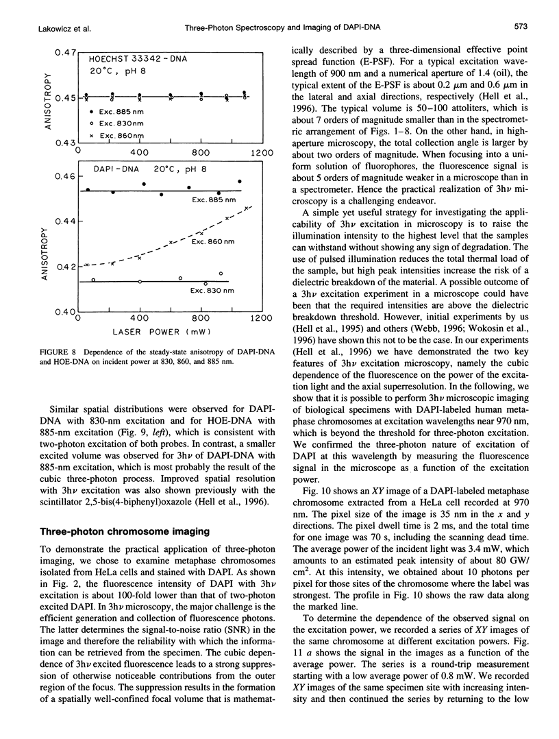
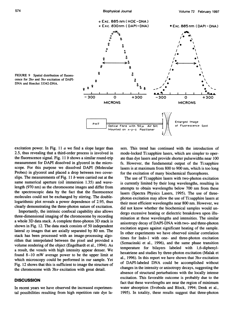
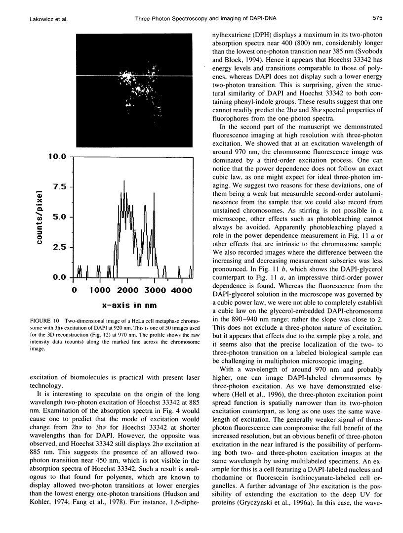
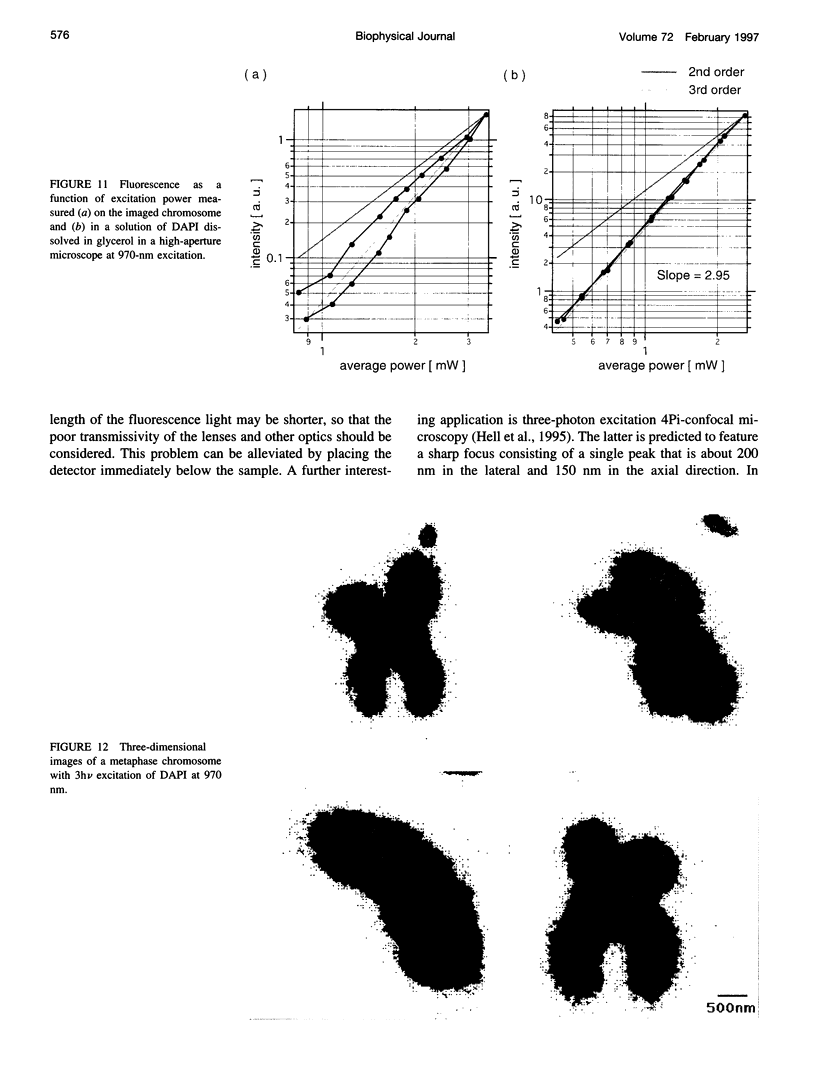
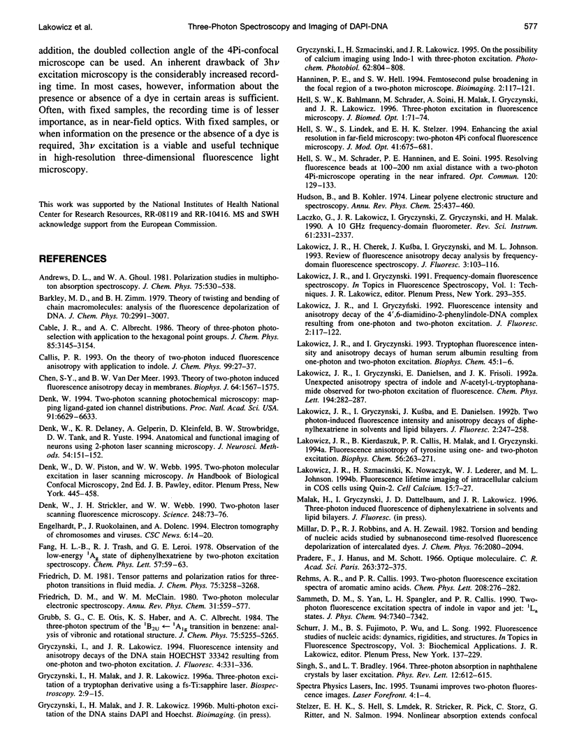
We examined the fluorescence spectral properties of the DNA stains DAPI (4′,6-diamidino-2-phenylindole, hydrochloride) and Hoechst 33342 (bis-benzimide, or 2,5′-bi-1H-benzimidazole2′-(4-ethoxyphenyl)-5-(4-methyl-1-piperazinyl)) with two-photon (2hν) and three-photon (3hν) excitation using femtosecond pulses from a Ti:sapphire laser from 830 to 885 nm. The mode of excitation of DAPI bound to DNA changed from two-photon at 830 nm to three-photon at 885 nm. In contrast, Hoechst 33342 displayed only two-photon excitation from 830 to 885 nm. DAPI-DNA displayed the same emission spectra and decay times for 2hν and 3hν excitation. Hoechst 33342-DNA displayed the same intensity decay for excitation at 830 and 885 nm. Both probes displayed higher anisotropies for multiphoton excitation as compared to one-photon excitation with ultraviolet wavelengths, and DAPI-DNA displays a higher anisotropy for 3hν at 885 nm than for 2hν at 830 nm. We used 970-nm excitation of DAPI-stained chromosomes to obtain the first three-dimensional images with three-photon excitation. Three-photon excitation of DAPI-stained chromosomes at 970 nm was demonstrated by the power dependence in the fluorescence microscope.
Full text
PDF

Images in this article
Selected References
These references are in PubMed. This may not be the complete list of references from this article.
- Chen S. Y., Van Der Meer B. W. Theory of two-photon induced fluorescence anisotropy decay in membranes. Biophys J. 1993 May;64(5):1567–1575. doi: 10.1016/S0006-3495(93)81526-1. [DOI] [PMC free article] [PubMed] [Google Scholar]
- Denk W., Delaney K. R., Gelperin A., Kleinfeld D., Strowbridge B. W., Tank D. W., Yuste R. Anatomical and functional imaging of neurons using 2-photon laser scanning microscopy. J Neurosci Methods. 1994 Oct;54(2):151–162. doi: 10.1016/0165-0270(94)90189-9. [DOI] [PubMed] [Google Scholar]
- Denk W., Strickler J. H., Webb W. W. Two-photon laser scanning fluorescence microscopy. Science. 1990 Apr 6;248(4951):73–76. doi: 10.1126/science.2321027. [DOI] [PubMed] [Google Scholar]
- Denk W. Two-photon scanning photochemical microscopy: mapping ligand-gated ion channel distributions. Proc Natl Acad Sci U S A. 1994 Jul 5;91(14):6629–6633. doi: 10.1073/pnas.91.14.6629. [DOI] [PMC free article] [PubMed] [Google Scholar]
- Gryczynski I., Szmacinski H., Lakowicz J. R. On the possibility of calcium imaging using Indo-1 with three-photon excitation. Photochem Photobiol. 1995 Oct;62(4):804–808. doi: 10.1111/j.1751-1097.1995.tb08733.x. [DOI] [PubMed] [Google Scholar]
- Lakowicz J. R., Gryczynski I. Tryptophan fluorescence intensity and anisotropy decays of human serum albumin resulting from one-photon and two-photon excitation. Biophys Chem. 1992 Nov;45(1):1–6. doi: 10.1016/0301-4622(92)87017-d. [DOI] [PubMed] [Google Scholar]
- Lakowicz J. R., Kierdaszuk B., Callis P., Malak H., Gryczynski I. Fluorescence anisotropy of tyrosine using one-and two-photon excitation. Biophys Chem. 1995 Nov;56(3):263–271. doi: 10.1016/0301-4622(95)00040-5. [DOI] [PubMed] [Google Scholar]
- Lakowicz J. R., Szmacinski H., Nowaczyk K., Lederer W. J., Kirby M. S., Johnson M. L. Fluorescence lifetime imaging of intracellular calcium in COS cells using Quin-2. Cell Calcium. 1994 Jan;15(1):7–27. doi: 10.1016/0143-4160(94)90100-7. [DOI] [PMC free article] [PubMed] [Google Scholar]
- Svoboda K., Block S. M. Biological applications of optical forces. Annu Rev Biophys Biomol Struct. 1994;23:247–285. doi: 10.1146/annurev.bb.23.060194.001335. [DOI] [PubMed] [Google Scholar]
- Szmacinski H., Gryczynski I., Lakowicz J. R. Three-photon induced fluorescence of the calcium probe Indo-1. Biophys J. 1996 Jan;70(1):547–555. doi: 10.1016/S0006-3495(96)79601-7. [DOI] [PMC free article] [PubMed] [Google Scholar]