Abstract
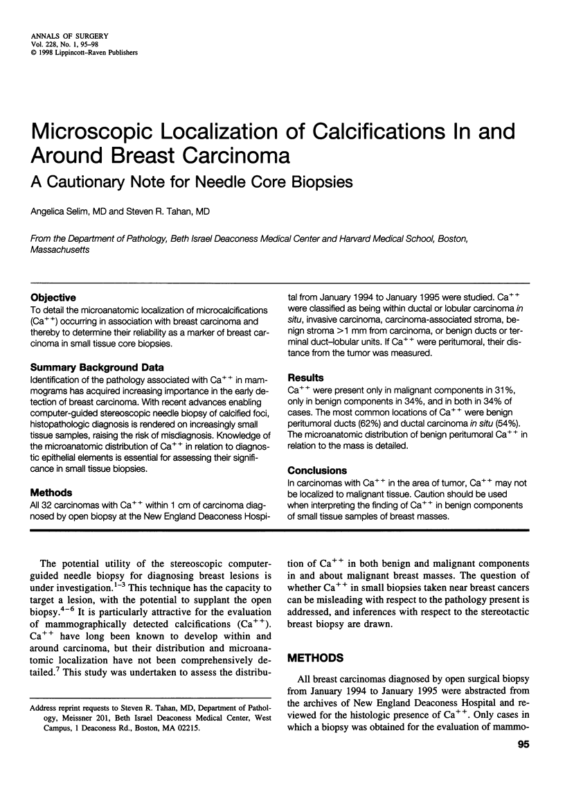
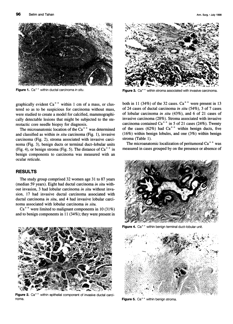
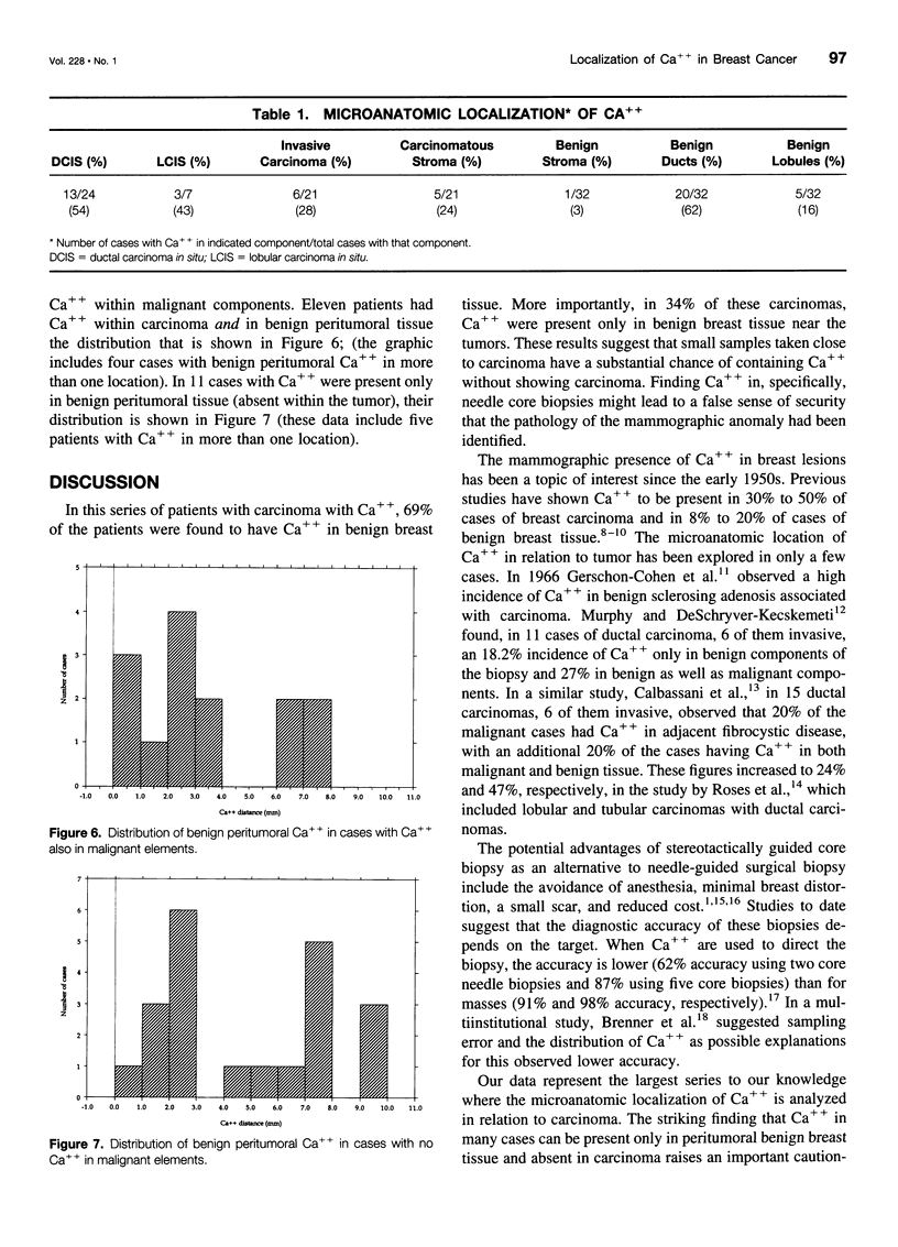
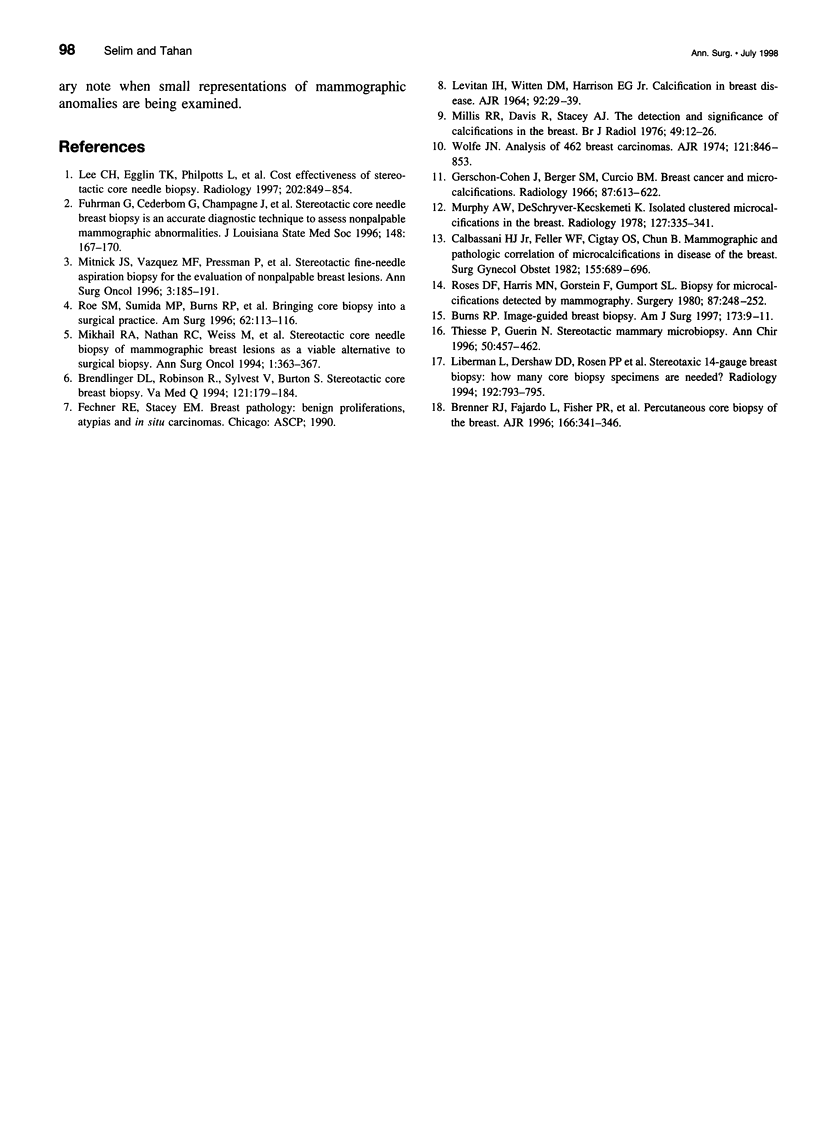
OBJECTIVE: To detail the microanatomic localization of microcalcifications (Ca++) occurring in association with breast carcinoma and thereby to determine their reliability as a marker of breast carcinoma in small tissue core biopsies. SUMMARY BACKGROUND DATA: Identification of the pathology associated with Ca++ in mammograms has acquired increasing importance in the early detection of breast carcinoma. With recent advances enabling computer-guided stereoscopic needle biopsy of calcified foci, histopathologic diagnosis is rendered on increasingly small tissue samples, raising the risk of misdiagnosis. Knowledge of the microanatomic distribution of Ca++ in relation to diagnostic epithelial elements is essential for assessing their significance in small tissue biopsies. METHODS: All 32 carcinomas with Ca++ within 1 cm of carcinoma diagnosed by open biopsy at the New England Deaconess Hospital from January 1994 to January 1995 were studied. Ca++ were classified as being within ductal or lobular carcinoma in situ, invasive carcinoma, carcinoma-associated stroma, benign stroma >1 mm from carcinoma, or benign ducts or terminal duct-lobular units. If Ca++ were peritumoral, their distance from the tumor was measured. RESULTS: Ca++ were present only in malignant components in 31%, only in benign components in 34%, and in both in 34% of cases. The most common locations of Ca++ were benign peritumoral ducts (62%) and ductal carcinoma in situ (54%). The microanatomic distribution of benign peritumoral Ca++ in relation to the mass is detailed. CONCLUSIONS: In carcinomas with Ca++ in the area of tumor, Ca++ may not be localized to malignant tissue. Caution should be used when interpreting the finding of Ca++ in benign components of small tissue samples of breast masses.
Full text
PDF


Images in this article
Selected References
These references are in PubMed. This may not be the complete list of references from this article.
- Brendlinger D. L., Robinson R., Sylvest V., Burton S. Stereotactic core breast biopsy. An alternative. Va Med Q. 1994 Summer;121(3):179–184. [PubMed] [Google Scholar]
- Brenner R. J., Fajardo L., Fisher P. R., Dershaw D. D., Evans W. P., Bassett L., Feig S., Mendelson E., Jackson V., Margolin F. R. Percutaneous core biopsy of the breast: effect of operator experience and number of samples on diagnostic accuracy. AJR Am J Roentgenol. 1996 Feb;166(2):341–346. doi: 10.2214/ajr.166.2.8553943. [DOI] [PubMed] [Google Scholar]
- Burns R. P. Image-guided breast biopsy. Am J Surg. 1997 Jan;173(1):9–13. doi: 10.1016/S0002-9610(96)00374-1. [DOI] [PubMed] [Google Scholar]
- Colbassani H. J., Jr, Feller W. F., Cigtay O. S., Chun B. Mammographic and pathologic correlation of microcalcification in disease of the breast. Surg Gynecol Obstet. 1982 Nov;155(5):689–696. [PubMed] [Google Scholar]
- Fuhrman G., Cederbom G., Champagne J., Farr G., McKinnon W., Bolton J., Ordoyne W. K. Stereotactic core needle breast biopsy is an accurate diagnostic technique to assess nonpalpable mammographic abnormalities. J La State Med Soc. 1996 Apr;148(4):167–170. [PubMed] [Google Scholar]
- Gershon-Cohen J., Berger S. M. Breast cancer with microcalcifications: diagnostic difficulties. Radiology. 1966 Oct;87(4):613–622. doi: 10.1148/87.4.613. [DOI] [PubMed] [Google Scholar]
- LEVITAN L. H., WITTEN D. M., HARRISON E. G., Jr CALCIFICATION IN BREAST DISEASE MAMMOGRAPHIC-PATHOLOGIC CORRELATION. Am J Roentgenol Radium Ther Nucl Med. 1964 Jul;92:29–39. [PubMed] [Google Scholar]
- Lee C. H., Egglin T. K., Philpotts L., Mainiero M. B., Tocino I. Cost-effectiveness of stereotactic core needle biopsy: analysis by means of mammographic findings. Radiology. 1997 Mar;202(3):849–854. doi: 10.1148/radiology.202.3.9051045. [DOI] [PubMed] [Google Scholar]
- Liberman L., Dershaw D. D., Rosen P. P., Abramson A. F., Deutch B. M., Hann L. E. Stereotaxic 14-gauge breast biopsy: how many core biopsy specimens are needed? Radiology. 1994 Sep;192(3):793–795. doi: 10.1148/radiology.192.3.8058949. [DOI] [PubMed] [Google Scholar]
- Mikhail R. A., Nathan R. C., Weiss M., Tummala R. M., Mullangi U. R., Lawrence L., Mukkamala A. Stereotactic core needle biopsy of mammographic breast lesions as a viable alternative to surgical biopsy. Ann Surg Oncol. 1994 Sep;1(5):363–367. doi: 10.1007/BF02303806. [DOI] [PubMed] [Google Scholar]
- Millis R. R., Davis R., Stacey A. J. The detection and significance of calcifications in the breast: a radiological and pathological study. Br J Radiol. 1976 Jan;49(577):12–26. doi: 10.1259/0007-1285-49-577-12. [DOI] [PubMed] [Google Scholar]
- Mitnick J. S., Vazquez M. F., Pressman P. I., Harris M. N., Roses D. F. Stereotactic fine-needle aspiration biopsy for the evaluation of nonpalpable breast lesions: report of an experience based on 2,988 cases. Ann Surg Oncol. 1996 Mar;3(2):185–191. doi: 10.1007/BF02305799. [DOI] [PubMed] [Google Scholar]
- Murphy W. A., DeSchryver-Kecskemeti K. Isolated clustered microcalcifications in the breast: radiologic-pathologic correlation. Radiology. 1978 May;127(2):335–341. doi: 10.1148/127.2.335. [DOI] [PubMed] [Google Scholar]
- Roe S. M., Sumida M. P., Burns R. P., Greer M. S., Clements J. B. Bringing core biopsy into a surgical practice. Am Surg. 1996 Feb;62(2):113–116. [PubMed] [Google Scholar]
- Roses D. F., Harris M. N., Gorstein F., Gumport S. L. Biopsy for microcalcification detected by mammography. Surgery. 1980 Mar;87(3):248–252. [PubMed] [Google Scholar]
- Thiesse P., Guérin N. La microbiopsie mammaire stéréotaxique. I.-Les aspects techniques. Ann Chir. 1996;50(6):457–462. [PubMed] [Google Scholar]
- Wolfe J. N. Analysis of 462 breast carcinomas. Am J Roentgenol Radium Ther Nucl Med. 1974 Aug;121(4):846–853. doi: 10.2214/ajr.121.4.846. [DOI] [PubMed] [Google Scholar]


