Abstract
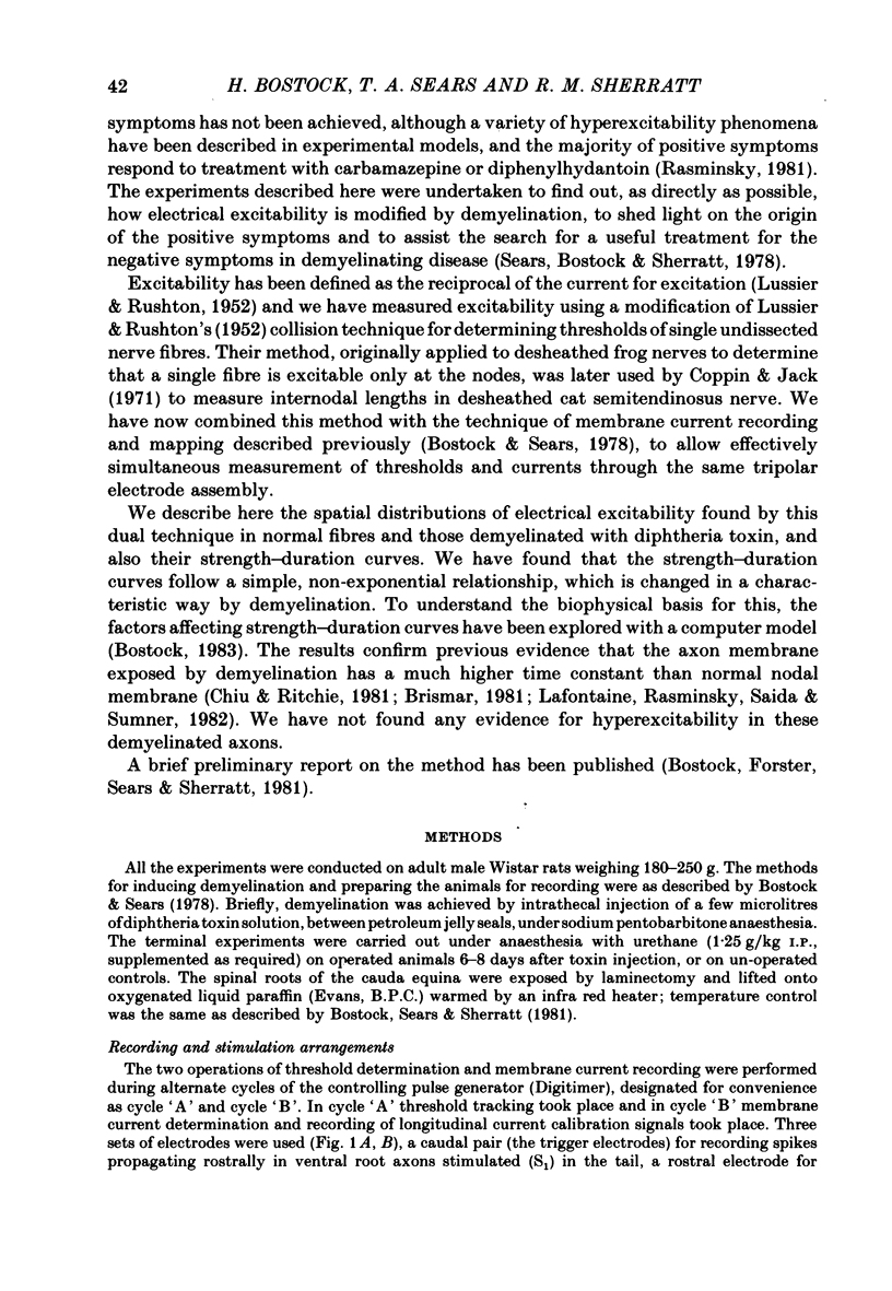
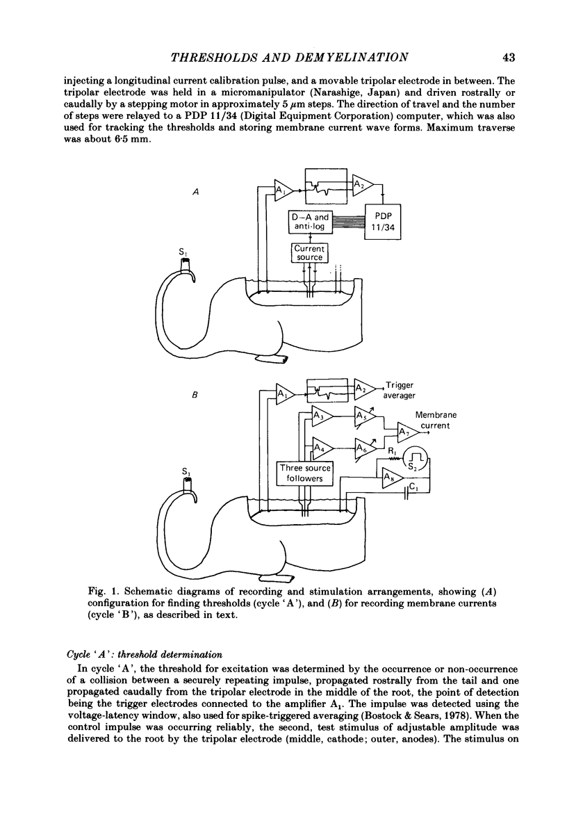
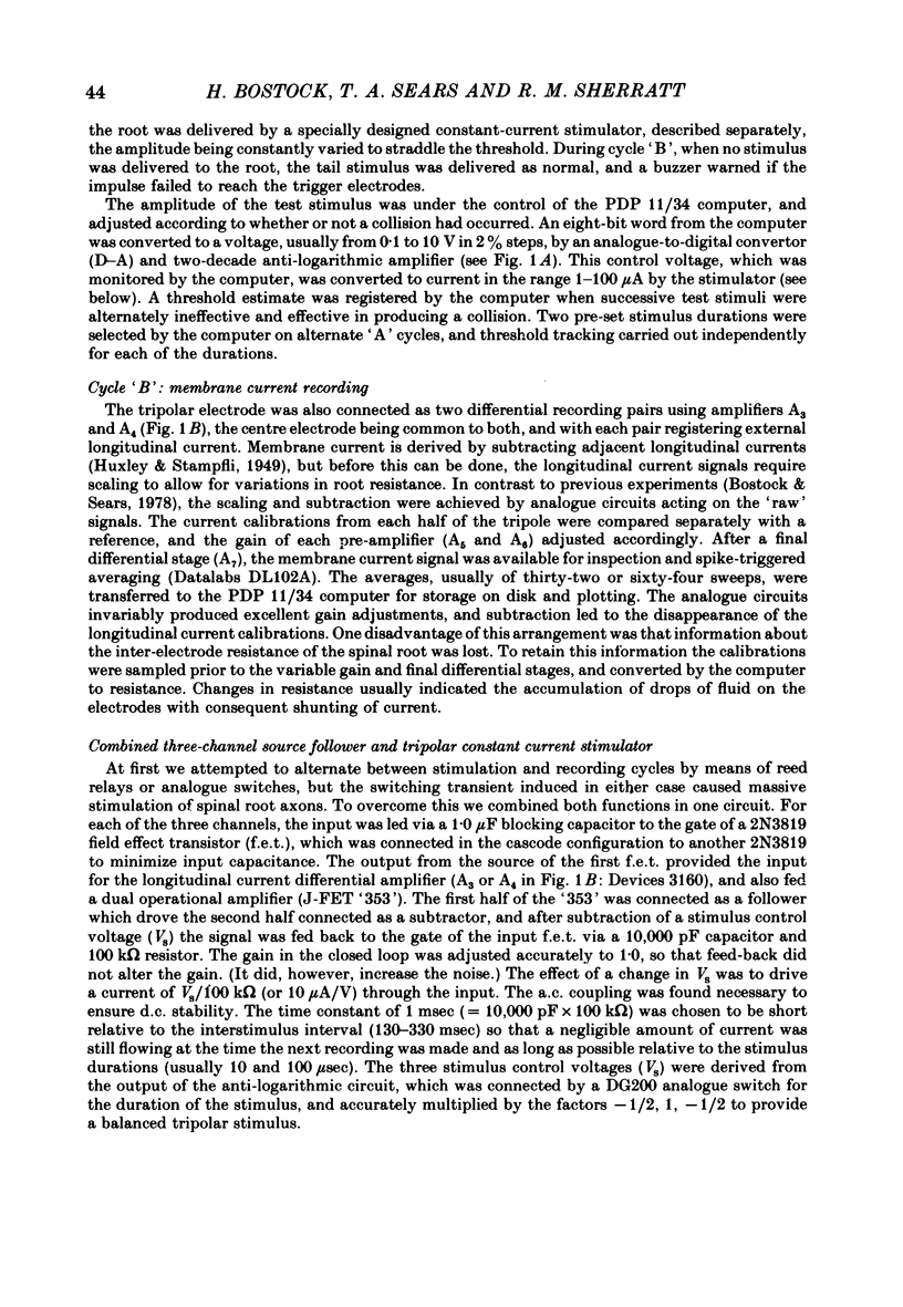
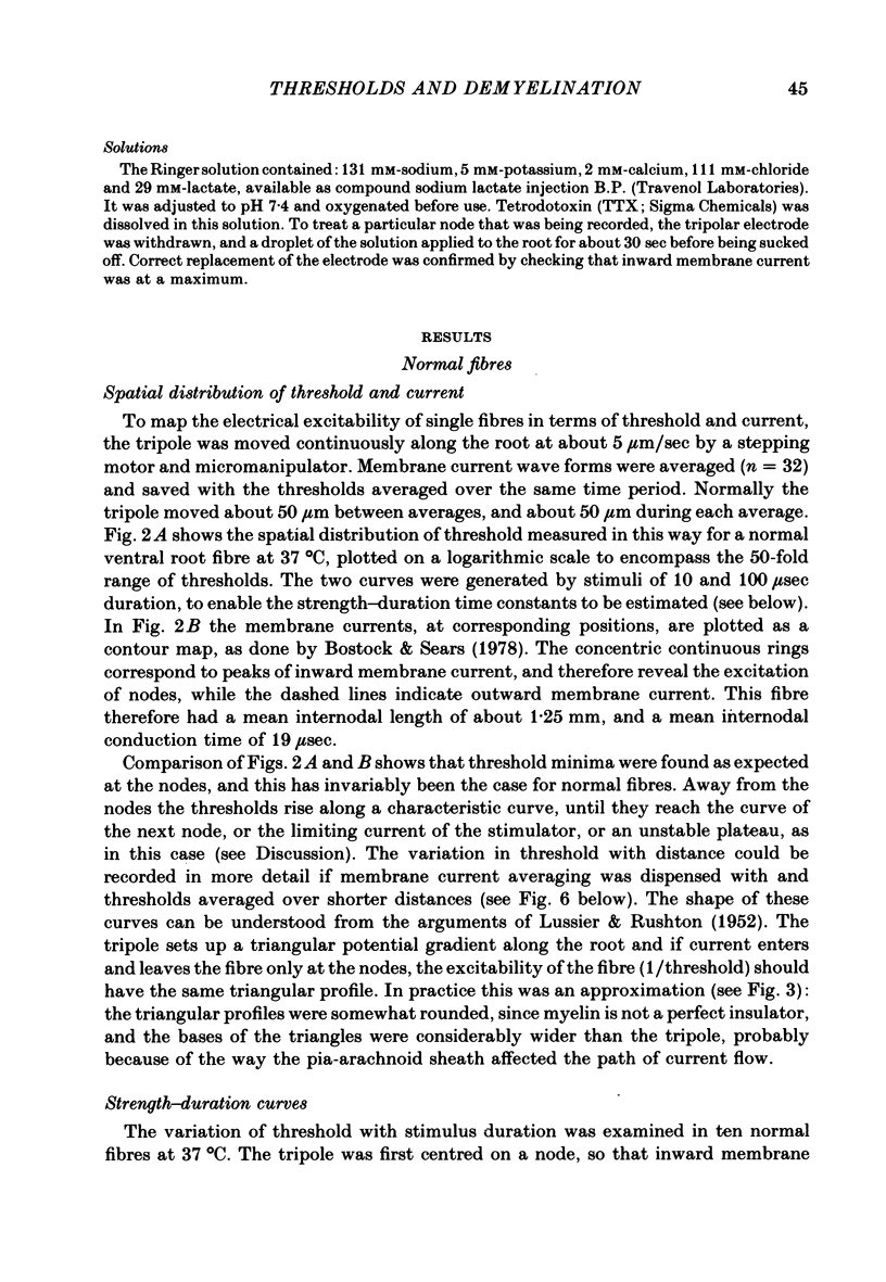
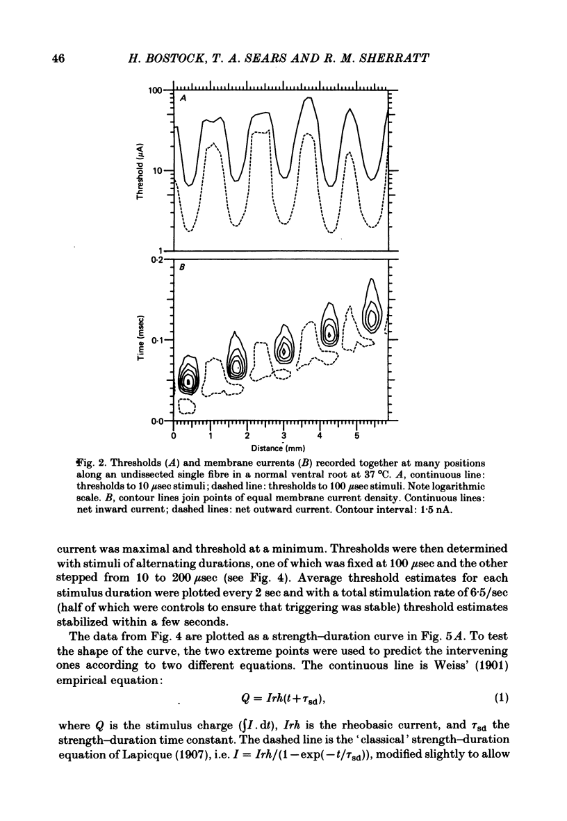
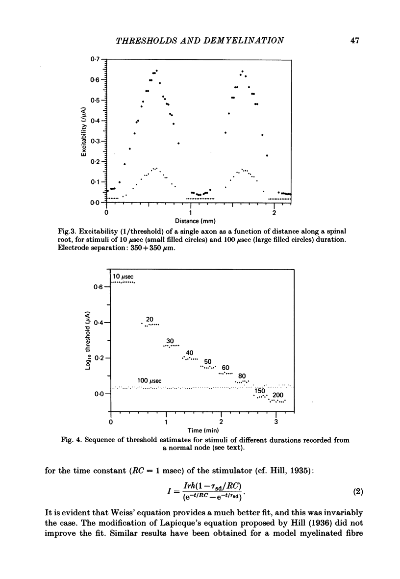
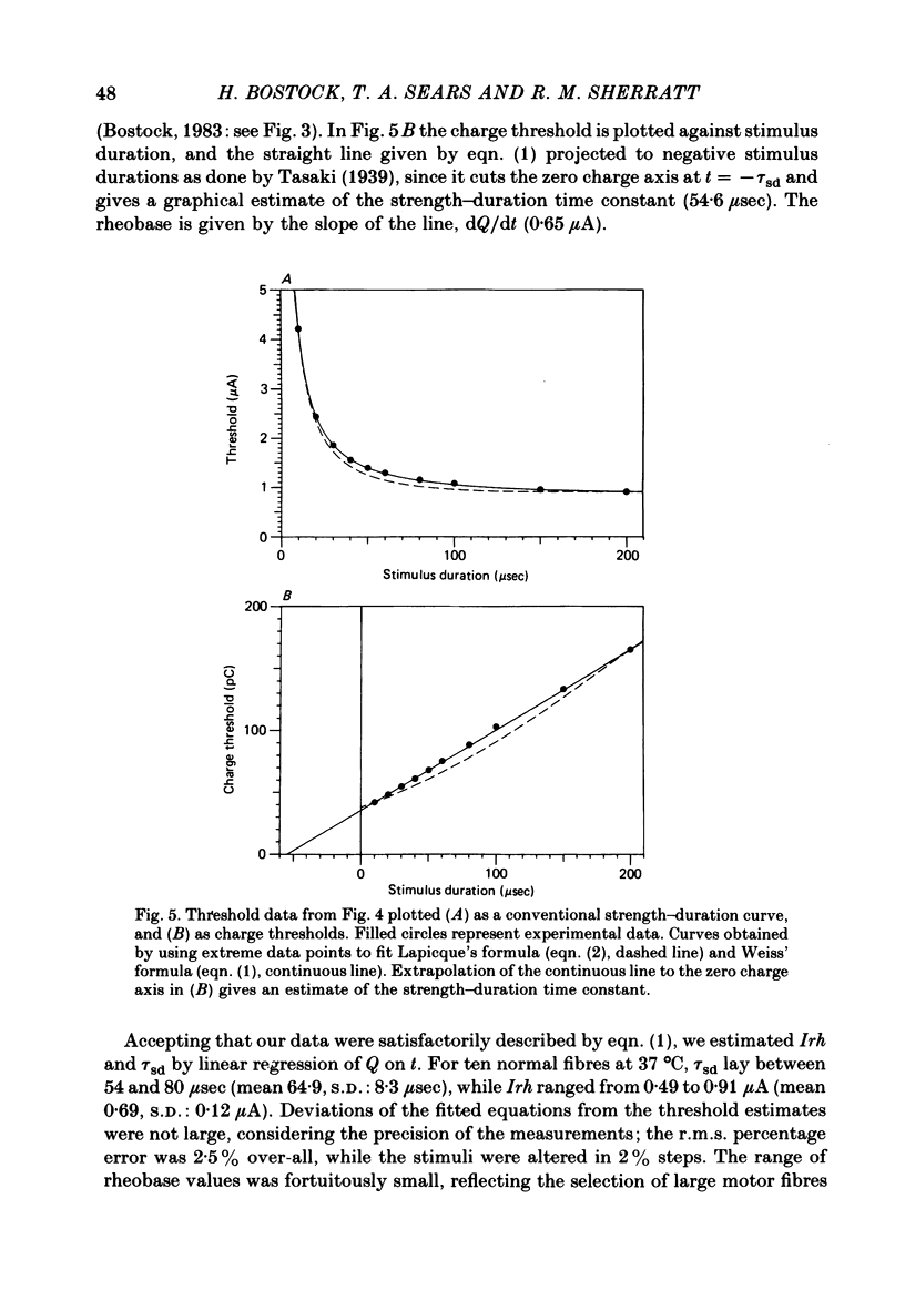
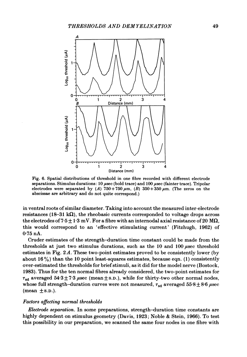
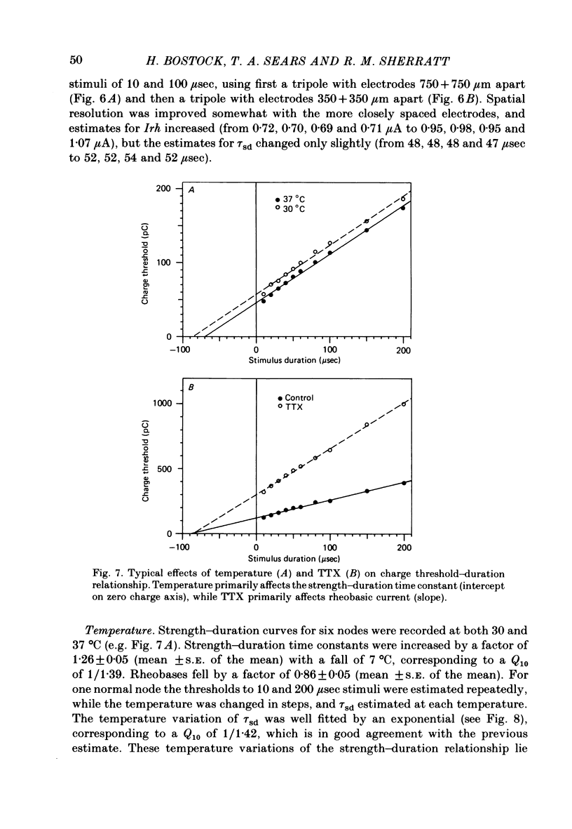
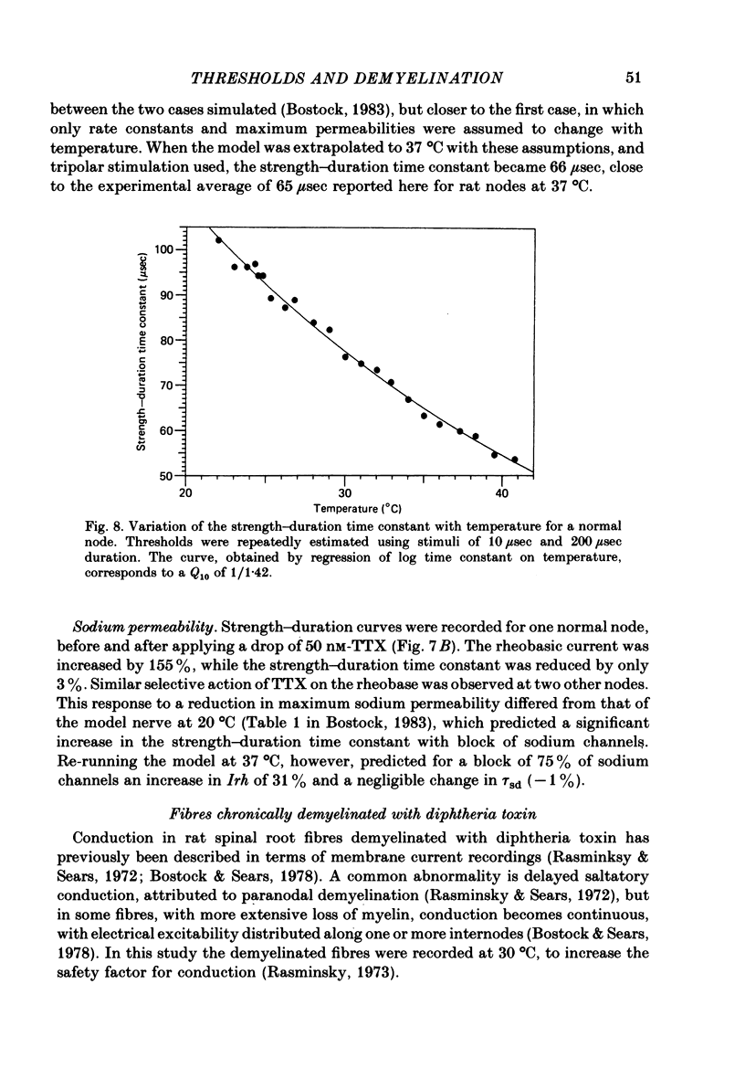
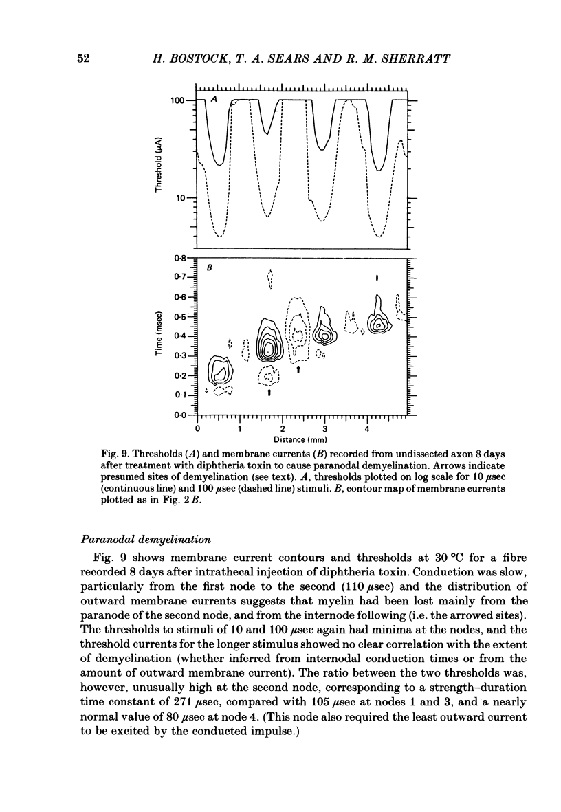
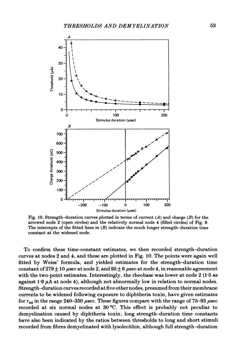
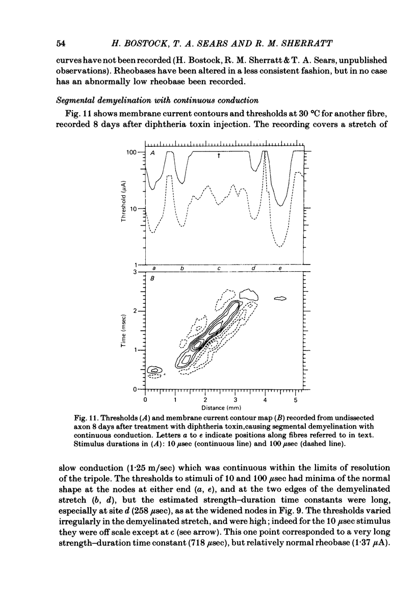
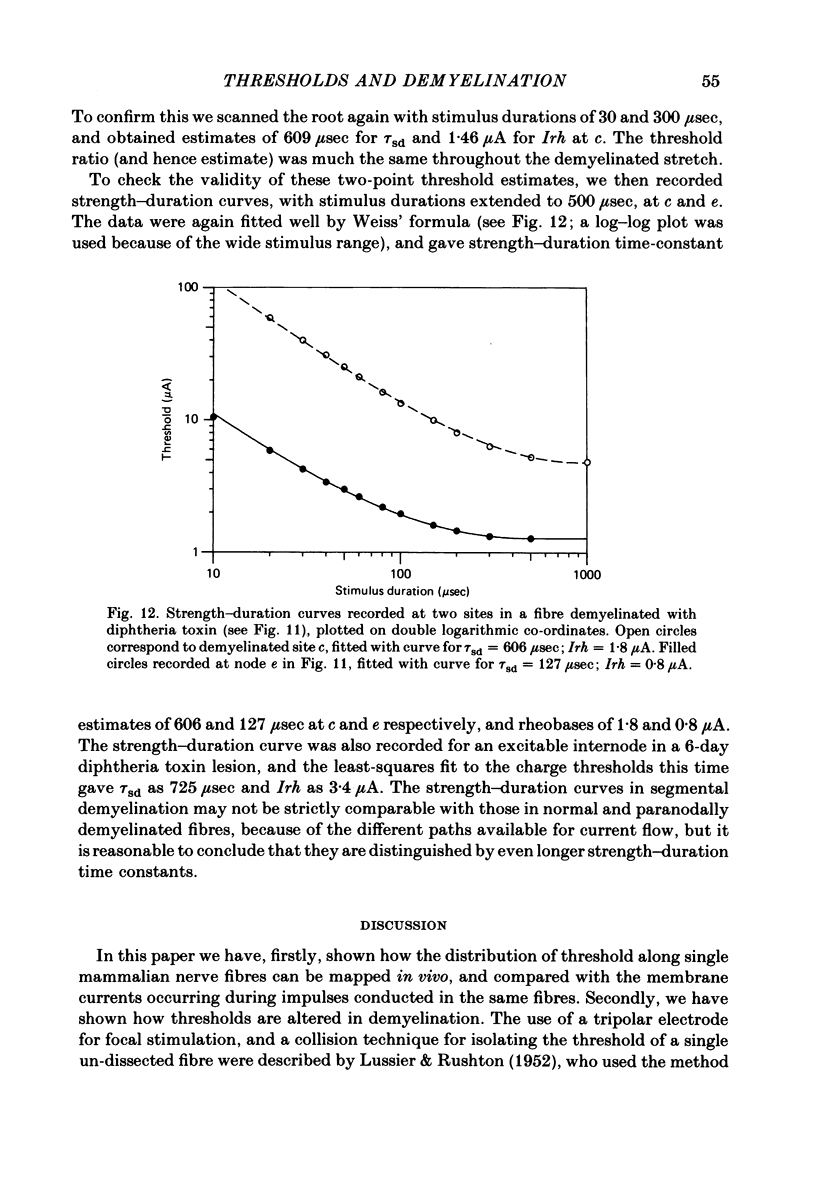
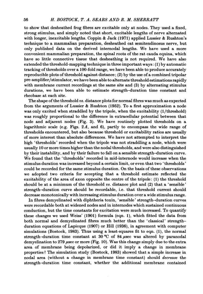
Thresholds to electrical stimulation have been recorded, concurrently with the membrane currents of conducted impulses, at many positions along undissected single fibres in rat spinal roots. In normal myelinated fibres, distinct threshold minima invariably coincided with sites of inward current generation, and were therefore identified as nodes of Ranvier. Between nodes, the thresholds rose by an order of magnitude. At normal nodes, the charge thresholds were linearly related to stimulus duration, as predicted by computer simulations of a model myelinated fibre (Bostock, 1983). The strength-duration time constants averaged 64.9 +/- 8.3 microseconds (mean +/- S.D.) at 37 degrees C, and had a Q10 of 1/1.39. They were relatively insensitive to changes in inter-electrode distance, or to partial anaesthetization with tetrodotoxin. In fibres treated with diphtheria toxin 6-8 days previously, to induce paranodal or segmental demyelination, threshold minima were found both at nodes and in internodal regions generating inward membrane current. In these fibres strength-duration curves were of the same general form as at normal nodes, but with strength-duration time constants increased at widened nodes (up to 350 microseconds) and at excitable internodes (600-725 microseconds). Comparison with the computer model indicated that these changes were most likely due to exposure of axon membrane with a time constant much longer than that of the normal nodal membrane. In none of the demyelinated fibres examined have we found any evidence of hyperexcitability.
Full text
PDF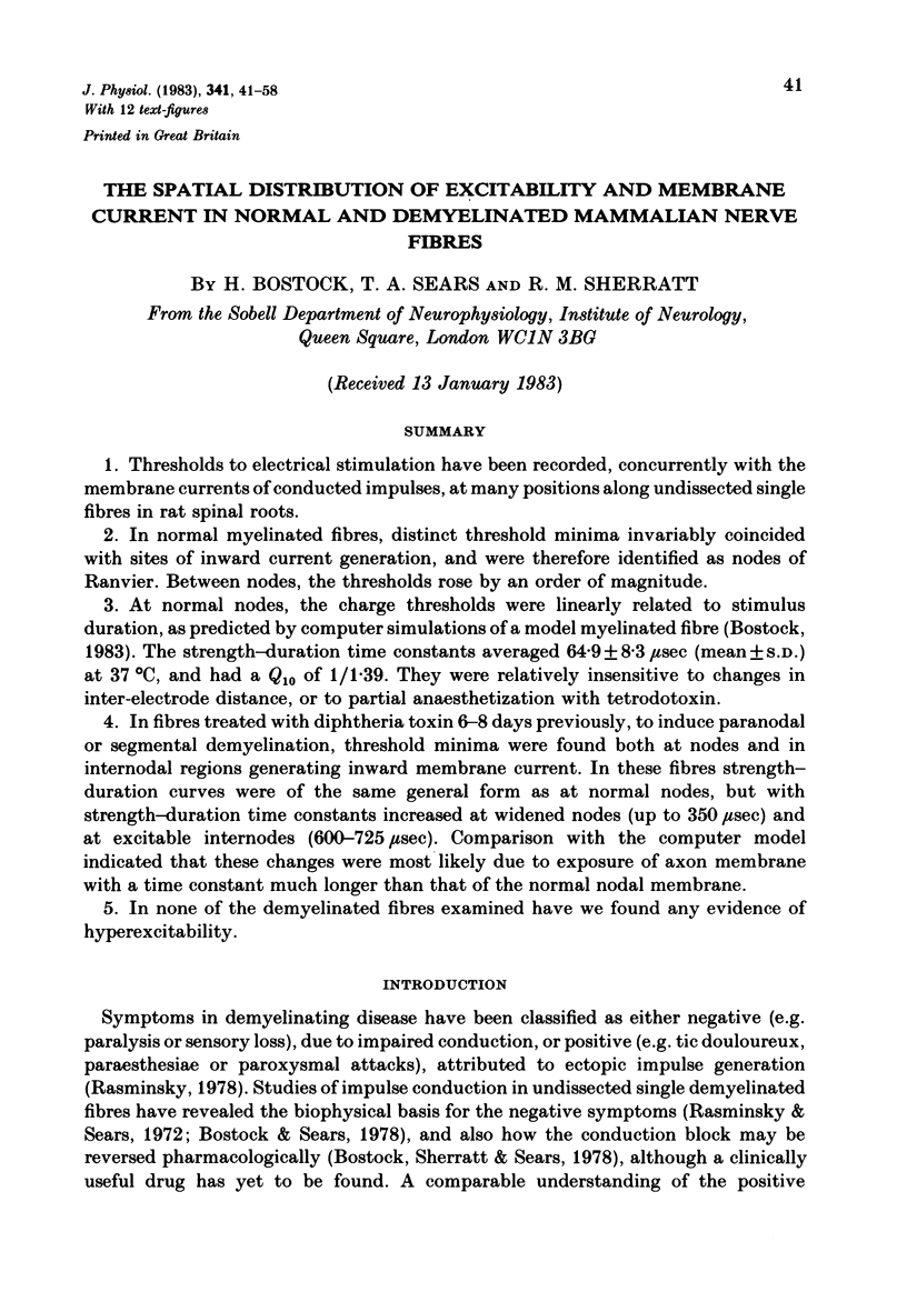
Selected References
These references are in PubMed. This may not be the complete list of references from this article.
- Bostock H., Sears T. A. The internodal axon membrane: electrical excitability and continuous conduction in segmental demyelination. J Physiol. 1978 Jul;280:273–301. doi: 10.1113/jphysiol.1978.sp012384. [DOI] [PMC free article] [PubMed] [Google Scholar]
- Bostock H., Sherratt R. M., Sears T. A. Overcoming conduction failure in demyelinated nerve fibres by prolonging action potentials. Nature. 1978 Jul 27;274(5669):385–387. doi: 10.1038/274385a0. [DOI] [PubMed] [Google Scholar]
- Bostock H. The strength-duration relationship for excitation of myelinated nerve: computed dependence on membrane parameters. J Physiol. 1983 Aug;341:59–74. doi: 10.1113/jphysiol.1983.sp014792. [DOI] [PMC free article] [PubMed] [Google Scholar]
- Brismar T. Electrical properties of isolated demyelinated rat nerve fibres. Acta Physiol Scand. 1981 Oct;113(2):161–166. doi: 10.1111/j.1748-1716.1981.tb06877.x. [DOI] [PubMed] [Google Scholar]
- Chiu S. Y., Ritchie J. M. Evidence for the presence of potassium channels in the paranodal region of acutely demyelinated mammalian single nerve fibres. J Physiol. 1981;313:415–437. doi: 10.1113/jphysiol.1981.sp013674. [DOI] [PMC free article] [PubMed] [Google Scholar]
- FITZHUGH R. Computation of impulse initiation and saltatory conduction in a myelinated nerve fiber. Biophys J. 1962 Jan;2:11–21. doi: 10.1016/s0006-3495(62)86837-4. [DOI] [PMC free article] [PubMed] [Google Scholar]
- Huxley A. F., Stämpfli R. Evidence for saltatory conduction in peripheral myelinated nerve fibres. J Physiol. 1949 May 15;108(3):315–339. [PMC free article] [PubMed] [Google Scholar]
- LUSSIER J. J., RUSHTON W. A. H. The excitability of a single fiber in a nerve trunk. J Physiol. 1952 May;117(1):87–108. [PMC free article] [PubMed] [Google Scholar]
- Lafontaine S., Rasminsky M., Saida T., Sumner A. J. Conduction block in rat myelinated fibres following acute exposure to anti-galactocerebroside serum. J Physiol. 1982 Feb;323:287–306. doi: 10.1113/jphysiol.1982.sp014073. [DOI] [PMC free article] [PubMed] [Google Scholar]
- Noble D., Stein R. B. The threshold conditions for initiation of action potentials by excitable cells. J Physiol. 1966 Nov;187(1):129–162. doi: 10.1113/jphysiol.1966.sp008079. [DOI] [PMC free article] [PubMed] [Google Scholar]
- Rasminsky M., Sears T. A. Internodal conduction in undissected demyelinated nerve fibres. J Physiol. 1972 Dec;227(2):323–350. doi: 10.1113/jphysiol.1972.sp010035. [DOI] [PMC free article] [PubMed] [Google Scholar]
- Sears T. A., Bostock H., Sheratt M. The pathophysiology of demyelination and its implications for the symptomatic treatment of multiple sclerosis. Neurology. 1978 Sep;28(9 Pt 2):21–26. doi: 10.1212/wnl.28.9_part_2.21. [DOI] [PubMed] [Google Scholar]


