Abstract
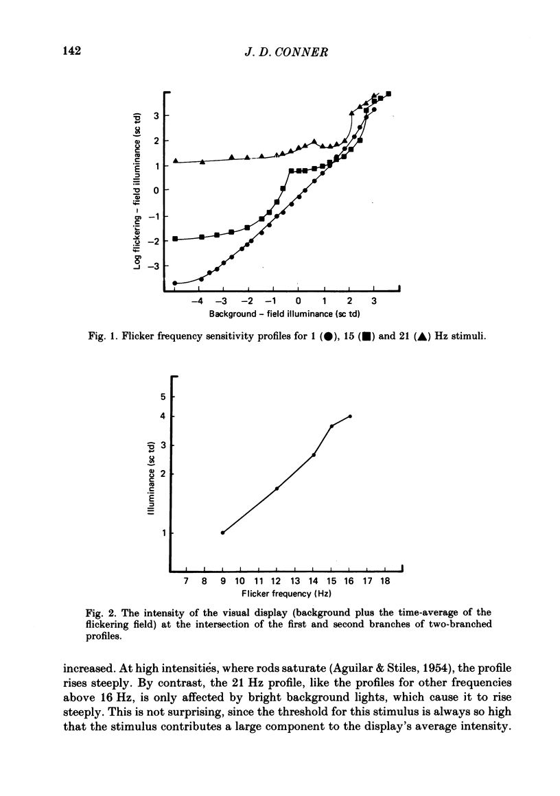
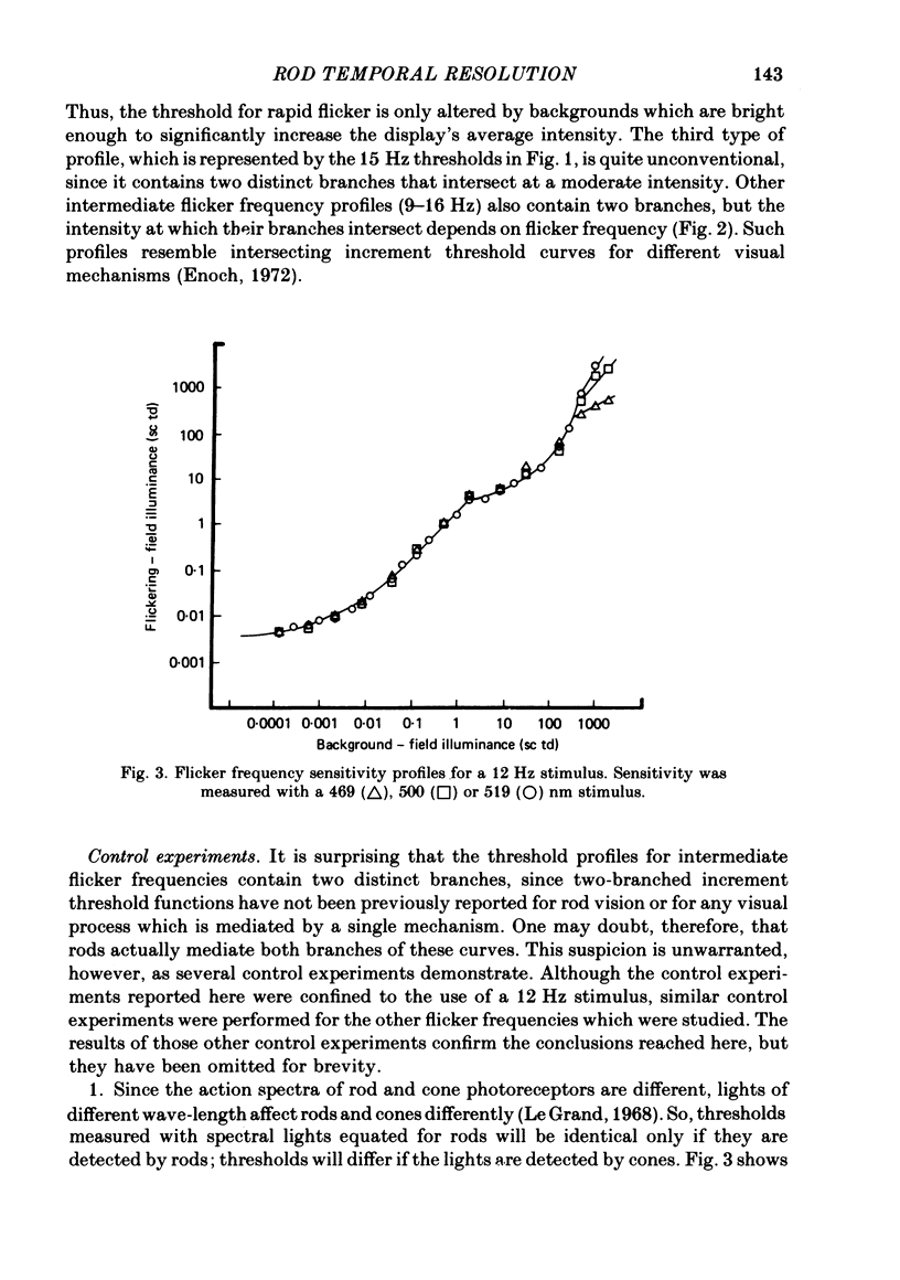
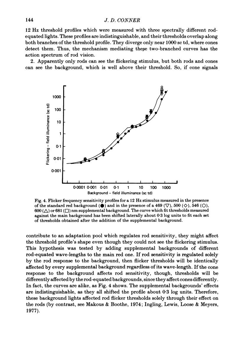
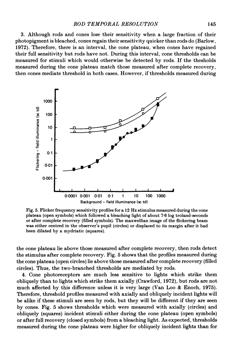
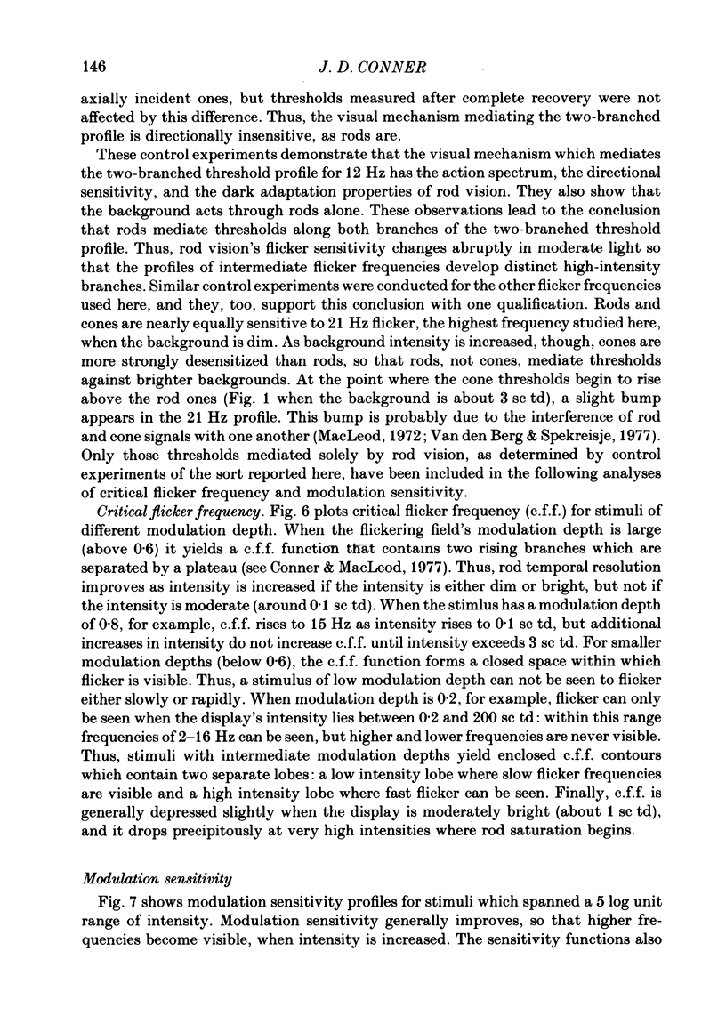
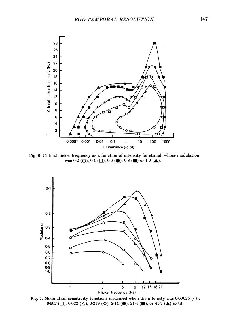
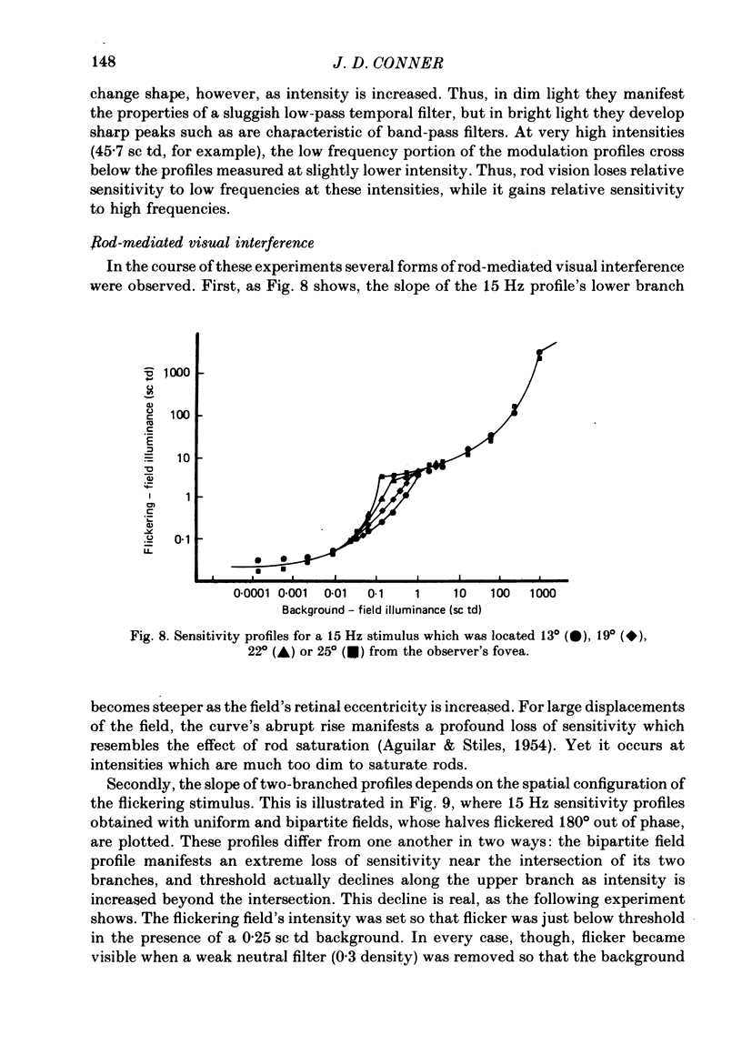
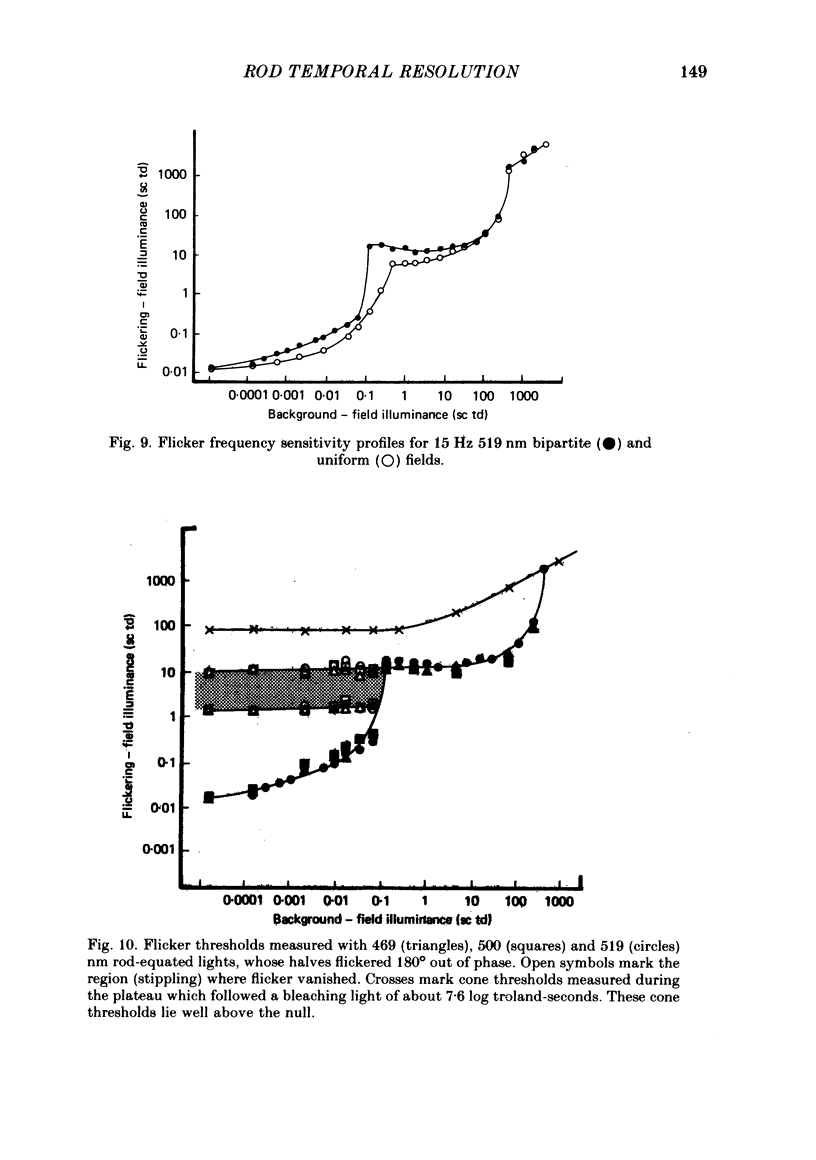
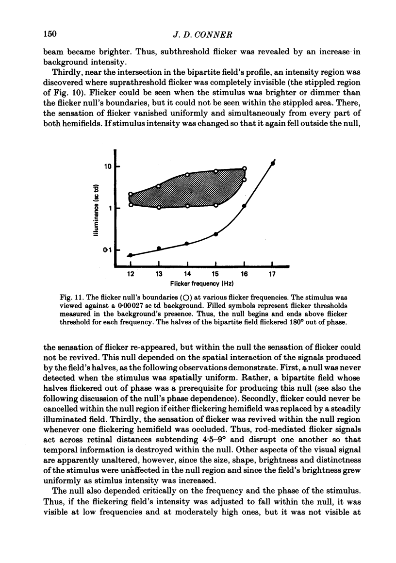
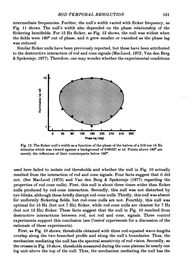
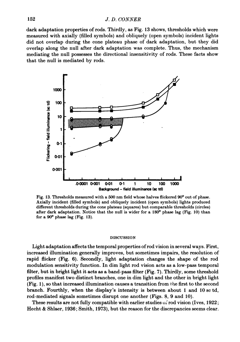
1. Profiles which represent rod thresholds for flickering fields seen against backgrounds of various intensity have shapes which depend on flicker frequency. Low frequency profiles rise smoothly as background intensity is increased. High frequency profiles are only affected by bright backgrounds, which cause them to rise steeply. Intermediate frequency profiles contain two distinct branches which resemble separate increment threshold functions. 2. The high intensity branches of two-branched threshold profiles cannot be attributed to cone intrusion. Instead, both branches of such profiles are mediated by visual mechanisms which have the spectral properties, the dark adaptation properties and the directional insensitivity of rod vision. Thus, the stimuli are detected by rods. 3. Plots of critical flicker frequency (c.f.f.) as a function of intensity contain two rising branches which are separated by a plateau (when modulation depth is large), or they form two enclosed lobes so that only intermediate frequencies, but neither high nor low ones, are seen (when modulation depth is small). C.f.f. is profoundly depressed by very bright light (above 100 scotopic trolands) which saturates rod vision. 4. In dim light rod modulation sensitivity functions resemble those of low-pass filters, but in bright light they resemble those of band-pass filters. 5. Several forms of rod mediated interference occur at moderate intensities, where rod vision's temporal properties ordinarily improve abruptly. With certain stimuli, rod signals conveying temporal information disrupt one another so completely that suprathreshold flicker cannot be seen within a ten-fold intensity range. 6. Many of these observations can be explained by the hypothesis that rod vision comprises two temporal channels which have different properties.
Full text
PDF
Selected References
These references are in PubMed. This may not be the complete list of references from this article.
- Baylor D. A., Lamb T. D., Yau K. W. The membrane current of single rod outer segments. J Physiol. 1979 Mar;288:589–611. [PMC free article] [PubMed] [Google Scholar]
- Bruch T. A. Rod-cone independence in dark adaptation. Vision Res. 1976;16(6):591–600. doi: 10.1016/0042-6989(76)90005-5. [DOI] [PubMed] [Google Scholar]
- Conner J. D., MacLeod D. I. Rod photoreceptors detect rapid flicker. Science. 1977 Feb 18;195(4279):698–699. doi: 10.1126/science.841308. [DOI] [PubMed] [Google Scholar]
- Ingling C. R., Jr, Lewis A. L., Loose D. R., Myers K. J. Cones change rod sensitivity. Vision Res. 1977;17(4):555–563. doi: 10.1016/0042-6989(77)90054-2. [DOI] [PubMed] [Google Scholar]
- MacLeod D. I., Hayhoe M. Rod origin of prolonged afterimages. Science. 1974 Sep 27;185(4157):1171–1172. doi: 10.1126/science.185.4157.1171. [DOI] [PubMed] [Google Scholar]
- MacLeod D. I. Rods cancel cones in flicker. Nature. 1972 Jan 21;235(5334):173–174. doi: 10.1038/235173a0. [DOI] [PubMed] [Google Scholar]
- Makous W., Boothe R. Cones block signals from rods. Vision Res. 1974 Apr;14(4):285–294. doi: 10.1016/0042-6989(74)90078-9. [DOI] [PubMed] [Google Scholar]
- Smith R. A., Jr Luminance-dependent changes in mesopic visual contrast sensitivity. J Physiol. 1973 Apr;230(1):115–135. doi: 10.1113/jphysiol.1973.sp010178. [DOI] [PMC free article] [PubMed] [Google Scholar]
- van den Berg T. J., Spekreijse H. Interaction between rod and cone signals studied with temporal sine wave stimulation. J Opt Soc Am. 1977 Sep;67(9):1210–1217. doi: 10.1364/josa.67.001210. [DOI] [PubMed] [Google Scholar]


