



Abstract
Fetal alcohol exposure at any stage of pregnancy can lead to fetal alcohol spectrum disorder (FASD), a group of life‐long conditions characterized by congenital malformations, as well as cognitive, behavioral, and emotional impairments. The teratogenic effects of alcohol have long been publicized; yet fetal alcohol exposure is one of the most common preventable causes of birth defects. Currently, alcohol abstinence during pregnancy is the best and only way to prevent FASD. However, alcohol consumption remains astoundingly prevalent among pregnant women; therefore, additional measures need to be made available to help protect the developing embryo before irreparable damage is done. Maternal nutritional interventions using methyl donors have been investigated as potential preventative measures to mitigate the adverse effects of fetal alcohol exposure. Here, we show that a single acute preimplantation (E2.5; 8‐cell stage) fetal alcohol exposure (2 × 2.5 g/kg ethanol with a 2h interval) in mice leads to long‐term FASD‐like morphological phenotypes (e.g. growth restriction, brain malformations, skeletal delays) in late‐gestation embryos (E18.5) and demonstrate that supplementing the maternal diet with a combination of four methyl donor nutrients, folic acid, choline, betaine, and vitamin B12, prior to conception and throughout gestation effectively reduces the incidence and severity of alcohol‐induced morphological defects without altering DNA methylation status of imprinting control regions and regulation of associated imprinted genes. This study clearly supports that preimplantation embryos are vulnerable to the teratogenic effects of alcohol, emphasizes the dangers of maternal alcohol consumption during early gestation, and provides a potential proactive maternal nutritional intervention to minimize FASD progression, reinforcing the importance of adequate preconception and prenatal nutrition.
Keywords: DNA methylation, DOHaD, embryonic development, environmental exposure, FASD, fetal alcohol exposure, maternal nutrition, methyl donors
Preventative supplementation of the maternal diet with the four main methyl donor nutrients, folic acid, choline, betaine and vitamin B12, before conception and throughout pregnancy can mitigate the adverse developmental effects of a single acute alcohol exposure during preimplantation (E2.5). At late gestation (E18.5), alcohol‐exposed embryos subjected to methyl donor supplementation had better long‐term health outcomes as evidenced by improved craniofacial dimensions, reduced incidence of morphological defects and fewer types of defects.

Abbreviations
- CpG
cytosine‐phosphate‐guanine dinucleotide
- Ctl
control
- DOHaD
developmental origins of health and disease
- E
embryonic day
- ENR
methyl donor‐enriched maternal diet
- EtOH
ethanol
- FASD
fetal alcohol spectrum disorder
- H19
H19 imprinted maternally expressed transcript
- ICR
imprinting control region
- IGF2
insulin‐like growth factor 2
- IGF2R
insulin‐like growth factor 2 receptor
- LTR
long terminal repeats
- SAM
S‐adenosylmethionine
- STD
standard maternal diet
1. INTRODUCTION
Fetal alcohol spectrum disorder (FASD) includes a broad range of adverse developmental conditions that result from maternal alcohol intake during any stage of pregnancy. 1 , 2 , 3 , 4 , 5 Even though alcohol is a well‐known teratogen and there are many awareness‐raising initiatives for healthcare providers and the general public, 6 , 7 , 8 an estimated 10% of pregnant women consume alcohol globally, of which over one quarter are reported to engage in binge drinking (single acute episode) specifically. 9 , 10 , 11 The global prevalence of FASD among children and youth is estimated at 0.8%, affecting approximately 0.9% in the Americas, 2% in European countries, and 11% in South Africa. 12 Individuals with FASD can exhibit a variety of mild to severe phenotypes including congenital anomalies (e.g. craniofacial malformations, ossification delays, intrauterine growth retardation, structural brain abnormalities), as well as cognitive (e.g. intellectual and learning disabilities), behavioral, and emotional disorders. 13 , 14 , 15 , 16 Various factors such as the amount of alcohol consumed and the timing, duration, and frequency of the exposure during embryonic development may contribute to the phenotypic variability of FASD. 3 , 17 , 18 For instance, studies suggest that a single acute (i.e., binge) exposure (peak blood alcohol level of ≥200 mg/dL) can be more damaging to the developing embryo than a chronic, milder exposure (blood alcohol level of ≤100 mg/dL). 19 , 20 , 21 , 22 Moreover, different inbred mouse strains have been shown to exhibit variable responses to fetal alcohol exposure, 23 , 24 , 25 , 26 suggesting that genetic differences play a part in interindividual susceptibility and resistance to the teratogenic effects of alcohol. Secondary factors, such as poor nutritional status of the mother 27 , 28 and exposure to early life stress, 29 have also been shown to contribute to the severity of FASD phenotypes.
Fetal alcohol spectrum disorder animal models have shown that alcohol exposure interferes with DNA methylation pathways in the developing embryo 30 , 31 , 32 , 33 , 34 , 35 and DNA methylation alterations in blood and buccal cells have been associated with FASD in cohorts of children. 36 , 37 , 38 The regulation of gene expression by DNA methylation mechanisms rely on one‐carbon metabolism, composed of the interdependent folate and methionine cycles, through which S‐adenosylmethionine (SAM) is produced, the universal methyl donor in cellular methylation reactions. 39 , 40 , 41 Folate, choline, betaine, and vitamin B12 are essential nutrients involved in SAM synthesis, and collectively referred to as methyl donors. Alcohol use can reduce bioavailability of many nutrients including folate and vitamin B12 42 , 43 , 44 , 45 , 46 as well as dysregulate one‐carbon metabolism pathways 47 , 48 , 49 , 50 and DNA methylation mechanisms. 51 , 52 , 53 , 54 During pregnancy, women are vulnerable to deficiencies in methyl donor nutrients as maternal metabolic demand for these nutrients increases to support fetal and placental development. 55 , 56 , 57 Consequently, many women do not consume the recommended dietary intake for several of these methyl donor nutrients during pregnancy. 58 , 59 , 60 , 61 Severe congenital abnormalities have been associated with maternal nutritional deficiencies in methyl donors. For example, maternal folate deficiency can lead to neural tube defects, 62 , 63 and maternal choline deficiency in mice has been shown to hinder development of the cerebral cortex 64 and hippocampus 65 and alter cognitive function in offspring. 66 Furthermore, studies have demonstrated that the teratogenic effects of alcohol in rats and mice can be intensified by suboptimal intake of methyl donors during pregnancy, worsening developmental outcomes in offspring. 67 , 68
Although we know that FASD can stem from fetal alcohol exposure taking place at any time during pregnancy, preimplantation, which occurs between fertilization and embryonic day 4.5 (E4.5) in mice or E7‐9 in humans, 69 , 70 , 71 is arguably the stage of development most at‐risk for unintended exposure. The likelihood of accidental fetal alcohol exposure during preimplantation is substantial considering that human chorionic gonadotropin (i.e., the hormone detected by pregnancy tests) is only produced after implantation of the embryo, 72 that 40% of pregnancies are unplanned, 73 and that there is an increasing rate of alcohol consumption and binge drinking behavior among women of reproductive age, 74 , 75 , 76 , 77 possibly even more so since the COVID‐19 pandemic. 78 During preimplantation, the embryo undergoes reprogramming of epigenetic modifications, notably DNA methylation, which is crucial for normal gene expression regulation during development 79 , 80 , 81 , 82 and is particularly vulnerable to unfavorable in utero environments, 83 for example, assisted reproductive technologies, 84 , 85 , 86 , 87 , 88 nutritional imbalances, 89 , 90 , 91 or exposures to chemical compounds (e.g. insecticides) 92 and alcohol. 30 , 33 , 35 Defective embryonic epigenetic reprogramming can result in DNA methylation dysregulation at imprinting control regions (ICRs), 93 , 94 , 95 , 96 , 97 which can lead to a myriad of developmental disorders 98 , 99 , 100 , 101 as ICRs control a cohort of genes (i.e., imprinted genes) that regulate growth and development of the fetus and placenta, 102 for example, H19 (H19 imprinted maternally expressed transcript), 103 IGF2 (insulin‐like growth factor 2), 104 and IGF2R (insulin‐like growth factor 2 receptor). 105 FASD in vivo models have shown that preimplantation alcohol exposure can induce permanent FASD‐like physical, behavioral, and molecular (i.e., epigenetic, transcriptomic) phenotypes observable throughout later stages of development. 30 , 33 , 106 , 107 , 108 , 109 In a previous study, we showed that a single acute preimplantation (E2.5) alcohol exposure in mice led to severe morphological defects (e.g. growth restriction, brain malformations) and DNA methylation dysregulation at some ICRs and neurodevelopmental loci in forebrain at midgestation (E10.5). 30 However, because of the all‐or‐nothing tenet, meaning the embryo would either die prior to implantation or survive unharmed, research has been mainly focused on the effects of alcohol exposure during postimplantation developmental stages. 20 , 23 , 24 , 25 , 26 , 34 , 110 , 111 , 112 , 113 Further research on the specific and long‐term teratogenic effects of alcohol exposure during preimplantation is necessary to have a more comprehensive understanding of FASD etiology.
Despite FASD being a serious widespread concern with lifelong health consequences, there are no proactive therapeutic measures in place to mitigate the effects of fetal alcohol exposure before birth. Studies in animal models and human subjects suggest that maternal nutritional supplementation of various methyl donor nutrients may have a protective effect, significantly minimizing the incidence of developmental defects and behavioral impairments as well as rescuing DNA methylation alterations in fetuses and offspring exposed to alcohol in utero. 110 , 111 , 112 , 113 , 114 , 115 , 116 , 117 , 118 However, in most of these studies, only one or two types of methyl donor nutrients were supplemented in the maternal diet, which could lead to unbalanced metabolism cycles, 119 and the amounts supplemented were strikingly higher than standard rodent chow (e.g. NIH‐31 rodent formula) levels (i.e., 7.5‐ to 30‐fold higher). Excess supplementation of various methyl donor nutrients in the maternal diet has been linked to DNA methylation alterations in cow and mice offspring 120 , 121 and high maternal intake of folic acid has been associated with insulin resistance in children 122 and rat offspring 123 and to altered synaptic transmission and increased susceptibility to seizures in rat offspring. 124 Since high‐dose supplementation can be harmful for the developing fetus and increase the risk of disease, we investigated if moderate supplementation of a combination of the four methyl donor nutrients folic acid, choline, betaine, and vitamin B12 in the maternal diet prior to and throughout pregnancy could mitigate late‐gestation (E18.5) detrimental effects (i.e., morphological defects) resulting from a single acute fetal alcohol exposure during mouse preimplantation (E2.5), without enhancing the risk of DNA methylation dysregulation at imprinting control regions.
2. MATERIALS AND METHODS
2.1. Maternal diets
Animal work was approved by the Comité institutionnel de bonnes pratiques animales en recherche of the Centre Hospitalier Universitaire Sainte‐Justine Research Center under the guidance of the Canadian Council on Animal Care. For all experiments, mice were maintained on a 12‐h light/dark cycle. At 4 weeks of age, female C57BL/6 mice (n = 8–9/condition) (Charles River, Canada) were fed ad libitum one of two defined diets for 4 weeks prior to mating, to allow stabilization of nutrients in serum, 125 and were fed the same diets throughout gestation until embryo collection (Figure 1). The NIH‐31 rodent formula (Envigo) containing 1.89 g/kg choline, 2 mg/kg folic acid and 0.06 mg/kg B12 was used as the standard diet, and a modified formula supplemented with methyl donors (5.76 g/kg choline, 5 g/kg betaine, 5 mg/kg folic acid, 0.5 mg/kg vitamin B12) as the enriched diet. After 4 weeks on their respective diet, females were mated with C57BL/6 males of same age (Charles River, Canada) that were kept on the standard diet. Females that showed copulatory plugs the next morning were considered pregnant with day 0.5 embryos (E0.5). These females were separated from the males and housed together with unlimited access to food (standard or enriched diet) and water.
FIGURE 1.

Mouse model combining a maternal methyl donor‐enriched diet and a single acute preimplantation alcohol exposure. CB57BL/6 females were fed a standard or a methyl donor‐enriched (folic acid, choline, betaine, and vitamin B12) diet for 4 weeks to stabilize serum nutrient levels prior to mating with CB57BL/6 males kept on a standard diet; Respective maternal diets were continued throughout gestation. Pregnant females were subjected to a single acute alcohol exposure (2.5 g/kg EtOH) or 0.15 M saline solution (control) by two subcutaneous injections of equivalent volumes with a 2‐h interval to target E2.5 stage embryos (~8‐cell stage). E18.5 embryos were harvested for morphological analysis, skeletal staining, and analysis of DNA methylation and gene expression of imprinted genes in forebrain tissue. Created with BioRender.com.
2.2. Acute preimplantation alcohol exposure
Using a recognized preimplantation acute alcohol exposure paradigm, 30 the pregnant mice were subcutaneously injected at E2.5 (8‐cell embryos) with two doses of 2.5 g/kg ethanol (EtOH‐exposed), or 0.15 M saline (control) with a 2‐h interval (Figure 1). Pregnant females under this paradigm reach a peak blood alcohol concentration (BAC) of 284.27 mg/dL (3 h post‐exposure) with an average of 158.31 mg/dL over a 4‐h window. 30 Females with the same diet regime or exposure treatment were housed together with negligible handling during gestation until embryo collection at E18.5 (late gestation).
2.3. Morphological analysis
At E18.5, embryos and their placenta were collected for morphological analysis. Placentae were weighed and then preserved at −80°C. Using a Leica stereo microscope and the Application Suite X (LAS X) software, embryo morphometric measurements were performed for; crown‐rump length (top of the head to the base of the tail), head length (occipital part of the head to the nasal process), head height (top of the head to the beginning of the torso), head width (width of the top of the head perpendicular to the ears), and distance between the eyes (space between the inner canthi). Morphological defects (e.g. growth restriction, eye anomaly, or any other abnormal feature) were evaluated. Next, whole E18.5 embryos were weighed, embryonic brains were extracted, weighed, and assessed for brain malformations, and forebrains were isolated for molecular analysis. Samples were kept at −80°C for further experiments. The sex of each embryo was determined by amplifying Kdm5c (X chromosome) and Kdm5d (Y chromosome) by qPCR (forward primer: CTGAAGCTTTTGGCTTTGAG, reverse primer: CCACTGCCAAATTCTTTGG) on yolk sac DNA extracted according to QIAamp DNA Mini Kit protocol (Qiagen). Number and sex of embryos in each condition group: STD‐Ctl (male: 39, female: 29, total: 68), ENR‐Ctl (male: 36, female: 33, total: 69), STD‐EtOH (male: 41, female: 29, total: 70), ENR‐EtOH (male: 44, female: 31, total: 75).
2.4. Skeletal preparation
The alcian blue and alizarin red staining protocol was adapted from Lambrot et al. 126 Embryos at E18.5 were fixed in 80% ethanol for 24 h. The skin and viscera were removed using forceps, and the fixing solution was replaced with 95% EtOH for 24 h. The embryo cartilages were stained for 24 h in alcian blue solution and washed in 95% EtOH for 5 h. To remove muscles, embryos were treated with 2% KOH solution for 24 h. The bones were then stained in 1% KOH/0.012% alizarin red S solution overnight. They were washed in 1% KOH/20% glycerol to clear the skeletons and then stored in 50% glycerol/50% EtOH. Imaging was done with a Leica stereo microscope in bright field. Number of embryos in each condition group: STD‐Ctl‐normal morphology (n = 6), ENR‐Ctl‐normal morphology (n = 6), STD‐EtOH‐normal morphology (n = 11), ENR‐EtOH‐normal morphology (n = 10), STD‐EtOH‐growth restricted (n = 7), ENR‐EtOH‐growth restricted (n = 6).
2.5. DNA extraction and DNA methylation analysis
Frozen E18.5 forebrains were first ground into powder using liquid nitrogen with a mortar and pestle and DNA was extracted according to QIAamp DNA Mini Kit protocol (Qiagen). Genomic DNA (1 μg) was then bisulfite‐converted using the EZ DNA methylation Gold kit (Zymo Research) according to the manufacturer's protocol and eluted in 100 μL EB buffer. The samples were then diluted to obtain a concentration of 1 ng/μL. Number of embryos in each condition group: STD‐Ctl‐normal morphology (n = 6), ENR‐Ctl‐normal morphology (n = 6), STD‐EtOH‐normal morphology (n = 6), ENR‐EtOH‐normal morphology (n = 6), STD‐EtOH‐growth restricted (n = 4), ENR‐EtOH‐growth restricted (n = 4).
DNA methylation levels of imprinted control regions (ICR) in forebrain samples were quantified using the Implicon technique adapted from Klobučar et al. 127 First, we designed ICR‐specific bisulfite sequencing primers and added the sequence “CTACACGACGCTCTTCCGATCT” to the forward primer and the sequence “TGCTGAACCGCTCTTCCGATCTNNNNNNNN” to the reverse primer (N corresponds to a random nucleotide) (Table S9). The random nucleotide sequences serve as unique molecular identifiers (UMI) for downstream bioinformatics analysis. For the first Implicon PCR reaction (PCR1), we used 1 ng of bisulfite‐converted DNA, 0.375 μM of pooled primers (forward and reverse), and KAPA Hifi Uracil+Ready Mix in a final volume of 8 μL for each sample and ICR. For each sample, the different amplified ICRs were pooled, cleaned up with 1.5× AMPure XP beads, and eluted in 20 μL EB buffer. This PCR pool was quantified by Qubit and diluted to a concentration of 0.40 ng/μL in a volume of 20 μL EB buffer. In the second PCR reaction, primers containing the barcoded Illumina adapters were used which ensures that each sample is amplified with a unique reverse barcoded adapter. The diluted PCR1 pool (20 μL) was amplified with 0.2 μM of Illumina PE1.0 primer, 0.2 μM of Illumina custom PE2.0 primer, and KAPA Hifi Uracil+Ready Mix in a final volume of 50 μL. After amplification, PCR2 reaction cleanup was done using 1X AMPure XP beads and eluted in 20 μL EB buffer. The libraries were sequenced with the Illumina Miseq platform to generate 250 bp paired‐end reads (62 500 reads minimum per library).
2.6. Bioinformatics analysis of DNA methylation
Quantification analysis of ICR DNA methylation levels was based on the Implicon bioinformatics pipeline. 127 First, the eight‐random nucleotide sequences serving as UMIs were filtered out and inserted into the readID of the two sequencing reads for deduplication afterward. Next, the lower quality sequences and the adaptor sequences were filtered out of the raw sequencing reads. These two steps were accomplished with Trim Galore (version 0.6.6). The sequencing reads were aligned to the mouse reference genome mm10 using Bismark (version 0.22) which is a specific program for mapping bisulfite‐treated sequencing reads and performing DNA methylation calls. After alignment, the reads were deduplicated and CpGs were extracted with Bismark. Only CpGs with a minimum sequencing coverage of 10× were retained and the average methylation level was calculated according to the coverage. To compare average CpG methylation levels between samples, we retained only the sequenced CpGs that were common among all samples.
2.7. Gene expression analysis
Gene expression levels of imprinted genes and Hprt1 (endogenous control) in E18.5 forebrains (frozen powder) of same embryos used for DNA methylation analysis were measured by quantitative PCR (qPCR) using total RNA extracted with the RNeasy Mini kit (Qiagen). Briefly, 1.5 μg of RNA for each sample was converted to cDNA using the SuperScript IV Reverse Transcriptase (Thermo Fisher). Primers specific to the regions of interest were designed with Primer3 (version 0.4.0) (Table S9) to obtain amplicons ranging from 60 to 150 bp. qPCR was performed using the SensiFAST™ SYBR® No‐ROX Kit on a LightCycler 96 (Roche Life Science). Expression levels were calculated with the 2−∆Cq method 128 using the Hprt1 gene for normalization. Mean normalized expression of the STD‐Ctl group was used to calculate relative expression. Number of embryos in each condition group: STD‐Ctl‐normal morphology (n = 6), ENR‐Ctl‐normal morphology (n = 6), STD‐EtOH‐normal morphology (n = 6), ENR‐EtOH‐normal morphology (n = 6), STD‐EtOH‐growth restricted (n = 4), ENR‐EtOH‐growth restricted (n = 4).
2.8. Statistical analysis
Statistical analysis of weight and weight gain of dams prior to mating was done using the Shapiro–Wilk normality test and Levene's test for homogeneity of variance followed by the two‐sample t test to compare weight means or the Mann–Whitney–Wilcoxon test for weight gain means. Statistical analysis of the frequencies of total morphological defects or total ossification delays in embryos was done using pairwise chi‐squared tests with Yates continuity correction. For all other assays, statistical analysis was done as follows. First, the Shapiro–Wilk test was used to verify normality. Then, to compare the different groups of conditions, parametric tests were used if all groups of conditions passed the normality test and non‐parametric tests were used if at least one condition group failed the normality test. Parametric tests: Levene's test was used to verify homogeneity of variance among the groups of conditions and then one‐way anova with pairwise comparisons and, if required, Welch's correction for unequal variances was performed with Benjamini–Hochberg p‐value adjustment. Non‐parametric tests: Kruskal–Wallis test and Pairwise Wilcoxon Rank Sum tests were performed with Benjamini–Hochberg p‐value adjustment.
3. RESULTS
3.1. Maternal diet enriched in methyl donors reduces morphological abnormalities associated with early fetal alcohol exposure
To avoid the increased risk of harmful outcomes associated with excessive methyl donor supplementation, we investigated if a moderate increase in nutrient availability of folic acid, choline, betaine, and vitamin B12 in the maternal diet could counteract the detrimental effects of early fetal alcohol exposure. Dams were fed either a standard (STD) or a methyl donor‐enriched diet (ENR) 4 weeks prior to mating 125 with males fed the standard diet (Figure 1). Females fed the standard or enriched diet showed no difference in weight gain curves (Figure S1, Table S1). Then, using our established preclinical mouse model of acute preimplantation (E2.5) alcohol exposure, 30 females on standard or enriched diets were subjected to a single acute level of ethanol (EtOH) or saline control solution and were kept on their respective diet until embryo collection at late gestation (E18.5) (Figure 1). The average number of embryos per litter was not significantly affected by either the ethanol exposure or the enriched diet (Figure S2, Table S5). Assessment of embryonic morphometric measurements (n = 68–75 embryos/condition, n = 8–9 litters/condition) showed that the maternal enriched diet alone did not significantly influence overall embryo morphometric measurements (Figure 2, Tables S2 and S3). In EtOH‐exposed embryos, the maternal enriched diet led to lower body weight and shorter crown‐rump distance (Figure 2A,G, Tables S2 and S3) compared to the maternal standard diet, but either measurement did not significantly differ from controls. Ethanol exposure of embryos subjected to the maternal standard diet led to significantly higher brain weight, longer head height, and longer head width (Figure 2B,D,E, Tables S2 and S3). The maternal enriched diet significantly safeguarded embryo head width against the effects of ethanol exposure but had no beneficial impact on embryo brain weight or head height. In both maternal diet conditions, ethanol exposure did not affect embryo body weight, brain to body weight ratio, head length, crown‐rump distance, distance between eyes, or placenta weight (Figure 2A,C,F–I, Tables S2and S3).
FIGURE 2.

Maternal methyl donor supplementation alleviates late‐gestation abnormal morphometrics induced by early fetal alcohol exposure. Morphometric measurements of E18.5 embryos subjected to a maternal standard diet (control; n = 68, ethanol‐exposed; n = 70) or a maternal methyl donor‐enriched diet (control; n = 69, ethanol‐exposed n = 75). Statistical analysis: Shapiro–Wilk test followed by either non‐parametric tests (Kruskal–Wallis test and Pairwise Wilcoxon Rank Sum tests) or parametric tests (Levene's test and one‐way anova with pairwise comparisons and, if required, Welch's correction) with Benjamini–Hochberg p‐value adjustment. *p ≤ .05, **p ≤ .01, ***p ≤ .001. See associated Tables S2 and S3.
To further explore if a methyl donor‐enriched maternal diet could have a protective effect against early fetal alcohol exposure, we performed a detailed assessment of visible morphological defects in late‐gestation embryos (E18.5) (Figure 3A–C). The types of defects included growth restriction (Figure 3C‐III, XI, XII), eye defects (e.g. anophthalmia, microphthalmia) (Figure 3C‐VII), brain anomalies (e.g. forebrain or midbrain malformations), and other defects (e.g. body dysmorphism, polydactyly; Figure 3C‐IV, VIII). The overall frequency of morphological defects was markedly highest in EtOH‐exposed embryos subjected to a maternal standard diet (Figure 3A, Table S4), which also displayed a broader frequency distribution among the different types of defects (Figure 3B, Table S4). Interestingly, early fetal alcohol exposure was particularly linked to brain malformations, which were exclusively identified in EtOH‐exposed embryos, and to growth restriction, which was prevalent in EtOH‐exposed embryos for both the standard and enriched maternal diet conditions (Figure 3B, Table S4). Compared to the standard maternal diet, the enriched maternal diet led to a considerably lower overall frequency of morphological defects in EtOH‐exposed embryos (STD‐EtOH: 0.19, ENR‐EtOH: 0.07) (Figure 3A, Table S4). The maternal enriched diet also eliminated certain types of ethanol‐induced defects; only brain malformations and growth restriction were observed in EtOH‐exposed embryos (Figure 3B, Table S4). To account for possible maternal effects in response to the various prenatal environmental conditions, we next examined the frequency of embryos with morphological defects per litter (Figure 3D, Table S5). The maternal enriched diet led to a lower proportion of affected litters compared to the maternal standard diet in both control (STD‐Ctl: 3/8 vs. ENR‐Ctl: 1/8) and ethanol exposure (STD‐EtOH: 5/9 vs. ENR‐EtOH: 3/9) groups. When looking at the range of frequencies of embryos with morphological defects in the affected litters, ethanol exposure led to more severely affected litters compared to controls for both maternal diets, but to a more moderate extent for the enriched diet (STD‐Ctl: 0.10–0.13, ENR‐Ctl: 0.17, STD‐EtOH: 0.17–0.43, ENR‐EtOH: 0.20–0.27) (Table S5).
FIGURE 3.

Maternal methyl donor‐enriched diet protects embryos from late‐gestation morphological defects associated with early fetal alcohol exposure. Total frequency of morphological defects (A) and of the specific types of morphological defects (growth restriction, brain anomalies, eye defects, and other defects) (B) in E18.5 embryos subjected to a maternal standard diet (control; n = 68, ethanol‐exposed; n = 70) or a maternal methyl donor‐enriched diet (control; n = 69, ethanol‐exposed n = 75). See associated Table S4. Statistical analysis (A): pairwise chi‐squared tests with Yates continuity correction *p ≤ .05, **p ≤ .01, ***p ≤ .001. (C) Images of E18.5 morphologically normal embryos and embryos with various morphological defects associated with ethanol exposure for both maternal diet conditions. (D) Comparison of the number of affected litters, number of E18.5 embryos per litter, and frequency of E18.5 embryos with morphological defects per litter between the four conditions. See associated Table S5.
3.2. Effect of early fetal alcohol exposure on skeletal development
Studies have shown that maternal alcohol intake during pregnancy can hinder skeletal development leading to ossification delays and craniofacial malformations in fetuses and offspring. 106 , 107 , 110 , 111 Therefore, we investigated if such delays and malformations would be visible at late gestation (E18.5) following early fetal alcohol exposure, if they would be particularly linked to the growth restriction phenotype prevalently observed in EtOH‐exposed E18.5 embryos, and if a maternal diet enriched in methyl donors would have a protective effect on skeletal development. To do so, we performed skeletal staining of E18.5 morphologically normal embryos (control and EtOH‐exposed) and growth‐restricted EtOH‐exposed embryos subjected to either the standard or the enriched maternal diet and completed a thorough analysis of primary ossification and craniofacial structure (Figure 4A–C, Table S6). The selection of growth‐restricted EtOH‐exposed E18.5 embryos was based on their abnormal stunted morphology and significantly lower body weight (Figure 4D, Tables S7 and S8). For all samples, we did not observe abnormal vertebral body count, limb defects, bone deformities, severe craniofacial structural anomalies or generalized skeletal dysplasia. We did however detect ossification delays exclusively in EtOH‐exposed embryos, most predominantly in growth‐restricted embryos for both maternal diets (Figure 4A, Table S6). All growth‐restricted EtOH‐exposed embryos under the standard maternal diet had ossification delays (7/7, frequency = 1.00) whereas a slightly lower frequency of ossification delays was observed in growth‐restricted EtOH‐exposed embryos under the enriched maternal diet (5/6, frequency = 0.83) (Figure 4A, Table S6). Morphologically normal EtOH‐exposed embryos under both maternal diets showed much lower frequencies of ossification delays (STD‐EtOH (normal): 1/11, frequency = 0.091; ENR‐EtOH (normal): 1/10, frequency = 0.10) (Figure 4A, Table S6). We observed delayed ossification of the occipital bone (Figure 4B,C‐III, XII) and 5th sternebrae (Figure 4B,C‐VI, XV), with several EtOH‐exposed embryos showing delays in both (Table S6). Growth‐restricted EtOH‐exposed embryos also had mild craniofacial hypoplasia demonstrated by significantly shorter head and snout length compared to morphologically normal control and EtOH‐exposed embryos (Figure 4C‐IX, XVIII, E, F, Tables S7 and S8). Overall, the maternal methyl donor‐enriched diet had little to no effect in preventing skeletal abnormalities associated with early fetal alcohol exposure, which were primarily linked to the ethanol‐induced growth restriction phenotype.
FIGURE 4.

Growth restriction linked to early fetal alcohol exposure is accompanied by ossification delays and mild craniofacial hypoplasia in both standard and enriched maternal diet conditions. Total frequency of ossification delays (A) and of occipital bone and 5th sternebrae ossification delays specifically (B) in E18.5 embryos with normal morphology (control and ethanol‐exposed) and with ethanol exposure‐associated growth restriction in both standard and enriched maternal diet conditions. See associated Table S6. Statistical analysis (A): pairwise chi‐squared tests with Yates continuity correction. (C) E18.5 embryo skeletal staining images showing examples of normal ossification, ossification delays (occipital bone (III, XII), 5th sternebrae (VI, XV)), and mild craniofacial hypoplasia (shorter head and snout length (IX, XVIII)). (D–F) Body weight, head length, and snout length of E18.5 embryos stained for skeletal analysis. See associated Tables S6 and S7. Statistical analysis (D–F): Shapiro–Wilk test followed by either non‐parametric tests (Kruskal–Wallis test and pairwise Wilcoxon rank sum tests) or parametric tests (Levene's test and one‐way anova with pairwise comparisons and, if required, Welch's correction) with Benjamini–Hochberg p‐value adjustment. Number of embryos: standard maternal diet; control (normal morphology: n = 6) and ethanol‐exposed embryos (normal morphology: n = 11, growth‐restricted: n = 7), enriched maternal diet; control (normal morphology: n = 5) and ethanol‐exposed embryos (normal morphology: n = 10, growth‐restricted: n = 6). *p ≤ .05, **p ≤ .01, ***p ≤ .001.
3.3. Effect of early fetal alcohol exposure on imprinted DNA methylation and gene expression
It has been well established that DNA methylation patterns must be faithfully maintained in imprinting control regions (ICRs) during early embryonic development 93 , 95 , 96 and that alcohol exposure as well as methyl donor intake (deficiency or excess) can interfere with these mechanisms. 30 , 32 , 33 , 35 Since many imprinted genes are involved in embryonic and placental growth regulation, 103 , 104 , 105 we questioned whether the growth restriction phenotype induced by early fetal alcohol exposure could be linked to dysregulation of ICR DNA methylation and expression of imprinted genes and, if so, whether the maternal methyl donor‐enriched diet would have a protective effect. Using a targeted approach (Implicon 127 ) that enables the detection of DNA methylation at single‐CpG resolution within a 250–500 bp DNA segment, we examined two regions within the H19 ICR associated with the H19‐Igf2 locus 129 (i.e., H19 ICR‐A, ICR‐B) and one ICR associated with Igf2r, 130 three imprinted genes with well‐defined growth regulatory roles during early development, as well as several other ICRs associated with Gpr1‐Zdbf2, Nesp, Gnas, Mest/Peg1, Snrpn, and Commd1‐Zrsr1. We compared ICR DNA methylation (Figure 5A, Figures S3 and S4) as well as expression levels (Figure 5B–D, Figure S5) among E18.5 morphologically normal control and EtOH‐exposed embryos, and growth‐restricted EtOH‐exposed embryos subjected to either the standard or the enriched maternal diet. Neither H19 nor Igf2r ICRs showed any significant differences in single‐CpG DNA methylation levels (Figure 5A, Figure S3A) or in average ICR DNA methylation levels (Figure S3B) among the different conditions. There were also no significant differences in average DNA methylation levels for the other six ICRs we examined (Figure S4). For all embryos, DNA methylation levels of all ICRs were within a standard range of 45%–55%. Although no changes in DNA methylation were detected, growth‐restricted EtOH‐exposed embryos subjected to the maternal enriched diet had a slight increase in H19 and Gnas expression (~1.3‐fold compared to STD‐Ctl) (Figure 5B, Figure S5D). No significant differences in gene expression were detected for any other imprinted gene (Figure 5C,D, Figure S5A–C,E–H). Altogether, neither early fetal alcohol exposure nor the maternal methyl donor‐enriched diet had any major impact on DNA methylation at these specific ICRs or on gene expression of associated imprinted genes in forebrain of late‐gestation embryos, even in those with an ethanol‐induced growth restriction phenotype.
FIGURE 5.
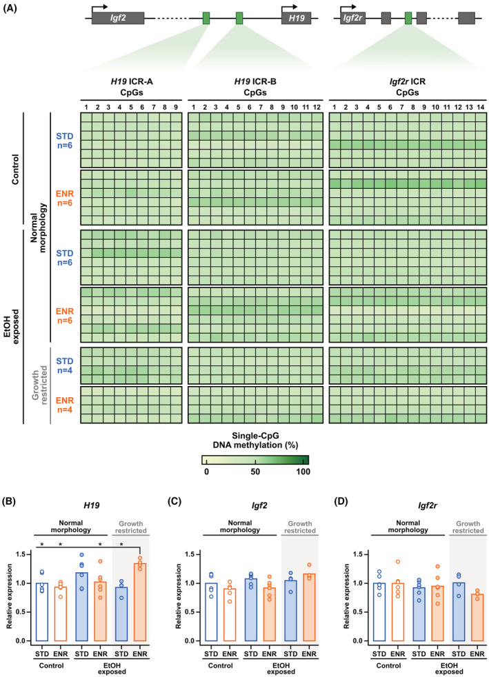
Early fetal alcohol exposure does not affect DNA methylation at the H19 and Igf2r ICRs in forebrain of late‐gestation embryos. (A) DNA methylation levels of single‐CpGs in two regions of the H19 ICR (H19 ICR‐A/B) and at the Igf2r ICR in E18.5 morphologically normal embryos (control or EtOH‐exposed) and growth‐restricted EtOH‐exposed embryos subjected to a standard (STD) or enriched (ENR) maternal diet. See associated Figure S3. (B–D) Relative expression levels (normalized to Hprt1 and STD‐Control) of H19, Igf2, and Igf2r, respectively, in same embryos used for DNA methylation analysis. Gene expression statistical analysis (B–D): Shapiro–Wilk test followed by Levene's Test and one‐way anova with pairwise comparisons, Welch's correction (if required), and Benjamini–Hochberg p‐value adjustment. *p ≤ .05. Number of embryos: Standard maternal diet: control (normal morphology: n = 6) and ethanol‐exposed embryos (normal morphology: n = 6, growth‐restricted: n = 4); Enriched maternal diet: control (normal morphology: n = 6) and ethanol‐exposed embryos (normal morphology: n = 6, growth‐restricted: n = 4).
4. DISCUSSION
It is now recognized that fetal alcohol exposure during any gestation period can have serious teratogenic effects on embryo development and lead to FASD, 1 , 2 , 3 , 4 , 5 , 14 yet the impact of alcohol exposure during the preimplantation period, a particularly vulnerable developmental window, 83 remains considerably understudied. Although FASD is a worldwide public health burden, 12 , 131 there are no tangible measures to proactively mitigate the effects of fetal alcohol exposure prior to birth. One way to prevent or attenuate the related adverse developmental outcomes could be through maternal diet interventions using methyl donors. 110 , 111 , 112 , 113 , 114 , 115 , 116 , 117 , 118 Here, we demonstrate that exposure to acute levels of alcohol during preimplantation (E2.5) leads to long‐term FASD‐like phenotypes in late‐gestation embryos (E18.5), and show that moderate enrichment of folic acid, choline, betaine, and vitamin B12 in the maternal diet prior to conception and throughout gestation has a protective effect, significantly reducing the incidence and severity of developmental defects in ethanol‐exposed embryos.
4.1. Lasting developmental impact of early preimplantation alcohol exposure
A growing body of evidence substantiates that the preimplantation embryo (i.e., fertilization to E4.5 in mice, E7‐9 in humans) is vulnerable to alcohol teratogenicity, 30 , 33 , 106 , 107 , 108 , 109 refuting the outdated all‐or‐nothing premise that preimplantation alcohol exposure leads to embryos either surviving unharmed or dying during early gestation. Here, acute ethanol exposure at E2.5 (i.e., two doses of 2.5 g/kg with a 2‐h interval) led to a variety of morphological malformations at late gestation (E18.5), including growth restriction, brain anomalies (e.g. forebrain or midbrain malformations), eye defects (e.g. anophthalmia, microphthalmia), and other defects such as polydactyly and body dysmorphism, as well as higher brain weight and larger head dimensions (i.e., head height and width). The frequency (19% of embryos) and types of late‐gestation malformations were highly similar to what we reported at midgestation (E10.5) in a previous study using the same exposure model, 30 suggesting that, in affected embryos, the adverse developmental effects of acute alcohol exposure during preimplantation are prompt and persist over time. Higher‐dose acute ethanol exposure during preimplantation can dramatically increase the severity of morphological malformations and the proportion of affected embryos. For instance, in a pioneer study where mouse dams were treated with a single acute dose of 5.8 g/kg ethanol at E2, there was a high incidence of fetal death occurring prior to E15 and severe malformations at E14 (e.g. hydrocephaly, craniofacial hypoplasia, delayed palatal closure, renal hypoplasia, exomphalos, severe growth restriction) were observed in 67%–100% of ethanol‐exposed embryos, with a considerable proportion of embryos having a combination of several malformations. 106 In our model, ~80% of ethanol‐exposed embryos were morphologically normal at mid‐ and late gestation, appearing to have either escaped or self‐restored the damage inflicted by alcohol toxicity. Though it is also possible that, despite being physically normal, neurobehavioral function in these embryos could be affected, which would only become apparent after birth. Although highly debilitating, physical defects only account for a small minority of FASD symptoms; children with FASD frequently have so‐called invisible symptoms such as intellectual, behavioral, and emotional disorders, which only manifest in childhood and adolescence. 132 , 133 , 134 At midgestation, we showed that forebrain of both morphologically normal and abnormal embryos exposed to alcohol during preimplantation had DNA methylation alterations in genomic regions linked to neurodevelopmental processes accompanied by some associated gene expression dysregulation, 30 which could eventually lead to defective cerebral cortex development and function, a main cause of neurodevelopmental and neurological disorders. 135 , 136 , 137
As with postimplantation alcohol exposure models, it is becoming clear that the alcohol exposure paradigm during preimplantation greatly influences teratogenesis. However, a prevalent developmental defect observed in fetal alcohol exposure studies regardless of dosage, duration, or developmental timing is intrauterine growth restriction, 106 , 107 , 110 , 111 which can be associated with long‐term physical ramifications, including skeletal malformations. 138 , 139 , 140 , 141 Although mammalian ossification of skeletal structures only begins at mid‐ to late gestation 142 (e.g. E13‐14 in mice) and studies identify gastrulation through neurulation (i.e., E6.25–E10 in mice) as a key developmental window for alcohol‐induced alteration of craniofacial development, 143 both preimplantation and postimplantation fetal alcohol exposure have been shown to hinder skeletal development. 106 , 107 , 110 , 111 In mouse models, acute high‐dose ethanol exposure (5.8 g/kg) at E2 106 or E9, 110 chronic moderate‐dose ethanol exposure (2.9 g/kg) from E0.5 to E8.5 107 and chronic high‐dose ethanol exposure (5.0 g/kg) from E6 to E15 111 led to growth restriction and to a variety of long‐term skeletal malformations ranging in severity (e.g. ossification delays, craniofacial abnormalities, vertebral anomalies, limb malformations). Here, our results further support that fetal alcohol exposure during preimplantation can have lasting adverse effects on skeletal development (i.e., ossification delays and mild craniofacial hypoplasia), and show a strong correlation between skeletal delays and alcohol‐induced growth restriction.
On a molecular level, epigenetic dysregulation of imprinted genes has been examined as a potential mechanism of intrauterine growth restriction, 102 , 144 particularly the H19‐Igf2 locus 145 , 146 , 147 as H19 suppresses, 103 while Igf2 promotes 104 placental and fetal growth. In a Japanese rice fish model of fetal alcohol exposure during the first 48 h of development, ethanol‐exposed embryos had decreased expression of DNA methyltransferase 1, 148 the enzyme responsible for maintaining DNA methylation at imprinting control regions (ICRs) in early embryonic development, 93 , 96 , 149 which could lead to permanent loss of DNA methylation at ICRs altering expression patterns of associated imprinted genes. H19‐Igf2 ICR DNA methylation (i.e., Igf2 ICR0‐3 and H19 ICR) 129 and transcriptomic dysregulation, often in a tissue‐specific manner, has been observed in various animal models of fetal alcohol exposure. 33 , 35 , 109 For instance, E16.5 mouse embryos chronically exposed to ethanol (7.25 g/kg) from E0.5 to E15.5 had tissue‐specific loss of DNA methylation at Igf2 ICR1, Igf2 ICR2, and H19 ICR in the heart, brain, and placenta, respectively, and increased Igf2 expression in the brain. 35 In another mouse model, chronic ethanol exposure (2.9 g/kg) from E0.5 to E8.5 induced H19 upregulation and downregulation in embryonic and placental tissues, respectively, and Igf2 downregulation only in the placenta at E9.5 and E16.5, but without any associated ICR DNA methylation alterations. 109 In the present study, growth restriction in embryos exposed to ethanol was not associated with DNA methylation or transcriptomic dysregulation of the H19‐Igf2 locus, or of several other imprinted loci (Igf2r, Gpr1‐Zdbf2, Nesp, Gnas, Mest/Peg1, Snrpn, and Commd1‐Zrsr1), except for a subtle (1.3‐fold) increase in H19 and Gnas expression, in fetal forebrain tissue at late gestation. However, we cannot rule out epigenetic dysregulation of H19‐Igf2, or other imprinted loci as a potential mechanism of alcohol‐induced growth restriction. In our previous study, using rRRBS (rapid reduced representation bisulfite sequencing), we observed partial loss of DNA methylation (>10% methylation difference, 100 bp tiles) at several ICRs in forebrain of ethanol‐exposed embryos at midgestation. 30 As the fetal forebrain matures from mid‐ to late gestation, the cell‐type composition is much more diversified, 150 therefore, perhaps DNA methylation dysregulation by alcohol is cell‐type specific. With recent advances in low‐input whole‐genome bisulfite sequencing and single‐cell approaches, a broader investigation of DNA methylation profiles subsequent to the time of exposure and at multiple developmental time points would be better suited to provide a more comprehensive conclusion. Moreover, since we and others have shown sex‐dependent DNA methylation alterations associated with fetal alcohol exposure, 30 , 151 , 152 increasing the number of samples would enable methodical sex‐specific analyses.
4.2. Mitigating the effects of early fetal alcohol exposure with a maternal diet enriched in methyl donor nutrients
Maternal nutritional interventions using methyl donor‐enriched diets have become increasingly promising for mitigating fetal alcohol exposure. 110 , 111 , 112 , 113 , 114 , 115 , 116 , 117 , 118 For instance, in a mouse (CD‐1) model of chronic fetal ethanol exposure (5.0 g/kg/day; E6–E15), maternal high‐dose folic acid (60.0 mg/kg; E1–E16) and vitamin B12 (1.0 mg/kg) combined supplementation mitigated developmental ethanol toxicity by reducing the incidence of skeletal abnormalities (E18). 111 However, similarly to how genetic differences can influence alcohol teratogenicity, 23 , 24 , 25 , 26 genetics may also contribute to the effectiveness of nutritional interventions during pregnancy. For example, supplementing only folic acid in the maternal diet (60.0 mg/kg; E1–E6) was able to prevent ethanol‐ (5.0 g/kg/day; E6–E15) induced defects in Balb/c mice, 112 but not in CD‐1 mice. 111 Most studies so far have used maternal diets supplemented with only one or two methyl donor nutrients at high concentration, which could potentially cause adverse effects in offspring due to metabolic imbalance or excess supplementation. 119 , 120 , 121 , 122 , 123 Here, we showed that moderate supplementation of several methyl donor nutrients (folic acid: 5 mg/kg, choline: 5.76 g/kg, betaine: 5 g/kg, and vitamin B12: 0.5 mg/kg) in the maternal diet prior to conception and throughout gestation was able to safeguard embryo head width, substantially reduce the incidence of morphological defects as well as eliminate specific types of morphological defects (e.g. dysmorphism, polydactyly, eye defects) in mouse embryos (C57BL/6) exposed to acute levels of ethanol during preimplantation (E2.5). Another multinutrient maternal supplementation intervention has also been shown to successfully mitigate the adverse effects of postimplantation acute fetal alcohol exposure (EtOH 5.8 g/kg at E9; C57BL/6) (e.g. lower prenatal mortality rate, improved prenatal growth, drastic reduction skeletal malformations) in late‐gestation embryos (E18). 110 However, their maternal methyl donor‐enriched diet (3SZM chow), in comparison to ours, had three‐fold higher levels of folic acid (15 mg/kg), choline (15 g/kg), betaine (15 g/kg) and vitamin B12 (1.5 mg/kg), and had additional supplementation of zinc (150 mg/kg) and L‐methionine (7.5 g/kg). This same 3SZM diet fed to pregnant yellow agouti (A vy ) females, in which expression of the agouti gene and coat color phenotype is driven by DNA methylation of a long terminal repeat (LTR), led to increased DNA methylation at the agouti LTR and altered coat color phenotype in pups, 121 indicating that excess supplementation of methyl donors can impact DNA methylation profiles. Thus, although we and others have demonstrated the advantages of methyl donor nutrient supplementation in the maternal diet for mitigating fetal alcohol exposure on a morphological level, we remain cautious about its long‐term impact on the embryonic epigenetic program. Sex‐, tissue‐, and cell‐specific genome‐wide analyses would be imperative to fully assess the impact of increased methyl donor nutrient levels in the maternal diet on the fetal methylome.
There have also been some studies in which postnatal nutritional interventions were used to mitigate the effects of fetal alcohol exposure. In animal models, memory and learning impairments in offspring exposed to alcohol during fetal development were moderately alleviated by postnatal choline supplementation, 153 , 154 , 155 and in humans, choline supplementation improved cognitive performance of children with FASD to some extent. 156 , 157 Overall, results of postnatal nutritional intervention studies are generally less conclusive than prenatal studies. The damage inflicted by fetal alcohol exposure may be too advanced after birth to be reversed by nutrient supplementation alone. Alternatively, the mechanisms driving the protective effect of methyl donor nutrients may occur mostly in the mother; perhaps methyl donors act on minimizing alcohol toxicity prior to it reaching the embryo rather than on correcting the aftermath.
5. CONCLUSION
Although the best way to prevent FASD remains to abstain from alcohol use during pregnancy, the results of this study suggest that supplementation of methyl donor nutrients in the maternal diet before and throughout pregnancy could be a potential proactive measure for FASD prevention, especially in cases where the mother has nutritional deficiencies, which could worsen the teratogenic effects of alcohol. 67 , 68 FASD and methyl donor nutrient deficiencies among pregnant women are particularly prevalent in lower income countries, 12 , 58 , 59 , 60 for instance, in sub‐Saharan Africa, where some regions have the highest prevalence of children and youth with FASD worldwide, 12 many women of reproductive age (≥15–49 years) are folate deficient. 61 Ultimately, our work emphasizes the dangers of alcohol consumption during early gestation and the need for proper preconception and prenatal nutrition.
AUTHOR CONTRIBUTIONS
Mélanie Breton‐Larrivée, Amanda J. MacFarlane, and Serge McGraw conceptualized the study. Mélanie Breton‐Larrivée and Lisa‐Marie Legault contributed to data acquisition. Mélanie Breton‐Larrivée, Elizabeth Elder, and Alexandra Langford‐Avelar participated in data analysis. Elizabeth Elder and Serge McGraw wrote the manuscript. All authors read and approved the final manuscript.
DISCLOSURES
The authors declare that they have no conflicts of interests.
Supporting information
Appendix S1.
ACKNOWLEDGMENTS
We thank the members of the McGraw lab for critical comments and suggestions. This work was supported by a research grant to SM from SickKids Foundation and Canadian Institutes of Health Research (CIHR). MBL is supported by a Fonds de Recherche du Québec en Santé (FRQS) Scholarship. EE is supported by a Natural Sciences and Engineering Research Council of Canada (NSERC) Canada Graduate Scholarship. LML is supported by a CIHR Canada Graduate Scholarship. ALA is supported by Université de Montréal and Réseau Québécois en reproduction (RQR) scholarships. SM is supported by a FRQS Junior 2 salary award.
Breton‐Larrivée M, Elder E, Legault L‐M, Langford‐Avelar A, MacFarlane AJ, McGraw S. Mitigating the detrimental developmental impact of early fetal alcohol exposure using a maternal methyl donor‐enriched diet. The FASEB Journal. 2023;37:e22829. doi: 10.1096/fj.202201564R
Mélanie Breton‐Larrivée and Elizabeth Elder contributed equally.
DATA AVAILABILITY STATEMENT
The DNA methylation data from this study have been submitted to GEO Expression Omnibus (GSE222974). All other data that support the findings of this study are available in the methods and supplementary material of this article.
REFERENCES
- 1. Jones KL, Smith DW. Recognition of the fetal alcohol syndrome in early infancy. Lancet. 1973;302(7836):999‐1001. [DOI] [PubMed] [Google Scholar]
- 2. Hoyme HE, Kalberg WO, Elliott AJ, et al. Updated clinical guidelines for diagnosing fetal alcohol spectrum disorders. Pediatrics. 2016;138(2):e20154256. [DOI] [PMC free article] [PubMed] [Google Scholar]
- 3. May PA, Blankenship J, Marais AS, et al. Maternal alcohol consumption producing fetal alcohol spectrum disorders (FASD): quantity, frequency, and timing of drinking. Drug Alcohol Depend. 2013;133(2):502‐512. [DOI] [PMC free article] [PubMed] [Google Scholar]
- 4. Wozniak JR, Riley EP, Charness ME. Clinical presentation, diagnosis, and management of fetal alcohol spectrum disorder. Lancet Neurol. 2019;18(8):760‐770. [DOI] [PMC free article] [PubMed] [Google Scholar]
- 5. Popova S, Dozet D, Shield K, Rehm J, Burd L. Alcohol's impact on the fetus. Nutrients. 2021;13(10):3452. [DOI] [PMC free article] [PubMed] [Google Scholar]
- 6. Lewis P. Australian guidelines to reduce health risks from drinking alcohol. National Health and Medical Research Council; 2009. [Google Scholar]
- 7. Centers for Disease Control and Prevention . Advisory on alcohol use during pregnancy. A 2005 message to women from the US surgeon general. Centers for Disease Control and Prevention; 2005. [Google Scholar]
- 8. World Health Organization . Guidelines for the identification and management of substance use and substance use disorders in pregnancy. World Health Organization; 2014. [PubMed] [Google Scholar]
- 9. Popova S, Lange S, Probst C, Gmel G, Rehm J. Estimation of national, regional, and global prevalence of alcohol use during pregnancy and fetal alcohol syndrome: a systematic review and meta‐analysis. Lancet Glob Health. 2017;5(3):e290‐e299. [DOI] [PubMed] [Google Scholar]
- 10. Popova S, Lange S, Probst C, Gmel G, Rehm J. Global prevalence of alcohol use and binge drinking during pregnancy, and fetal alcohol spectrum disorder. Biochem Cell Biol. 2018;96(2):237‐240. [DOI] [PubMed] [Google Scholar]
- 11. Lange S, Probst C, Rehm J, Popova S. Prevalence of binge drinking during pregnancy by country and World Health Organization region: systematic review and meta‐analysis. Reprod Toxicol. 2017;73:214‐221. [DOI] [PubMed] [Google Scholar]
- 12. Lange S, Probst C, Gmel G, Rehm J, Burd L, Popova S. Global prevalence of fetal alcohol spectrum disorder among children and youth: a systematic review and meta‐analysis. JAMA Pediatr. 2017;171(10):948‐956. [DOI] [PMC free article] [PubMed] [Google Scholar]
- 13. Alati R, Davey Smith G, Lewis SJ, et al. Effect of prenatal alcohol exposure on childhood academic outcomes: contrasting maternal and paternal associations in the ALSPAC study. PLoS One. 2013;8(10):e74844. [DOI] [PMC free article] [PubMed] [Google Scholar]
- 14. Feldman HS, Jones KL, Lindsay S, et al. Prenatal alcohol exposure patterns and alcohol‐related birth defects and growth deficiencies: a prospective study. Alcohol Clin Exp Res. 2012;36(4):670‐676. [DOI] [PubMed] [Google Scholar]
- 15. Larkby CA, Goldschmidt L, Hanusa BH, Day NL. Prenatal alcohol exposure is associated with conduct disorder in adolescence: findings from a birth cohort. J Am Acad Child Adolesc Psychiatry. 2011;50(3):262‐271. [DOI] [PMC free article] [PubMed] [Google Scholar]
- 16. Muggli E, Matthews H, Penington A, et al. Association between prenatal alcohol exposure and craniofacial shape of children at 12 months of age. JAMA Pediatr. 2017;171(8):771‐780. [DOI] [PMC free article] [PubMed] [Google Scholar]
- 17. Lunde ER, Washburn SE, Golding MC, Bake S, Miranda RC, Ramadoss J. Alcohol‐induced developmental origins of adult‐onset diseases. Alcohol Clin Exp Res. 2016;40(7):1403‐1414. [DOI] [PMC free article] [PubMed] [Google Scholar]
- 18. Kleiber ML, Mantha K, Stringer RL, Singh SM. Neurodevelopmental alcohol exposure elicits long‐term changes to gene expression that alter distinct molecular pathways dependent on timing of exposure. J Neurodev Disord. 2013;5(1):6. [DOI] [PMC free article] [PubMed] [Google Scholar]
- 19. Pierce DR, West JR. Blood alcohol concentration: a critical factor for producing fetal alcohol effects. Alcohol. 1986;3(4):269‐272. [DOI] [PubMed] [Google Scholar]
- 20. Bonthius DJ, West JR. Alcohol‐induced neuronal loss in developing rats: increased brain damage with binge exposure. Alcohol Clin Exp Res. 1990;14(1):107‐118. [DOI] [PubMed] [Google Scholar]
- 21. Jacobson JL, Jacobson SW. Drinking moderately and pregnancy. Effects on child development. Alcohol Res Health. 1999;23(1):25‐30. [PMC free article] [PubMed] [Google Scholar]
- 22. Jacobson JL, Jacobson SW, Sokol RJ, Ager JW Jr. Relation of maternal age and pattern of pregnancy drinking to functionally significant cognitive deficit in infancy. Alcohol Clin Exp Res. 1998;22(2):345‐351. [DOI] [PubMed] [Google Scholar]
- 23. Downing C, Balderrama‐Durbin C, Broncucia H, Gilliam D, Johnson TE. Ethanol teratogenesis in five inbred strains of mice. Alcohol Clin Exp Res. 2009;33(7):1238‐1245. [DOI] [PMC free article] [PubMed] [Google Scholar]
- 24. Downing C, Gilliam D. Cytoplasmic factors do not contribute to a maternal effect on ethanol teratogenesis. Behav Genet. 1999;29(1):31‐39. [DOI] [PubMed] [Google Scholar]
- 25. Ogawa T, Kuwagata M, Ruiz J, Zhou FC. Differential teratogenic effect of alcohol on embryonic development between C57BL/6 and DBA/2 mice: a new view. Alcohol Clin Exp Res. 2005;29(5):855‐863. [DOI] [PubMed] [Google Scholar]
- 26. Boehm SL 2nd, Lundahl KR, Caldwell J, Gilliam DM. Ethanol teratogenesis in the C57BL/6J, DBA/2J, and A/J inbred mouse strains. Alcohol. 1997;14(4):389‐395. [DOI] [PubMed] [Google Scholar]
- 27. Shankar K, Ronis MJ, Badger TM. Effects of pregnancy and nutritional status on alcohol metabolism. Alcohol Res Health. 2007;30(1):55‐59. [PMC free article] [PubMed] [Google Scholar]
- 28. Abel EL, Hannigan JH. Maternal risk factors in fetal alcohol syndrome: provocative and permissive influences. Neurotoxicol Teratol. 1995;17(4):445‐462. [DOI] [PubMed] [Google Scholar]
- 29. Alberry B, Laufer BI, Chater‐Diehl E, Singh SM. Epigenetic impacts of early life stress in fetal alcohol spectrum disorders shape the neurodevelopmental continuum. Front Mol Neurosci. 2021;14:671891. [DOI] [PMC free article] [PubMed] [Google Scholar]
- 30. Legault LM, Doiron K, Breton‐Larrivée M, et al. Pre‐implantation alcohol exposure induces lasting sex‐specific DNA methylation programming errors in the developing forebrain. Clin Epigenetics. 2021;13(1):164. [DOI] [PMC free article] [PubMed] [Google Scholar]
- 31. Chen Y, Ozturk NC, Zhou FC. DNA methylation program in developing hippocampus and its alteration by alcohol. PLoS One. 2013;8(3):e60503. [DOI] [PMC free article] [PubMed] [Google Scholar]
- 32. Stouder C, Somm E, Paoloni‐Giacobino A. Prenatal exposure to ethanol: a specific effect on the H19 gene in sperm. Reprod Toxicol. 2011;31(4):507‐512. [DOI] [PubMed] [Google Scholar]
- 33. Haycock PC, Ramsay M. Exposure of mouse embryos to ethanol during preimplantation development: effect on DNA methylation in the h19 imprinting control region. Biol Reprod. 2009;81(4):618‐627. [DOI] [PubMed] [Google Scholar]
- 34. Garro AJ, McBeth DL, Lima V, Lieber CS. Ethanol consumption inhibits fetal DNA methylation in mice: implications for the fetal alcohol syndrome. Alcohol Clin Exp Res. 1991;15(3):395‐398. [DOI] [PubMed] [Google Scholar]
- 35. Zhou J, Qian Y, Zhou Q, et al. Tissue‐specific differential DNA methylation at Igf2/H19 locus in a mouse model for embryonal ethanol exposure. Int J Clin Exp Pathol. 2016;9(4):4862‐4871. [Google Scholar]
- 36. Portales‐Casamar E, Lussier AA, Jones MJ, et al. DNA methylation signature of human fetal alcohol spectrum disorder. Epigenetics Chromatin. 2016;9:25. [DOI] [PMC free article] [PubMed] [Google Scholar]
- 37. Laufer BI, Kapalanga J, Castellani CA, Diehl EJ, Yan L, Singh SM. Associative DNA methylation changes in children with prenatal alcohol exposure. Epigenomics. 2015;7(8):1259‐1274. [DOI] [PubMed] [Google Scholar]
- 38. Masemola ML, van der Merwe L, Lombard Z, Viljoen D, Ramsay M. Reduced DNA methylation at the PEG3 DMR and KvDMR1 loci in children exposed to alcohol in utero: a South African Fetal Alcohol Syndrome cohort study. Front Genet. 2015;6:85. [DOI] [PMC free article] [PubMed] [Google Scholar]
- 39. Clare CE, Brassington AH, Kwong WY, Sinclair KD. One‐carbon metabolism: linking nutritional biochemistry to epigenetic programming of long‐term development. Annu Rev Anim Biosci. 2019;7:263‐287. [DOI] [PubMed] [Google Scholar]
- 40. Mentch SJ, Locasale JW. One‐carbon metabolism and epigenetics: understanding the specificity. Ann N Y Acad Sci. 2016;1363(1):91‐98. [DOI] [PMC free article] [PubMed] [Google Scholar]
- 41. Friso S, Udali S, De Santis D, Choi SW. One‐carbon metabolism and epigenetics. Mol Aspects Med. 2017;54:28‐36. [DOI] [PubMed] [Google Scholar]
- 42. Lieber CS. Relationships between nutrition, alcohol use, and liver disease. Alcohol Res Health. 2003;27(3):220‐231. [PMC free article] [PubMed] [Google Scholar]
- 43. Hamid A, Kaur J. Kinetic characteristics of folate binding to rat renal brush border membrane in chronic alcoholism. Mol Cell Biochem. 2005;280(1–2):219‐225. [DOI] [PubMed] [Google Scholar]
- 44. Hamid A, Wani NA, Rana S, Vaiphei K, Mahmood A, Kaur J. Down‐regulation of reduced folate carrier may result in folate malabsorption across intestinal brush border membrane during experimental alcoholism. FEBS J. 2007;274(24):6317‐6328. [DOI] [PubMed] [Google Scholar]
- 45. Halsted CH, Robles EA, Mezey E. Decreased jejunal uptake of labeled folic acid (3 H‐PGA) in alcoholic patients: roles of alcohol and nutrition. N Engl J Med. 1971;285(13):701‐706. [DOI] [PubMed] [Google Scholar]
- 46. Fragasso A, Mannarella C, Ciancio A, et al. Holotranscobalamin is a useful marker of vitamin B12 deficiency in alcoholics. ScientificWorldJournal. 2012;2012:128182. [DOI] [PMC free article] [PubMed] [Google Scholar]
- 47. Auta J, Zhang H, Pandey SC, Guidotti A. Chronic alcohol exposure differentially alters one‐carbon metabolism in rat liver and brain. Alcohol Clin Exp Res. 2017;41(6):1105‐1111. [DOI] [PMC free article] [PubMed] [Google Scholar]
- 48. Fowler AK, Hewetson A, Agrawal RG, et al. Alcohol‐induced one‐carbon metabolism impairment promotes dysfunction of DNA base excision repair in adult brain. J Biol Chem. 2012;287(52):43533‐43542. [DOI] [PMC free article] [PubMed] [Google Scholar]
- 49. Ji C, Shinohara M, Vance D, et al. Effect of transgenic extrahepatic expression of betaine‐homocysteine methyltransferase on alcohol or homocysteine‐induced fatty liver. Alcohol Clin Exp Res. 2008;32(6):1049‐1058. [DOI] [PMC free article] [PubMed] [Google Scholar]
- 50. Song Z, Zhou Z, Chen T, et al. S‐adenosylmethionine (SAMe) protects against acute alcohol induced hepatotoxicity in mice. J Nutr Biochem. 2003;14(10):591‐597. [DOI] [PubMed] [Google Scholar]
- 51. Kruman II, Fowler AK. Impaired one carbon metabolism and DNA methylation in alcohol toxicity. J Neurochem. 2014;129(5):770‐780. [DOI] [PubMed] [Google Scholar]
- 52. Mandal C, Halder D, Jung KH, Chai YG. Gestational alcohol exposure altered DNA methylation status in the developing fetus. Int J Mol Sci. 2017;18(7):1386. [DOI] [PMC free article] [PubMed] [Google Scholar]
- 53. Varela‐Rey M, Woodhoo A, Martinez‐Chantar ML, Mato JM, Lu SC. Alcohol, DNA methylation, and cancer. Alcohol Res. 2013;35(1):25‐35. [DOI] [PMC free article] [PubMed] [Google Scholar]
- 54. Nagre NN, Subbanna S, Shivakumar M, Psychoyos D, Basavarajappa BS. CB1‐receptor knockout neonatal mice are protected against ethanol‐induced impairments of DNMT1, DNMT3A, and DNA methylation. J Neurochem. 2015;132(4):429‐442. [DOI] [PMC free article] [PubMed] [Google Scholar]
- 55. Nnam NM. Improving maternal nutrition for better pregnancy outcomes. Proc Nutr Soc. 2015;74(4):454‐459. [DOI] [PubMed] [Google Scholar]
- 56. Zeisel SH. Importance of methyl donors during reproduction. Am J Clin Nutr. 2009;89(2):673s‐677s. [DOI] [PMC free article] [PubMed] [Google Scholar]
- 57. Dominguez‐Salas P, Cox SE, Prentice AM, Hennig BJ, Moore SE. Maternal nutritional status, C(1) metabolism and offspring DNA methylation: a review of current evidence in human subjects. Proc Nutr Soc. 2012;71(1):154‐165. [DOI] [PMC free article] [PubMed] [Google Scholar]
- 58. Jiang T, Christian P, Khatry SK, Wu L, West KP Jr. Micronutrient deficiencies in early pregnancy are common, concurrent, and vary by season among rural Nepali pregnant women. J Nutr. 2005;135(5):1106‐1112. [DOI] [PubMed] [Google Scholar]
- 59. Gossell‐Williams M, Fletcher H, McFarlane‐Anderson N, Jacob A, Patel J, Zeisel S. Dietary intake of choline and plasma choline concentrations in pregnant women in Jamaica. West Indian Med J. 2005;54(6):355‐359. [DOI] [PMC free article] [PubMed] [Google Scholar]
- 60. Lindsay KL, Gibney ER, McAuliffe FM. Maternal nutrition among women from Sub‐Saharan Africa, with a focus on Nigeria, and potential implications for pregnancy outcomes among immigrant populations in developed countries. J Hum Nutr Diet. 2012;25(6):534‐546. [DOI] [PubMed] [Google Scholar]
- 61. Harika R, Faber M, Samuel F, Kimiywe J, Mulugeta A, Eilander A. Micronutrient status and dietary intake of iron, vitamin A, iodine, folate and zinc in women of reproductive age and pregnant women in Ethiopia, Kenya, Nigeria and South Africa: a systematic review of data from 2005 to 2015. Nutrients. 2017;9(10):1096. [DOI] [PMC free article] [PubMed] [Google Scholar]
- 62. Prevention of neural tube defects: results of the Medical Research Council Vitamin Study. MRC Vitamin Study Research Group. Lancet. 1991;338(8760):131‐137. [PubMed] [Google Scholar]
- 63. Czeizel AE, Dudás I. Prevention of the first occurrence of neural‐tube defects by periconceptional vitamin supplementation. N Engl J Med. 1992;327(26):1832‐1835. [DOI] [PubMed] [Google Scholar]
- 64. Wang Y, Surzenko N, Friday WB, Zeisel SH. Maternal dietary intake of choline in mice regulates development of the cerebral cortex in the offspring. FASEB J. 2016;30(4):1566‐1578. [DOI] [PMC free article] [PubMed] [Google Scholar]
- 65. Albright CD, Tsai AY, Friedrich CB, Mar MH, Zeisel SH. Choline availability alters embryonic development of the hippocampus and septum in the rat. Brain Res Dev Brain Res. 1999;113(1–2):13‐20. [DOI] [PubMed] [Google Scholar]
- 66. Meck WH, Williams CL. Simultaneous temporal processing is sensitive to prenatal choline availability in mature and aged rats. Neuroreport. 1997;8(14):3045‐3051. [DOI] [PubMed] [Google Scholar]
- 67. Gutierrez CM, Ribeiro CN, de Lima GA, Yanaguita MY, Peres LC. An experimental study on the effects of ethanol and folic acid deficiency, alone or in combination, on pregnant Swiss mice. Pathology. 2007;39(5):495‐503. [DOI] [PubMed] [Google Scholar]
- 68. Idrus NM, Breit KR, Thomas JD. Dietary choline levels modify the effects of prenatal alcohol exposure in rats. Neurotoxicol Teratol. 2017;59:43‐52. [DOI] [PMC free article] [PubMed] [Google Scholar]
- 69. Fujimori T. Preimplantation development of mouse: a view from cellular behavior. Dev Growth Differ. 2010;52(3):253‐262. [DOI] [PubMed] [Google Scholar]
- 70. Wang H, Dey SK. Roadmap to embryo implantation: clues from mouse models. Nat Rev Genet. 2006;7(3):185‐199. [DOI] [PubMed] [Google Scholar]
- 71. Niakan KK, Han J, Pedersen RA, Simon C, Pera RA. Human pre‐implantation embryo development. Development. 2012;139(5):829‐841. [DOI] [PMC free article] [PubMed] [Google Scholar]
- 72. Cole LA. New discoveries on the biology and detection of human chorionic gonadotropin. Reprod Biol Endocrinol. 2009;7:8. [DOI] [PMC free article] [PubMed] [Google Scholar]
- 73. Sedgh G, Singh S, Hussain R. Intended and unintended pregnancies worldwide in 2012 and recent trends. Stud Fam Plann. 2014;45(3):301‐314. [DOI] [PMC free article] [PubMed] [Google Scholar]
- 74. Tan CH, Denny CH, Cheal NE, Sniezek JE, Kanny D. Alcohol use and binge drinking among women of childbearing age – United States, 2011‐2013. MMWR Morb Mortal Wkly Rep. 2015;64(37):1042‐1046. [DOI] [PubMed] [Google Scholar]
- 75. Wilsnack SC, Wilsnack RW, Kantor LW. Focus on: women and the costs of alcohol use. Alcohol Res. 2013;35(2):219‐228. [DOI] [PMC free article] [PubMed] [Google Scholar]
- 76. Lim SS, Vos T, Flaxman AD, et al. A comparative risk assessment of burden of disease and injury attributable to 67 risk factors and risk factor clusters in 21 regions, 1990‐2010: a systematic analysis for the Global Burden of Disease Study 2010. Lancet. 2012;380(9859):2224‐2260. [DOI] [PMC free article] [PubMed] [Google Scholar]
- 77. Grucza RA, Norberg K, Bucholz KK, Bierut LJ. Correspondence between secular changes in alcohol dependence and age of drinking onset among women in the United States. Alcohol Clin Exp Res. 2008;32(8):1493‐1501. [DOI] [PMC free article] [PubMed] [Google Scholar]
- 78. Castaldelli‐Maia JM, Segura LE, Martins SS. The concerning increasing trend of alcohol beverage sales in the U.S. during the COVID‐19 pandemic. Alcohol. 2021;96:37‐42. [DOI] [PMC free article] [PubMed] [Google Scholar]
- 79. Reik W, Dean W, Walter J. Epigenetic reprogramming in mammalian development. Science. 2001;293(5532):1089‐1093. [DOI] [PubMed] [Google Scholar]
- 80. Messerschmidt DM, Knowles BB, Solter D. DNA methylation dynamics during epigenetic reprogramming in the germline and preimplantation embryos. Genes Dev. 2014;28(8):812‐828. [DOI] [PMC free article] [PubMed] [Google Scholar]
- 81. Guo H, Zhu P, Yan L, et al. The DNA methylation landscape of human early embryos. Nature. 2014;511(7511):606‐610. [DOI] [PubMed] [Google Scholar]
- 82. Zhu P, Guo H, Ren Y, et al. Single‐cell DNA methylome sequencing of human preimplantation embryos. Nat Genet. 2018;50(1):12‐19. [DOI] [PubMed] [Google Scholar]
- 83. Breton‐Larrivée M, Elder E, McGraw S. DNA methylation, environmental exposures and early embryo development. Anim Reprod. 2019;16(3):465‐474. [DOI] [PMC free article] [PubMed] [Google Scholar]
- 84. Choufani S, Turinsky AL, Melamed N, et al. Impact of assisted reproduction, infertility, sex and paternal factors on the placental DNA methylome. Hum Mol Genet. 2019;28(3):372‐385. [DOI] [PMC free article] [PubMed] [Google Scholar]
- 85. Choux C, Binquet C, Carmignac V, et al. The epigenetic control of transposable elements and imprinted genes in newborns is affected by the mode of conception: ART versus spontaneous conception without underlying infertility. Hum Reprod. 2018;33(2):331‐340. [DOI] [PubMed] [Google Scholar]
- 86. White CR, Denomme MM, Tekpetey FR, Feyles V, Power SG, Mann MR. High frequency of imprinted methylation errors in human preimplantation embryos. Sci Rep. 2015;5:17311. [DOI] [PMC free article] [PubMed] [Google Scholar]
- 87. Market‐Velker BA, Fernandes AD, Mann MR. Side‐by‐side comparison of five commercial media systems in a mouse model: suboptimal in vitro culture interferes with imprint maintenance. Biol Reprod. 2010;83(6):938‐950. [DOI] [PubMed] [Google Scholar]
- 88. Whidden L, Martel J, Rahimi S, Chaillet JR, Chan D, Trasler JM. Compromised oocyte quality and assisted reproduction contribute to sex‐specific effects on offspring outcomes and epigenetic patterning. Hum Mol Genet. 2016;25(21):4649‐4660. [DOI] [PubMed] [Google Scholar]
- 89. D'Fonseca NMM, Gibson CME, van Doorn DA, Roelfsema E, de Ruijter‐Villani M, Stout TAE. Effect of overfeeding shetland pony mares on embryonic glucose and lipid accumulation, and expression of imprinted genes. Animals. 2021;11(9):2504. [DOI] [PMC free article] [PubMed] [Google Scholar]
- 90. Mahajan A, Sapehia D, Rahat B, Kaur J. Altered dietary ratio of folic acid and vitamin B(12) during pregnancy influences the expression of imprinted H19/IGF2 locus in C57BL/6 mice. Br J Nutr. 2022;128(8):1470‐1489. [DOI] [PubMed] [Google Scholar]
- 91. Wang L, Chang S, Wang Z, et al. Altered GNAS imprinting due to folic acid deficiency contributes to poor embryo development and may lead to neural tube defects. Oncotarget. 2017;8(67):110797‐110810. [DOI] [PMC free article] [PubMed] [Google Scholar]
- 92. Amstislavsky SY, Kizilova EA, Golubitsa AN, Vasilkova AA, Eroschenko VP. Preimplantation exposures of murine embryos to estradiol or methoxychlor change postnatal development. Reprod Toxicol. 2004;18(1):103‐108. [DOI] [PubMed] [Google Scholar]
- 93. McGraw S, Zhang JX, Farag M, et al. Transient DNMT1 suppression reveals hidden heritable marks in the genome. Nucleic Acids Res. 2015;43(3):1485‐1497. [DOI] [PMC free article] [PubMed] [Google Scholar]
- 94. McGraw S, Oakes CC, Martel J, et al. Loss of DNMT1o disrupts imprinted X chromosome inactivation and accentuates placental defects in females. PLoS Genet. 2013;9(11):e1003873. [DOI] [PMC free article] [PubMed] [Google Scholar]
- 95. Howell CY, Steptoe AL, Miller MW, Chaillet JR. cis‐acting signal for inheritance of imprinted DNA methylation patterns in the preimplantation mouse embryo. Mol Cell Biol. 1998;18(7):4149‐4156. [DOI] [PMC free article] [PubMed] [Google Scholar]
- 96. Howell CY, Bestor TH, Ding F, et al. Genomic imprinting disrupted by a maternal effect mutation in the Dnmt1 gene. Cell. 2001;104(6):829‐838. [DOI] [PubMed] [Google Scholar]
- 97. Liang G, Chan MF, Tomigahara Y, et al. Cooperativity between DNA methyltransferases in the maintenance methylation of repetitive elements. Mol Cell Biol. 2002;22(2):480‐491. [DOI] [PMC free article] [PubMed] [Google Scholar]
- 98. Plasschaert RN, Bartolomei MS. Genomic imprinting in development, growth, behavior and stem cells. Development. 2014;141(9):1805‐1813. [DOI] [PMC free article] [PubMed] [Google Scholar]
- 99. Mackay DJG, Temple IK. Human imprinting disorders: principles, practice, problems and progress. Eur J Med Genet. 2017;60(11):618‐626. [DOI] [PubMed] [Google Scholar]
- 100. Elhamamsy AR. Role of DNA methylation in imprinting disorders: an updated review. J Assist Reprod Genet. 2017;34(5):549‐562. [DOI] [PMC free article] [PubMed] [Google Scholar]
- 101. Kalish JM, Jiang C, Bartolomei MS. Epigenetics and imprinting in human disease. Int J Dev Biol. 2014;58(2–4):291‐298. [DOI] [PubMed] [Google Scholar]
- 102. Abu‐Amero S, Monk D, Apostolidou S, Stanier P, Moore G. Imprinted genes and their role in human fetal growth. Cytogenet Genome Res. 2006;113(1–4):262‐270. [DOI] [PubMed] [Google Scholar]
- 103. Bartolomei MS, Zemel S, Tilghman SM. Parental imprinting of the mouse H19 gene. Nature. 1991;351(6322):153‐155. [DOI] [PubMed] [Google Scholar]
- 104. DeChiara TM, Robertson EJ, Efstratiadis A. Parental imprinting of the mouse insulin‐like growth factor II gene. Cell. 1991;64(4):849‐859. [DOI] [PubMed] [Google Scholar]
- 105. Barlow DP, Stöger R, Herrmann BG, Saito K, Schweifer N. The mouse insulin‐like growth factor type‐2 receptor is imprinted and closely linked to the Tme locus. Nature. 1991;349(6304):84‐87. [DOI] [PubMed] [Google Scholar]
- 106. Padmanabhan R, Hameed MS. Effects of acute doses of ethanol administered at pre‐implantation stages on fetal development in the mouse. Drug Alcohol Depend. 1988;22(1–2):91‐100. [DOI] [PubMed] [Google Scholar]
- 107. Kaminen‐Ahola N, Ahola A, Maga M, et al. Maternal ethanol consumption alters the epigenotype and the phenotype of offspring in a mouse model. PLoS Genet. 2010;6(1):e1000811. [DOI] [PMC free article] [PubMed] [Google Scholar]
- 108. Marjonen H, Sierra A, Nyman A, et al. Early maternal alcohol consumption alters hippocampal DNA methylation, gene expression and volume in a mouse model. PLoS One. 2015;10(5):e0124931. [DOI] [PMC free article] [PubMed] [Google Scholar]
- 109. Marjonen H, Toivonen M, Lahti L, Kaminen‐Ahola N. Early prenatal alcohol exposure alters imprinted gene expression in placenta and embryo in a mouse model. PLoS One. 2018;13(5):e0197461. [DOI] [PMC free article] [PubMed] [Google Scholar]
- 110. Downing C, Johnson TE, Larson C, et al. Subtle decreases in DNA methylation and gene expression at the mouse Igf2 locus following prenatal alcohol exposure: effects of a methyl‐supplemented diet. Alcohol. 2011;45(1):65‐71. [DOI] [PMC free article] [PubMed] [Google Scholar]
- 111. Xu Y, Li Y, Tang Y, et al. The maternal combined supplementation of folic acid and Vitamin B(12) suppresses ethanol‐induced developmental toxicity in mouse fetuses. Reprod Toxicol. 2006;22(1):56‐61. [DOI] [PubMed] [Google Scholar]
- 112. Xu Y, Tang Y, Li Y. Effect of folic acid on prenatal alcohol‐induced modification of brain proteome in mice. Br J Nutr. 2008;99(3):455‐461. [DOI] [PubMed] [Google Scholar]
- 113. Thomas JD, Abou EJ, Dominguez HD. Prenatal choline supplementation mitigates the adverse effects of prenatal alcohol exposure on development in rats. Neurotoxicol Teratol. 2009;31(5):303‐311. [DOI] [PMC free article] [PubMed] [Google Scholar]
- 114. Bastons‐Compta A, Astals M, Andreu‐Fernandez V, Navarro‐Tapia E, Garcia‐Algar O. Postnatal nutritional treatment of neurocognitive deficits in fetal alcohol spectrum disorder. Biochem Cell Biol. 2018;96(2):213‐221. [DOI] [PubMed] [Google Scholar]
- 115. Shrestha U, Singh M. Effect of folic acid in prenatal alcohol induced behavioral impairment in Swiss albino mice. Ann Neurosci. 2013;20(4):134‐138. [DOI] [PMC free article] [PubMed] [Google Scholar]
- 116. Kable JA, Coles CD, Keen CL, et al. The impact of micronutrient supplementation in alcohol‐exposed pregnancies on information processing skills in Ukrainian infants. Alcohol. 2015;49(7):647‐656. [DOI] [PMC free article] [PubMed] [Google Scholar]
- 117. Jacobson SW, Carter RC, Molteno CD, et al. Efficacy of maternal choline supplementation during pregnancy in mitigating adverse effects of prenatal alcohol exposure on growth and cognitive function: a randomized, double‐blind, placebo‐controlled clinical trial. Alcohol Clin Exp Res. 2018;42(7):1327‐1341. [DOI] [PMC free article] [PubMed] [Google Scholar]
- 118. Warton FL, Molteno CD, Warton CMR, et al. Maternal choline supplementation mitigates alcohol exposure effects on neonatal brain volumes. Alcohol Clin Exp Res. 2021;45(9):1762‐1774. [DOI] [PMC free article] [PubMed] [Google Scholar]
- 119. Castaño‐Moreno E, Castillo V, Peñailillo R, Llanos MN, Valenzuela R, Ronco AM. Fatty acid and lipid metabolism in liver of pregnant mice and their offspring is influenced by unbalanced folates/vitamin B12 diets. Prostaglandins Leukot Essent Fatty Acids. 2020;154:102057. [DOI] [PubMed] [Google Scholar]
- 120. Bach A, Aris A, Guasch I. Consequences of supplying methyl donors during pregnancy on the methylome of the offspring from lactating and non‐lactating dairy cattle. PLoS One. 2017;12(12):e0189581. [DOI] [PMC free article] [PubMed] [Google Scholar]
- 121. Cooney CA, Dave AA, Wolff GL. Maternal methyl supplements in mice affect epigenetic variation and DNA methylation of offspring. J Nutr. 2002;132(8):2393S‐2400S. [DOI] [PubMed] [Google Scholar]
- 122. Krishnaveni GV, Veena SR, Karat SC, Yajnik CS, Fall CH. Association between maternal folate concentrations during pregnancy and insulin resistance in Indian children. Diabetologia. 2014;57(1):110‐121. [DOI] [PMC free article] [PubMed] [Google Scholar]
- 123. Keating E, Correia‐Branco A, Araújo JR, et al. Excess perigestational folic acid exposure induces metabolic dysfunction in post‐natal life. J Endocrinol. 2015;224(3):245‐259. [DOI] [PubMed] [Google Scholar]
- 124. Girotto F, Scott L, Avchalumov Y, et al. High dose folic acid supplementation of rats alters synaptic transmission and seizure susceptibility in offspring. Sci Rep. 2013;3:1465. [DOI] [PMC free article] [PubMed] [Google Scholar]
- 125. Ly L, Chan D, Aarabi M, et al. Intergenerational impact of paternal lifetime exposures to both folic acid deficiency and supplementation on reproductive outcomes and imprinted gene methylation. Mol Hum Reprod. 2017;23(7):461‐477. [DOI] [PMC free article] [PubMed] [Google Scholar]
- 126. Lambrot R, Xu C, Saint‐Phar S, et al. Low paternal dietary folate alters the mouse sperm epigenome and is associated with negative pregnancy outcomes. Nat Commun. 2013;4:2889. [DOI] [PMC free article] [PubMed] [Google Scholar]
- 127. Klobučar T, Kreibich E, Krueger F, et al. IMPLICON: an ultra‐deep sequencing method to uncover DNA methylation at imprinted regions. Nucleic Acids Res. 2020;48(16):e92. [DOI] [PMC free article] [PubMed] [Google Scholar]
- 128. Livak KJ, Schmittgen TD. Analysis of relative gene expression data using real‐time quantitative PCR and the 2(‐Delta Delta C(T)) Method. Methods. 2001;25(4):402‐408. [DOI] [PubMed] [Google Scholar]
- 129. Lopes S, Lewis A, Hajkova P, et al. Epigenetic modifications in an imprinting cluster are controlled by a hierarchy of DMRs suggesting long‐range chromatin interactions. Hum Mol Genet. 2003;12(3):295‐305. [DOI] [PubMed] [Google Scholar]
- 130. Wutz A, Barlow DP. Imprinting of the mouse Igf2r gene depends on an intronic CpG island. Mol Cell Endocrinol. 1998;140(1–2):9‐14. [DOI] [PubMed] [Google Scholar]
- 131. May PA, Chambers CD, Kalberg WO, et al. Prevalence of fetal alcohol spectrum disorders in 4 US communities. JAMA. 2018;319(5):474‐482. [DOI] [PMC free article] [PubMed] [Google Scholar]
- 132. Chudley AE, Conry J, Cook JL, Loock C, Rosales T, LeBlanc N. Fetal alcohol spectrum disorder: Canadian guidelines for diagnosis. CMAJ. 2005;172(5 Suppl):S1‐S21. [DOI] [PMC free article] [PubMed] [Google Scholar]
- 133. Kodituwakku P, Kodituwakku E. Cognitive and behavioral profiles of children with fetal alcohol spectrum disorders. Curr Dev Disord Rep. 2014;1(3):149‐160. [Google Scholar]
- 134. May PA, Gossage JP, Kalberg WO, et al. Prevalence and epidemiologic characteristics of FASD from various research methods with an emphasis on recent in‐school studies. Dev Disabil Res Rev. 2009;15(3):176‐192. [DOI] [PubMed] [Google Scholar]
- 135. Rubenstein JL. Annual Research Review: development of the cerebral cortex: implications for neurodevelopmental disorders. J Child Psychol Psychiatry. 2011;52(4):339‐355. [DOI] [PMC free article] [PubMed] [Google Scholar]
- 136. Bast T, Pezze M, McGarrity S. Cognitive deficits caused by prefrontal cortical and hippocampal neural disinhibition. Br J Pharmacol. 2017;174(19):3211‐3225. [DOI] [PMC free article] [PubMed] [Google Scholar]
- 137. Shaw P, Eckstrand K, Sharp W, et al. Attention‐deficit/hyperactivity disorder is characterized by a delay in cortical maturation. Proc Natl Acad Sci USA. 2007;104(49):19649‐19654. [DOI] [PMC free article] [PubMed] [Google Scholar]
- 138. Fritz H. Prenatal ossification in rabbits as indicative of fetal maturity. Teratology. 1975;11(3):313‐319. [DOI] [PubMed] [Google Scholar]
- 139. Namgung R, Tsang RC, Specker BL, Sierra RI, Ho ML. Reduced serum osteocalcin and 1,25‐dihydroxyvitamin D concentrations and low bone mineral content in small for gestational age infants: evidence of decreased bone formation rates. J Pediatr. 1993;122(2):269‐275. [DOI] [PubMed] [Google Scholar]
- 140. Akcakus M, Kurtoglu S, Koklu E, Kula M, Koklu S. The relationship between birth weight leptin and bone mineral status in newborn infants. Neonatology. 2007;91(2):101‐106. [DOI] [PubMed] [Google Scholar]
- 141. Beltrand J, Alison M, Nicolescu R, et al. Bone mineral content at birth is determined both by birth weight and fetal growth pattern. Pediatr Res. 2008;64(1):86‐90. [DOI] [PubMed] [Google Scholar]
- 142. DeSesso JM, Scialli AR. Bone development in laboratory mammals used in developmental toxicity studies. Birth Defects Res. 2018;110(15):1157‐1187. [DOI] [PubMed] [Google Scholar]
- 143. Sulik KK, Cook CS, Webster WS. Teratogens and craniofacial malformations: relationships to cell death. Development. 1988;103(Suppl):213‐231. [DOI] [PubMed] [Google Scholar]
- 144. Piedrahita JA. The role of imprinted genes in fetal growth abnormalities. Birth Defects Res A Clin Mol Teratol. 2011;91(8):682‐692. [DOI] [PMC free article] [PubMed] [Google Scholar]
- 145. Gonzalez‐Rodriguez P, Cantu J, O'Neil D, et al. Alterations in expression of imprinted genes from the H19/IGF2 loci in a multigenerational model of intrauterine growth restriction (IUGR). Am J Obstet Gynecol. 2016;214(5):625.e1‐625.e11. [DOI] [PMC free article] [PubMed] [Google Scholar]
- 146. Bourque DK, Avila L, Peñaherrera M, von Dadelszen P, Robinson WP. Decreased placental methylation at the H19/IGF2 imprinting control region is associated with normotensive intrauterine growth restriction but not preeclampsia. Placenta. 2010;31(3):197‐202. [DOI] [PubMed] [Google Scholar]
- 147. Tabano S, Colapietro P, Cetin I, et al. Epigenetic modulation of the IGF2/H19 imprinted domain in human embryonic and extra‐embryonic compartments and its possible role in fetal growth restriction. Epigenetics. 2010;5(4):313‐324. [DOI] [PubMed] [Google Scholar]
- 148. Dasmahapatra AK, Khan IA. DNA methyltransferase expressions in Japanese rice fish (Oryzias latipes) embryogenesis is developmentally regulated and modulated by ethanol and 5‐azacytidine. Comp Biochem Physiol C Toxicol Pharmacol. 2015;176–177:1‐9. [DOI] [PubMed] [Google Scholar]
- 149. Hermann A, Goyal R, Jeltsch A. The Dnmt1 DNA‐(cytosine‐C5)‐methyltransferase methylates DNA processively with high preference for hemimethylated target sites. J Biol Chem. 2004;279(46):48350‐48359. [DOI] [PubMed] [Google Scholar]
- 150. Dennis D, Picketts D, Slack RS, Schuurmans C. Forebrain neurogenesis: from embryo to adult. Trends Dev Biol. 2016;9(1):77‐90. [PMC free article] [PubMed] [Google Scholar]
- 151. Loke YJ, Muggli E, Nguyen L, et al. Time‐ and sex‐dependent associations between prenatal alcohol exposure and placental global DNA methylation. Epigenomics. 2018;10(7):981‐991. [DOI] [PubMed] [Google Scholar]
- 152. Loke YJ, Muggli E, Saffery R, et al. Sex‐ and tissue‐specific effects of binge‐level prenatal alcohol consumption on DNA methylation at birth. Epigenomics. 2021;13(24):1921‐1938. [DOI] [PubMed] [Google Scholar]
- 153. Thomas JD, La Fiette MH, Quinn VR, Riley EP. Neonatal choline supplementation ameliorates the effects of prenatal alcohol exposure on a discrimination learning task in rats. Neurotoxicol Teratol. 2000;22(5):703‐711. [DOI] [PubMed] [Google Scholar]
- 154. Schneider RD, Thomas JD. Adolescent choline supplementation attenuates working memory deficits in rats exposed to alcohol during the third trimester equivalent. Alcohol Clin Exp Res. 2016;40(4):897‐905. [DOI] [PMC free article] [PubMed] [Google Scholar]
- 155. Waddell J, Mooney SM. Choline and working memory training improve cognitive deficits caused by prenatal exposure to ethanol. Nutrients. 2017;9(10):1080. [DOI] [PMC free article] [PubMed] [Google Scholar]
- 156. Wozniak JR, Fuglestad AJ, Eckerle JK, et al. Choline supplementation in children with fetal alcohol spectrum disorders: a randomized, double‐blind, placebo‐controlled trial. Am J Clin Nutr. 2015;102(5):1113‐1125. [DOI] [PMC free article] [PubMed] [Google Scholar]
- 157. Wozniak JR, Fink BA, Fuglestad AJ, et al. Four‐year follow‐up of a randomized controlled trial of choline for neurodevelopment in fetal alcohol spectrum disorder. J Neurodev Disord. 2020;12(1):9. [DOI] [PMC free article] [PubMed] [Google Scholar]
Associated Data
This section collects any data citations, data availability statements, or supplementary materials included in this article.
Supplementary Materials
Appendix S1.
Data Availability Statement
The DNA methylation data from this study have been submitted to GEO Expression Omnibus (GSE222974). All other data that support the findings of this study are available in the methods and supplementary material of this article.