Abstract
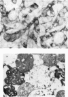
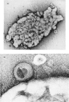
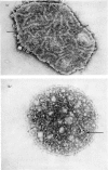
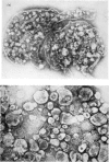
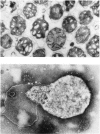
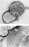
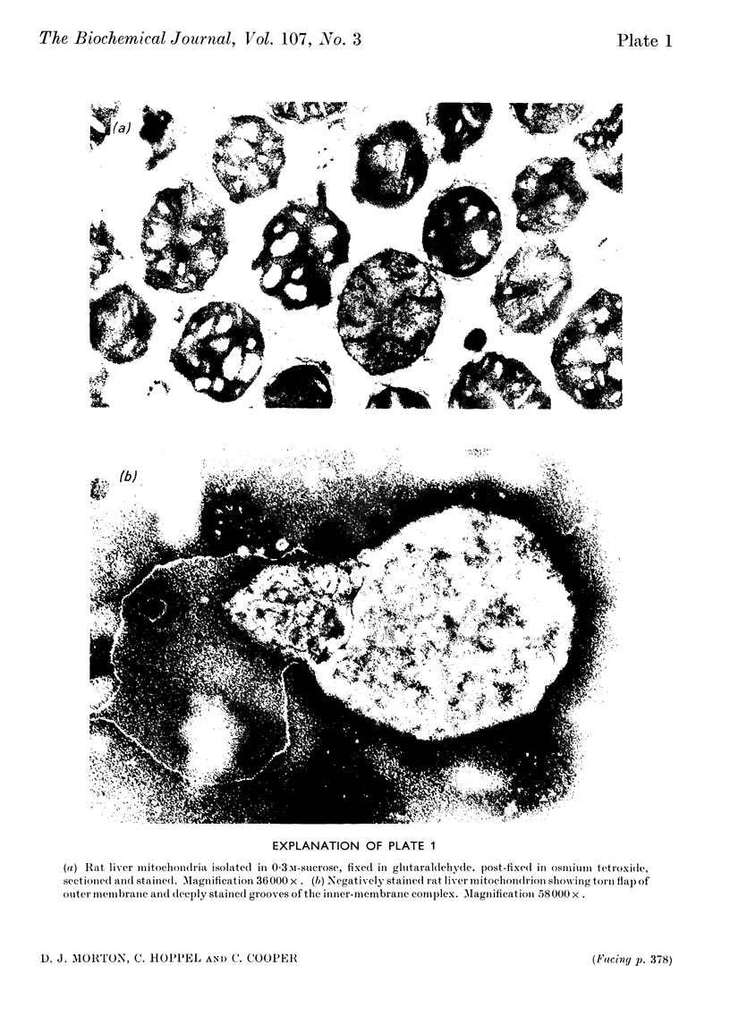
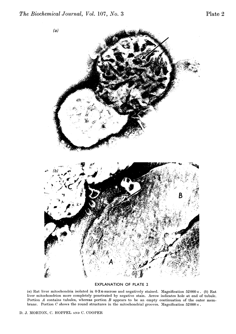
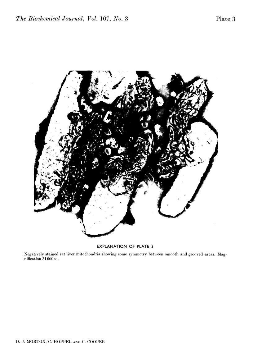
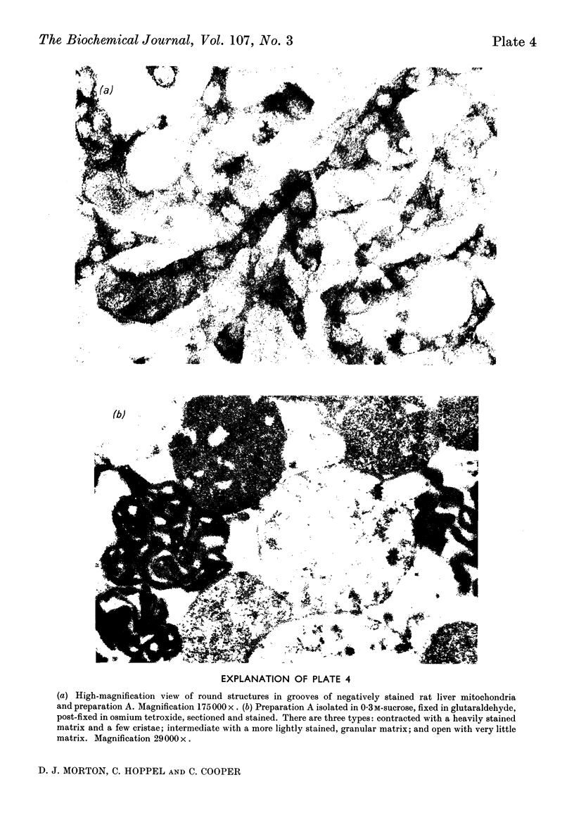
1. Rat liver mitochondria were examined in the electron microscope by using negative staining in the presence of 0·3m-sucrose. The intact outer membrane does not appear to be freely permeable to the stain. Where the stain penetrated through a tear it was seen that the inner membrane had randomly oriented grooves, many of which contained round structures varying between 200 and 900å in diameter. Laminar structures containing two to five layers of approx. 50å each were found at the periphery. 2. When the outer membrane was removed by treating the mitochondria with digitonin several types of inner-membrane complexes were formed and they showed a general correlation with those observed in sectioned samples of the same preparations. The main types were: (a) a condensed form looking very much like the intact mitochondrion without the outer membrane (this still showed the grooves, some of which contained the round structures, and the laminar whirls at the edges); (b) a more transparent form containing tubules of uniform width and various lengths (some of these appeared to terminate in a hole at the surface of the inner membrane); (c) a large torn sac, probably the inner membrane, containing some tubules and vesicles. 3. When the inner-membrane complex was further treated with digitonin it was disrupted and the resulting material consisted of pieces of membrane, doughnut-shaped units and lamellar structures. Most of these pieces varied in size between 500 and 1000å.
Full text
PDF
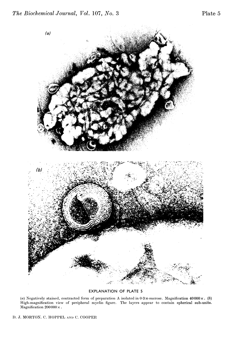
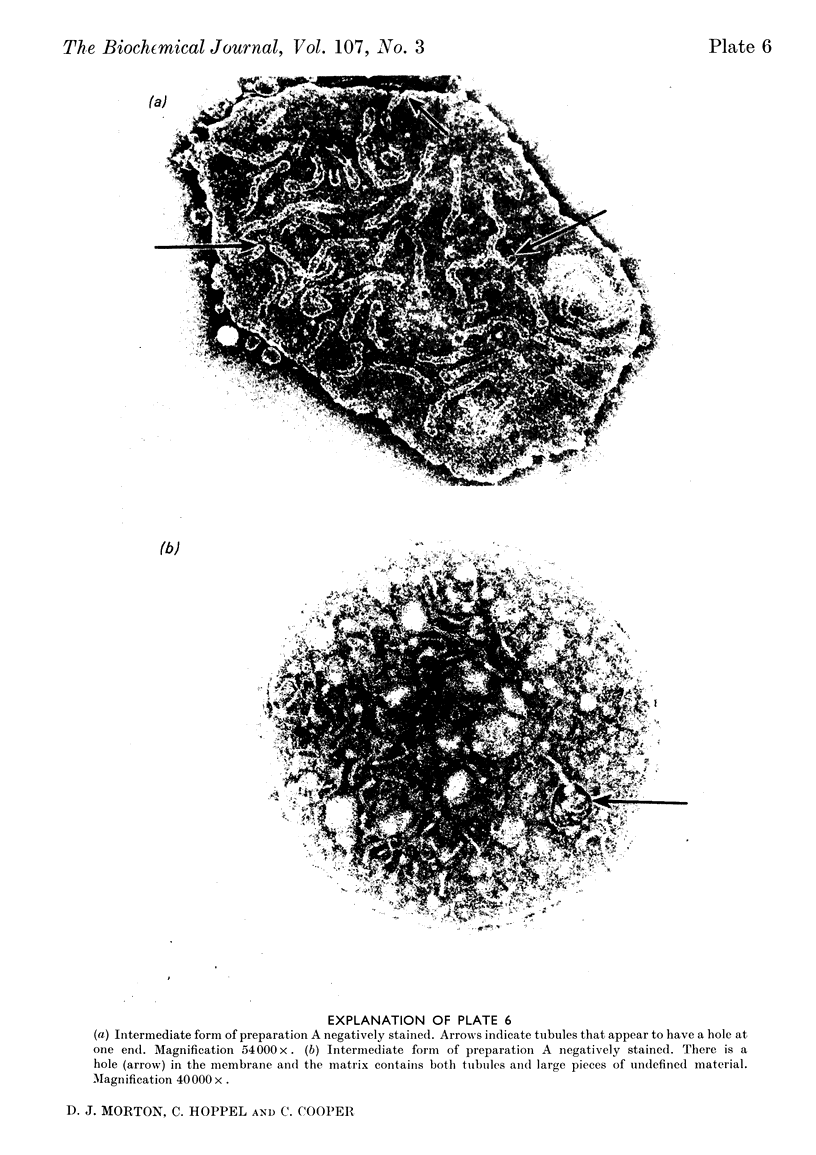
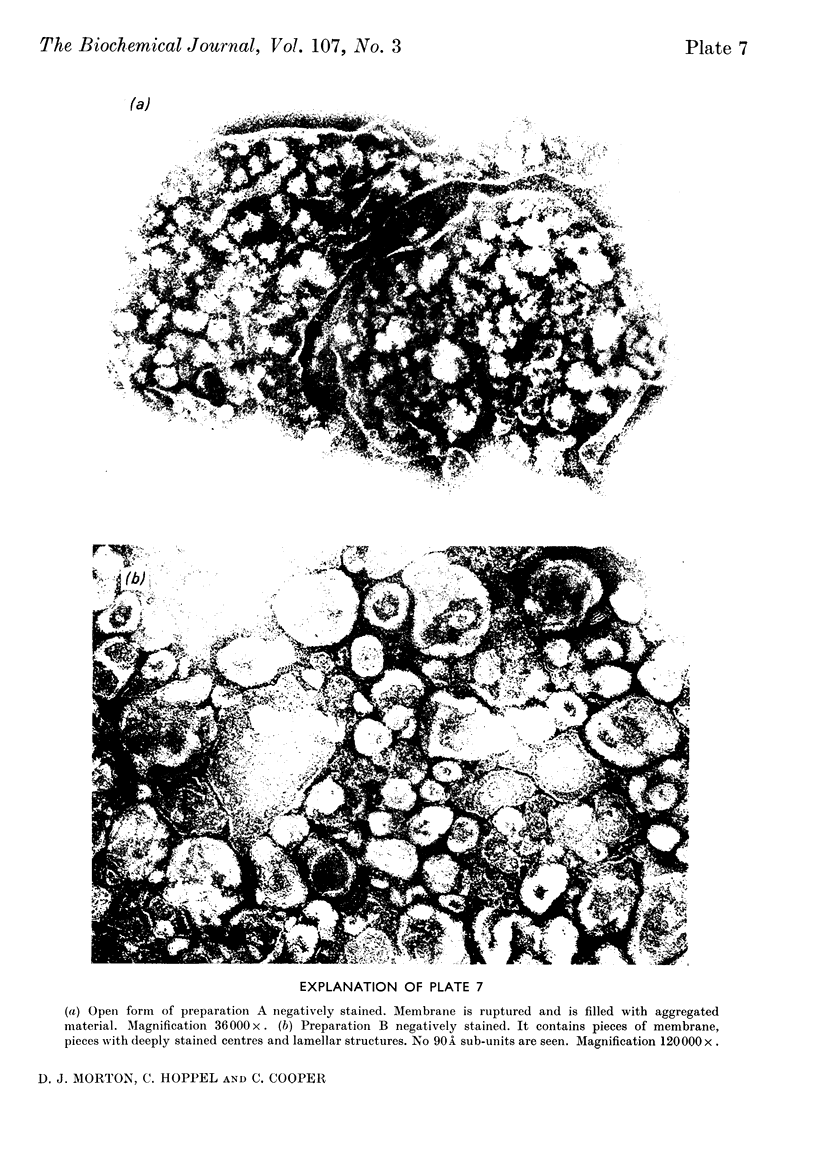
Images in this article
Selected References
These references are in PubMed. This may not be the complete list of references from this article.
- COOPER C., LEHNINGER A. L. Oxidative phosphorylation by an enzyme complex from extracts of mitochondria. I. The span beta-hydroxybutyrate to oxygen. J Biol Chem. 1956 Mar;219(1):489–506. [PubMed] [Google Scholar]
- FERNANDEZ-MORAN H. Cell-membrane ultrastructure. Low-temperature electron microsopy and x-ray diffraction studies of lipoprotein components in lamellar systems. Circulation. 1962 Nov;26:1039–1065. doi: 10.1161/01.cir.26.5.1039. [DOI] [PubMed] [Google Scholar]
- Hackenbrock C. R. Ultrastructural bases for metabolically linked mechanical activity in mitochondria. I. Reversible ultrastructural changes with change in metabolic steady state in isolated liver mitochondria. J Cell Biol. 1966 Aug;30(2):269–297. doi: 10.1083/jcb.30.2.269. [DOI] [PMC free article] [PubMed] [Google Scholar]
- Hoppel C., Cooper C. The action of digitonin on rat liver mitochondria. The effects on enzyme content. Biochem J. 1968 Apr;107(3):367–375. doi: 10.1042/bj1070367. [DOI] [PMC free article] [PubMed] [Google Scholar]
- Kagawa Y., Racker E. Partial resolution of the enzymes catalyzing oxidative phosphorylation. X. Correlation of morphology and function in submitochondrial particles. J Biol Chem. 1966 May 25;241(10):2475–2482. [PubMed] [Google Scholar]
- LUCY J. A., GLAUERT A. M. STRUCTURE AND ASSEMBLY OF MACROMOLECULAR LIPID COMPLEXES COMPOSED OF GLOBULAR MICELLES. J Mol Biol. 1964 May;8:727–748. doi: 10.1016/s0022-2836(64)80121-2. [DOI] [PubMed] [Google Scholar]
- Lévy M., Toury R., André J. Essai de séparation des deux membranes mitochondriales. C R Acad Sci Hebd Seances Acad Sci D. 1966 Apr 4;262(14):1593–1596. [PubMed] [Google Scholar]
- MILLONIG G. A modified procedure for lead staining of thin sections. J Biophys Biochem Cytol. 1961 Dec;11:736–739. doi: 10.1083/jcb.11.3.736. [DOI] [PMC free article] [PubMed] [Google Scholar]
- Parsons D. F. Recent advances correlating structure and function in mitochondria. Int Rev Exp Pathol. 1965;4:1–54. [PubMed] [Google Scholar]
- Parsons D. F., Williams G. R., Chance B. Characteristics of isolated and purified preparations of the outer and inner membranes of mitochondria. Ann N Y Acad Sci. 1966 Jul 14;137(2):643–666. doi: 10.1111/j.1749-6632.1966.tb50188.x. [DOI] [PubMed] [Google Scholar]
- SIEKEVITZ P., WATSON M. L. Some cytochemical characteristics of a phosphorylating digitonin preparation of mitochondria. Biochim Biophys Acta. 1957 Aug;25(2):274–279. doi: 10.1016/0006-3002(57)90469-9. [DOI] [PubMed] [Google Scholar]
- SJOESTRAND F. S., CEDERGREN E. A., KARLSSON U. MYELIN-LIKE FIGURES FORMED FROM MITOCHONDRIAL MATERIAL. Nature. 1964 Jun 13;202:1075–1078. doi: 10.1038/2021075a0. [DOI] [PubMed] [Google Scholar]
- Schnaitman C., Erwin V. G., Greenawalt J. W. The submitochondrial localization of monoamine oxidase. An enzymatic marker for the outer membrane of rat liver mitochondria. J Cell Biol. 1967 Mar;32(3):719–735. doi: 10.1083/jcb.32.3.719. [DOI] [PMC free article] [PubMed] [Google Scholar]
- Sottocasa G. L., Kuylenstierna B., Ernster L., Bergstrand A. An electron-transport system associated with the outer membrane of liver mitochondria. A biochemical and morphological study. J Cell Biol. 1967 Feb;32(2):415–438. doi: 10.1083/jcb.32.2.415. [DOI] [PMC free article] [PubMed] [Google Scholar]
- WATSON M. L. Staining of tissue sections for electron microscopy with heavy metals. II. Application of solutions containing lead and barium. J Biophys Biochem Cytol. 1958 Nov 25;4(6):727–730. doi: 10.1083/jcb.4.6.727. [DOI] [PMC free article] [PubMed] [Google Scholar]


