



Abstract
Sepsis is a major contributor to global health loss, yet effective therapeutic options remain scarce. This study aims to identify potential therapeutic targets for sepsis. We integrated data from the druggable genome, expression quantitative trait loci (eQTLs) from human blood, and genome-wide association studies on sepsis. Mendelian randomization (MR) was employed to investigate causal relationships between drug target genes and sepsis. The eQTLGen Consortium data served as the discovery set and was validated using genotype-tissue expression (GTEx) eQTLs. Sensitivity and colocalization analyses were conducted to support causal inferences. Additionally, phenome-wide MR (Phe-MR) was used to assess potential side effects of druggable genes. The expression levels of the target genes were validated using the GSE154918 dataset. In the discovery MR analysis phase, we identified 26 potential targets with significant expression in blood (PFDR < 0.05). PDGFB and BPI were further validated in the replication MR analysis. Colocalization analysis provided strong evidence (PPH4 > 0.75) supporting PDGFB as a therapeutic candidate for sepsis. Phe-MR analysis suggested that targeting PDGFB is unlikely to cause adverse effects. PDGFB downregulation was confirmed in sepsis groups via the GEO dataset. PDGFB is identified as a promising druggable target for sepsis treatment, supported by strong evidence of its therapeutic potential.
Supplementary Information
The online version contains supplementary material available at 10.1038/s41598-025-96655-7.
Keywords: Sepsis, PDGFB, Genomic, Mendelian randomization, GEO database
Subject terms: Data mining, Genome informatics, Genetics, Immunology, Molecular biology, Biomarkers
Introduction
Sepsis is a life-threatening organ dysfunction caused by a dysregulated host response to infection1 and is considered a major contributor to global health loss and disease burden. Epidemiological data worldwide indicate that sepsis patients have an intensive care unit and hospital mortality rate of 26% and 35%2, respectively. In the United States, sepsis is the leading cause of in-hospital mortality, costing more than $24 billion annually3. Moreover, survivors of sepsis continue to exhibit a high mortality rate within 5 years after the event4. To date, there is no specific therapeutic measure for sepsis, with treatments largely based on supportive care. While numerous clinical trials are underway, currently, there is no FDA-approved drug for the treatment of sepsis. Therefore, exploring the pathogenesis of sepsis and identifying new therapeutic targets are both urgent and necessary tasks.
Large-scale human genetic studies have been widely applied to drug development for many complex diseases. Drug targets which are supported by genetic evidence are considered more likely to succeed in clinical trials5. The “druggable genome” refers to a subset of the genome that encodes genes with potential drug targets. These genes typically encode proteins with drug-binding sites, increasing the success rate of drug target discovery6. Although Genome-Wide Association Studies (GWAS) have identified many single nucleotide polymorphisms (SNPs) associated with the risk of sepsis7, GWAS cannot directly identify the causative genes or provide effective information for drug development, as many identified SNPs reside in non-coding or intergenic regions.
Mendelian randomization (MR) is a genetic technique that can predict drug efficacy by mimicking randomized controlled trials. SNPs associated with gene expression levels (expression quantitative trait loci, eQTL) may be analogous to lifelong exposure to drugs targeting the encoded proteins8,9. Genetic variations from GWAS can be extracted to investigate their association with disease (outcome). MR integrates eQTLs data (SNP gene expression) and GWAS (SNP disease association) to infer the causal effect of exposure on the outcome, identifying potential drug targets. Recently, MR of the druggable genome has identified potential drug targets for several diseases, including COVID-19 and Alzheimer’s disease10,11.
Several studies have examined genetic genomic variations that may influence sepsis, but very few employed MR to investigate the causal association between these variations and the risk of sepsis. Moreover, the application of drug targets may be limited due to the lack of cross-validation with different independent datasets and the influence of potential pleiotropic effects8,12. To date, a systematic establishment of drug targets specifically for sepsis has not been achieved.
Here, we performed a systematic druggable genome-wide MR to identify therapeutic targets for sepsis. Initially, we utilized druggable eQTLs data from human blood sourced from the eQTLGen Consortium as the discovery dataset for MR analysis, aiming to identify significant causal genes linked to sepsis risk. Subsequently, we replicated and confirmed the identified genes using blood eQTLs data from the Genotype-Tissue Expression (GTEx) project. We then conducted colocalization analyses on key druggable target genes to further assess their causal relationships with sepsis. Finally, we employed a phenome-wide MR (Phe-MR) approach to evaluate the potential side effects of the identified drug targets. Additionally, the expression levels of these druggable targets were validated using the GSE154918 dataset. Our results offer important insights into the underlying pathophysiology of sepsis and provide a foundation for further in vitro and in vivo studies to validate these targets and evaluate the therapeutic effects of related drug interventions.
Methods
The overall study design is depicted in Fig. 1. This study adheres to the “Strengthening the Reporting of Observational Studies in Epidemiology using Mendelian Randomization (STROBE-MR) " checklist13, as provided in Supplementary Material 1: Table S1.
Fig. 1.

Flow diagram of the study.
Firstly, we obtained 5,883 druggable genes from the DGIdb database and the study by Finan et al. Secondly, we cross-referenced these identified 5,883 druggable genes with the blood eQTL dataset and extracted genetic variants significantly associated with the expression of these druggable genes (within a 1 Mb range on either side of the coding sequences) as instrument variables (IVs). Subsequently, we performed a two-sample MR analysis to assess the causal impact of blood eQTLs on sepsis and performed replication and validation studies. Thirdly, we used Bayesian colocalization to detect shared causal genetic variants for significant drug target genes identified in the MR analysis. Finally, we evaluated the potential adverse effects of previously identified druggable genes for sepsis treatment using a Phe-MR analysis. Additionally, the expression levels of these druggable targets were validated using the GSE154918 dataset.
Identify the druggable genome
Interaction data were downloaded from the Drug-Gene Interaction database (DGIdb v4.2.0, https://www.dgidb.org/downloads)14, as of the February 2022 release, resulting in the identification of 3,953 potential drug target genes (Supplementary Material 1: Table S3). DGIdb serves as an online compendium that aggregates information on drug-gene interactions from literature, databases, and various web-based sources. In addition, from the study conducted by Finan et al., we extracted a list of 4,463 druggable genes6 (Supplementary Material 1: Table S4). Collating the druggable genes from both sources resulted in a final tally of 5,883 unique druggable genes, all with official Human Genome Organisation Gene Nomenclature Committee (HGNC) nomenclature.
eQTL datasets
Cis-expression Quantitative Trait Loci (cis-eQTLs) refer to genetic variations near the drug target gene’s genome that can directly affect the expression of that gene15. Compared to trans-eQTLs, cis-eQTLs have more direct and specific biological effects and better reflect the genetic mechanisms controlling gene expression regulation16.
In this study, we selected cis-eQTL data from human blood tissue, focusing on genetic variations within a 1 Mb window flanking the coding sequence of the target gene. Previous studies have shown that significant regulatory effects of cis-eQTLs are typically concentrated within this range. This window effectively captures biologically relevant genetic variants while minimizing the inclusion of unrelated regions17–19.
The discovery blood cis-eQTL dataset was sourced from the eQTLGen Consortium (https://eqtlgen.org/), comprising cis-eQTLs for 16,987 genes across 31,684 European individuals20. Cis-eQTL results with statistical significance (False Discovery Rate (FDR) < 0.05) were selected.
To ensure the identified drug target genes’ utility and reliability, we verified our findings using cis-eQTL data from the GTEx project Version 821. Detailed descriptions of these eQTL datasets can be found in the original publications (Supplementary Material 1: Table S2).
Sepsis GWAS dataset
In this study, the sources of sepsis GWAS summary data were derived from two independent cohorts of European ancestry: UK Biobank and FinnGen Release 12. In the discovery phase, we utilized the most recent GWAS summary data from UK Biobank, which included 11,643 cases and 474,841 controls. In the replication phase, we incorporated additional sepsis summary data from the FinnGen consortium, comprising 17,133 cases and 439,048 controls. Both datasets were analyzed separately to assess the consistency of the target gene findings across different cohorts. The diagnosis of the sepsis cases conformed to the standards outlined in the International Classification of Diseases (ICD-10) codes22. All participants were of European descent.
Instrument selection
To ensure the robustness and validity of the MR analysis, we meticulously selected genetic instruments based on rigorous criteria aimed at maximizing statistical power and minimizing bias. Initially, a cross-analysis of the 5,883 potential druggable genes with the human blood eQTL dataset was carried out to obtain an eQTL dataset for druggable genes. Genetic variants closely associated with the expression of druggable genes (within a 1 Mb range flanking the coding sequences of the druggable genes) were extracted as IVs for MR analysis. These variants were first filtered based on a genome-wide significance threshold of p < 5 × 10⁻⁸ to ensure strong associations with the exposure of interest. Additionally, an F-statistic threshold of > 10 was applied to exclude weak instruments, thereby enhancing the reliability of the MR analysis. To address potential confounding from linkage disequilibrium (LD), clumping was performed with a pairwise LD threshold of r2 < 0.1. This threshold was carefully chosen to maintain a balance between retaining independent SNPs while avoiding the exclusion of relevant causal variants. More stringent LD thresholds, such as r2 < 0.001, could have led to the loss of informative variants and reduced statistical power, making the chosen threshold of r2 < 0.1 more suitable for ensuring comprehensive inclusion of potentially causal variants23,24. Furthermore, we queried the GWAS Catalog (https://www.ebi.ac.uk/gwas/) to retrieve phenotype information for all selected SNPs25. Phenotypes directly related to the outcome, such as smoking, obesity, diabetes, and others, were classified as confounding SNPs, and their corresponding rs numbers were excluded (threshold of p = 1 × 10⁻⁵)26,27. SNPs linked to polygenic traits were also excluded to prevent pleiotropic effects that could distort the causal inference28. These exclusions were critical for ensuring that the final set of SNPs were specific to the exposure of interest. Finally, allele frequencies of the effect were harmonized between exposure and outcome datasets to remove palindromic SNPs.
Mendelian randomization analysis
In the initial analysis, if a druggable gene set feature comprises only one IV, the Wald ratio test is applied in MR analysis. When a druggable gene set feature encompasses multiple IVs, the Inverse Variance Weighted (IVW) method is utilized as the primary analytical approach, supplemented by MR-Egger and Weighted Median methods (WME). To ensure the robustness of our results, the inferred causal effect direction from the IVW, MR-Egger, and WME must be consistent. To ensure the reliability and robustness of the results, a series of sensitivity analyses and quality controls were employed. Cochran’s Q test was used to assess heterogeneity among genetic instrumental variables, with the presence of heterogeneity indicated by P < 0.05, in which case a random effects model was adopted. Otherwise, a fixed-effects model was utilized. Pleiotropy was tested using MR-PRESSO and the MR-Egger intercept approaches. It is noteworthy that, although traditional MR-Egger regression and MR-PRESSO methods reasonably address horizontal pleiotropy issues, they do not account for potential biases introduced by related pleiotropy. Therefore, we employed the latest constrained Maximum Likelihood and Model Averaging MR method (cML-MA) to control for both related and unrelated pleiotropic effects29. Additionally, the MR Steiger directionality test was conducted to ensure the validity of the inferred causal direction30.
In the discovery analysis, the Benjamini-Hochberg false discovery rate (FDR) was employed to correct for multiple hypothesis testing results31, with FDR < 0.05 defined as significant. In validation analyses, p < 0.05 was considered statistically significant.
For genes identified as significant in the discovery phase of MR analysis, we applied more stringent LD thresholds (r2 < 0.01 and r2 < 0.001) to further test the robustness of our findings. This additional step aims to mitigate any potential bias introduced by LD between the genetic variants, ensuring that our observations are not confounded by correlated SNPs.
All statistical analyses were performed using the “TwoSampleMR (version 0.5.8),” “MR-PRESSO (version 0.5.8),” and “MR cML (version 0.0.0.9)” packages in R (version 4.3.2).
Colocalization analysis
Bayesian colocalization analysis is a powerful statistical tool used to explore whether druggable genes and sepsis share the same causal genetic variants. Specifically, this method evaluates five posterior probability hypotheses32: (1) PPH0: no association for both traits; (2) PPH1: association only for druggable genes with the genetic variant; (3) PPH2: association only for sepsis with the genetic variant; (4) PPH3: both traits are associated with the genetic variant, but the associations are caused by different genetic variants; (5) PPH4: both traits are associated with the genetic variant, and the associations are caused by the same genetic variant. If the posterior probability PPH4 > 0.75, it is considered that druggable genes and sepsis share the same genetic variant32. The analysis was conducted using the “coloc” R package (version 5.2.3).
Phenome-wide MR
To evaluate potential side effects or other indications of identified druggable genes, we conducted a Phe-MR study. Zhou et al. analyzed over 1400 binary phenotypes from 408,961 participants of European ancestry from the UK Biobank using the scalable accurate implementation of generalized mixed models (SAIGE V.0.29)33. To ensure statistical power, we selected 775 non-sepsis diseases or traits with sample sizes greater than 500 from the SAIGE GWAS (https://www.leelabsg.org/resource)34. Additionally, we applied a stricter LD threshold (r2 < 0.001) while keeping other parameters consistent with previous studies for MR analysis using cis-eQTLs of druggable genes identified in blood samples. FDR < 0.05 was considered statistically significant.
Differential expression analysis
We utilized the GSE154918 dataset from the Gene Expression Omnibus (GEO) repository to evaluate the differential expression of druggable genes between sepsis patients and healthy controls. This analysis included blood transcriptome data from 20 sepsis patients and 40 healthy control individuals. To identify differentially expressed genes (DEGs), we used GEO2R, an R-based online tool within the GEO database35. The Benjamini and Hochberg method was applied to adjust p-values, minimizing the false positive rate, and DEGs were filtered using the criteria of ∣log2 FC∣≥ 1 and adjusted p-value < 0.05.
Results
Candidate druggable genes for Sepsis
We intersected a dataset of 5,883 potential druggable genes with human blood eQTL data, identifying 3972 druggable genes. Genetic variants within 1 Mb of the coding regions of these genes were extracted, and after selecting IVs, 3,466 genes were retained. Following quality control to remove confounding and duplicate SNPs, 3,398 genes were included in the MR analysis (Supplementary Material 1: Table S6–S9).
In the discovery MR analysis, we identified 26 potential drug targets that exhibited significant expression levels in blood (PFDR < 0.05) (Fig. 2 and Supplementary Material 1: Table S10). Of these, the genetically predicted expression levels of 15 drug target genes were significantly inversely correlated with the risk of sepsis, which we have labeled as ‘protective’ in Fig. 3. Conversely, 11 drug target genes showed a significant positive correlation with sepsis risk, which are labeled as ‘risk’ in Fig. 3.
Fig. 2.
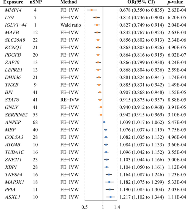
Forest plots displaying the findings from the discovery phase for 26 significant genes.
Fig. 3.

Volcano map of MR analysis in discovery phase. Significant genes are annotated and highlighted in green and pink. Genes with an OR greater than 1 are marked in pink and identified as risk genes for sepsis, while those with an OR less than 1 are marked in green and considered protective genes against sepsis. The horizontal dashed line indicates the p-value corresponding to FDR of 0.05.
To assess the robustness of these findings, we applied more stringent LD thresholds (r2 < 0.01 and r2 < 0.001) to the 26 drug target genes. The results indicated that, with stricter LD thresholds, the number of instrumental variables for these genes decreased, and their causal associations with sepsis weakened (Supplementary Material 1: Table S11). At r2 < 0.01, 14 genes retained significant causal associations with sepsis, whereas at r2 < 0.001, only 9 genes remained associated (Supplementary Material 1: Table S12). Notably, seven genes (PDGFB, IGLV1-44, LY9, MMP14, PPIA, BPI, and ZAP70) consistently exhibited causal associations with sepsis across all three LD thresholds (Supplementary Material 1: Table S13).
In the replication MR analysis, three potential drug targets were initially identified. However, ZNF211 was excluded from further analysis due to a reversal in the direction of the OR between the discovery and replication phases. As a result, two drug targets, PDGFB and BPI, were successfully validated in the replication phase. (p < 0.05) (Supplementary Material 1: Table S14).
Colocalization analysis
Subsequently, we performed colocalization analysis on the candidate drug target genes to further ascertain the probability of shared causal genetic variation between druggable genes and sepsis. The colocalization results indicated strong evidence of colocalization for PDGFB with sepsis (PPH4 = 0.81) (Fig. 4 and Supplementary Material 1: Table S16). This suggests that PDGFB is potential drug target for sepsis, with increased expression of PDGFB(OR: 0.864, 95% CI: 0.816–0.915, p = 6.02 × 10⁻⁷) gene in blood negatively correlated with susceptibility to sepsis. Targeting PDGFB may reduce the risk of sepsis. Sensitivity analysis and cML-MA test showed that the result was robust and reliable (Supplementary Material 1: Table S15). MR Steiger directionality test showed that the direction of inferred causality was correct. (p < 0.05; Supplementary Material 1: Table S10).
Fig. 4.
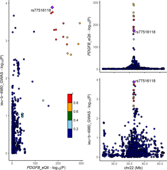
The results of colocalization. A strong colocalization signal between PDGFB and sepsis is shown. Instrumental variables and their chromosomal positions are visualized.
Phenome-wide MR analysis
Given that most drugs act through the bloodstream, we assessed whether the expression of PDGFB gene had beneficial or detrimental effects on other indications. Therefore, we made Phe-MR analysis on 775 diseases and traits within the UK Biobank (Supplementary Material 1: Table S17). Post-SNP aggregation, 2 SNPs encompassing one potential drug target genes were included. Causal effects with an FDR < 0.05 were considered statistically significant. Phe-MR results indicated that increased expression of PDGFB in blood may reduce the risk of Senile cataract (OR: 0.827, 95% CI: 0.749–0.914, p = 5.70 × 10⁻⁵, FDR = 0.044) (Supplementary Material 1: Table S18). Moreover, no causal effects of PDGFB gene expression in blood on other traits or diseases were observed (Fig. 5), suggesting that drugs targeting this gene do not have potential adverse reactions.
Fig. 5.

Manhattan plot for MR results of whole phenome with blood PDGFB. The vertical axis shows the p-values in phenome-wide MR results. The horizontal axis represents the categorization of 775 disease traits from the UK Biobank, clustered into 17 categories. Points represent disease traits, with different colors indicating different MR results of expression. Disease traits with significant causal effects (FDR <0.05) are highlighted and marked.
Differential expression verification
Differential Expression Verification in the GSE154918 dataset, we identified 4,505 DEGs between the sepsis and healthy control groups, including 2,658 upregulated genes and 1,847 downregulated genes. Among these downregulated genes, PDGFB ranked 164th based on a combination of adjusted p-values and log2 FC (Supplementary Material 1: Table S19).
While DEG ranking is undoubtedly important in many studies, the primary focus of our research is to identify and explore genes with a causal relationship to sepsis, rather than simply ranking genes based on statistical significance. Although PDGFB did not rank among the top 10 or top 20 genes in terms of p-value or log2FC, its biological relevance to sepsis makes it a key target for further validation. This underscores that gene ranking alone cannot fully capture the biological significance of a gene, especially in the complex pathophysiological context of sepsis.
In conclusion, based on the GEO dataset analysis, we validated the reduced expression of PDGFB in sepsis patients, further supporting its potential therapeutic value in sepsis.
Discussion
By integrating druggable genome data, eQTLs from human blood, and sepsis GWAS, we used MR to explore causal links between druggable genes and sepsis. In the UKB cohort, we identified 26 potential drug targets, validating 2 genes (PDGFB and BPI) in the FinnGen cohort. Colocalization analysis further supported PDGFB as a therapeutic target. To assess the safety of PDGFB as a therapeutic target, we conducted Phe-MR analysis to evaluate its potential effects on various phenotypes. Verification through the GEO dataset confirmed the downregulation of PDGFB in sepsis patients, further strengthening its association with sepsis pathophysiology. Overall, our findings suggest that PDGFB is a promising target for sepsis treatment, warranting further exploration.
Platelet-derived growth factor subunit B (PDGFB) is a member of the PDGF family that plays a crucial regulatory role in wound healing, cell proliferation, survival, and migration36,37. PDGFB is expressed in endothelial cells and exists in its active form as the homodimer PDGF-BB. PDGF-BB exerts its biological effects by binding to platelet-derived growth factor receptor β (PDGFR-β) expressed on pericytes, activating a series of signaling pathways, including PI3K/Akt, MAPK/ERK, PLC-γ1/PKC, and JAK/STAT pathways.
The PI3K/Akt pathway is essential for cell survival and vascular homeostasis38. PDGF-BB activates this pathway to enhance endothelial cell survival, reduce apoptosis, and promote endothelial function recovery39. This effect is particularly important in diseases like sepsis, where endothelial cell damage occurs, as activation of the PI3K/Akt pathway can alleviate vascular leakage and restore microcirculation, improving organ perfusion. The MAPK/ERK pathway, which regulates cell proliferation and migration, plays a key role in the repair of damaged vessels40. In sepsis-induced microvascular injury, activation of the MAPK/ERK pathway helps restore vascular wall stability and alleviate microcirculatory dysfunction caused by increased vascular permeability. The PLC-γ1/PKC pathway regulates cell motility and cytoskeletal rearrangement, maintaining vascular barrier integrity41. Through this pathway, PDGF-BB promotes interactions between vascular smooth muscle cells and endothelial cells, facilitating vascular repair. The JAK/STAT pathway, which regulates immune responses, influences immune cell recruitment and cytokine release, thereby modulating the inflammatory response in sepsis42.
Preclinical and animal studies have demonstrated the critical role of PDGF-BB in vascular repair and blood flow regulation. Mice lacking the PDGFB gene exhibit perinatal lethality due to widespread hemorrhage and edema43,44. Furthermore, disruption of PDGFBB signaling leads to a significant reduction in pericytes within the central nervous system, resulting in decreased expression of tight junction proteins in capillary endothelial cells, increased transcellular transport, reduced glial membrane coverage, and ultimately, leakage of the blood-brain barrier45. These findings suggest that PDGF-BB therapy may help mitigate vascular dysfunction and microcirculatory disturbances associated with sepsis. Further studies support our view. The study by Liu et al. demonstrated that recombinant PDGF-BB treatment improved survival rates and reduced tissue damage in mouse models of sepsis induced by CLP (cecal ligation and puncture) and LPS (lipopolysaccharide) by decreasing the production of pro-inflammatory cytokines (such as TNF-α, IL-6, IL-1β, and IL-8) and chemokines (such as CXCL-1 and CCL2)46. Additionally, PDGF-BB (1–15 µg/kg, intravenous injection) significantly improved hemodynamics, blood perfusion, and vascular reactivity in key organs (such as the liver and kidneys) of rats with hemorrhagic shock, thereby increasing survival rates47.
Beyond animal models, emerging evidence suggests that PDGFB signaling may have translational potential in clinical settings. Recombinant human activated protein C (rhAPC) stimulates the transcription of PDGF-BB messenger RNA and the release of PDGF-BB in human endothelial cells in vitro, promoting the structural integrity of the vascular wall and enhancing the tissue healing capacity in patients with severe sepsis48. A novel mesoporous bioactive glass fiber scaffold loaded with PDGF-B adenovirus (AdPDGF) has been shown to recruit mesenchymal stem cells (MSCs) for tissue repair in vitro and in mice49.
Unfortunately, despite the compounds related to the PDGFB gene (such as AXITINIB and SUNITINIB) being authorized by the FDA50,51, they are used as inhibitors. It is necessary to develop agonists related to the PDGFB gene for therapeutic intervention in sepsis. It is worth noting that recombinant human platelet-derived growth factor-BB (rhPDGF-BB) has been approved by the U.S. FDA and is widely used in various clinical indications, including the treatment of periodontal and orthopedic bone defects, as well as the promotion of dermal wound healing52,53. Therefore, rhPDGF-BB holds great promise for the treatment of sepsis. More clinical trials are needed in the future to verify the efficacy and safety of rhPDGF-BB in sepsis treatment.
The present study has several strengths. Firstly, we employed a two-stage MR analysis to detect the expression of potentially druggable genes associated with sepsis in different blood tissues, and utilized multiple correction and colocalization analyses to identify potential targets. Secondly, various methods were employed to mitigate the effects of horizontal pleiotropy, including utilizing the GWAS Catalog database to exclude potentially confounding SNPs that may affect the association between druggable genes and sepsis, employing traditional MR-Egger regression and MR-PRESSO to control unrelated horizontal pleiotropy, and utilizing the latest cML-MA method to control for correlated pleiotropic effects. Thirdly, the safety of druggable genes was evaluated through Phe-MR analysis, providing insights for subsequent drug development. Fourthly, differential expression analysis using the GEO dataset confirmed the downregulation of PDGFB in sepsis patients, underscoring its relevance to sepsis pathophysiology.
Nevertheless, this study has its limitations. Firstly, the integrated eQTL dataset for druggable genes and the GWAS dataset for sepsis were both derived from European populations. Caution is warranted when generalizing conclusions to other non-European populations, and further studies on more diverse populations are warranted in the future. Secondly, MR cannot fully replicate clinical trials or predict drug effects entirely, as standard MR studies typically ascertain lifespan and low-dose exposure, whereas clinical trials often investigate relatively high drug doses over a short period. Thirdly, only potential adverse reactions of PDGFB in the disease cohort from the UK Biobank were explored. Given the broad nature of drug-target interactions, many off-target effects were not captured by MR alone. Additional basic and clinical research is needed to better understand these effects. Finally, although both the UK Biobank and FinnGen cohorts consist mainly of individuals of European ancestry, the UK Biobank includes subpopulations such as British White and British Non-white groups, which may exhibit genetic or environmental differences. This could introduce potential confounding effects not addressed here due to the lack of subgroup-level data. Future studies should consider stratified analyses to minimize biases associated with population structure.
In the discovery phase of our analysis, we identified 26 potential druggable genes associated with sepsis. However, in the replication phase using the FinnGen cohort, only two genes were successfully validated. The lack of replication for other genes could be attributed to several factors, including differences in population characteristics between the UK Biobank and FinnGen cohorts, as well as potential gene-environment interactions that may affect gene expression and its association with sepsis. Additionally, statistical power limitations, such as smaller sample sizes or the potential for a lower effect size in the FinnGen cohort, could have affected the ability to replicate other druggable genes. These findings emphasize the need for future studies to examine the generalizability of these results across different populations and to consider potential subpopulation effects.
While our study primarily investigates PDGFB as a therapeutic target for sepsis, we also observed its potential therapeutic role in senile cataract (Supplementary Material 1: Table S20 and S21). PDGF-BB promotes the proliferation and migration of lens epithelial cells through its interaction with PDGFR, which is essential for maintaining lens metabolism and transparency54. The gene’s involvement in tissue repair, vascular integrity, and inflammation regulation may explain its relevance to both conditions55,56. Given the shared biological mechanisms, such as tissue damage and inflammation, PDGFB’s broader role in cellular function suggests its therapeutic potential across multiple diseases. Future research should further explore PDGFB’s therapeutic scope, including its effects beyond sepsis.
Conclusion
In this study, we identified PDGFB as a potential therapeutic target for sepsis and evaluated its safety using a Phe-MR analysis. The genetic evidence suggests that targeting PDGFB could be beneficial in reducing the risk of sepsis. Future assessments through adequately powered randomized trials are necessary to further investigate the efficacy and safety of PDGFB in the early management of sepsis.
Electronic supplementary material
Below is the link to the electronic supplementary material.
Acknowledgements
We want to acknowledge all the GWASs for making the summary data publicly available, and we appreciate all the investigators and participants who contributed to those studies.
Abbreviations
- cis-eQTL
Cis-expression quantitative trait loci
- MR
Mendelian randomization
- Phe-MR
Phenome-wide mendelian randomization
- GWAS
Genome-wide association study
- SNP
Single nucleotide polymorphism
- IVW
Inverse variance weighted
- WME
Weighted median
- MR-PRESSO
Mendelian randomization pleiotropy residual sum and outlier
- cML-MA
Constrained maximum likelihood and model averaging
- LD
Linkage disequilibrium
- PDGFB
Plateletderived growth factor subunit B
- GEO
Gene expression omnibus
Author contributions
MJG conceived the study, analyzed and interpreted the data, and drafted the manuscript. LS analyzed the data. ZHH conceived the study and critically revised the manuscript. All authors read and approved the final manuscript.
Funding
This work was supported by the National Natural Science Foundation of China (82272216).
Data availability
All data generated or analyzed during this study are included in this published article and its additional files.
Declarations
Competing interests
The authors declare no competing interests.
Footnotes
Publisher’s note
Springer Nature remains neutral with regard to jurisdictional claims in published maps and institutional affiliations.
References
- 1.Singer, M. et al. The third international consensus definitions for sepsis and septic shock (Sepsis-3). Jama315(8), 801–810 (2016). [DOI] [PMC free article] [PubMed] [Google Scholar]
- 2.Vincent, J. L. et al. Assessment of the worldwide burden of critical illness: the intensive care over nations (ICON) audit. Lancet Respir Med.2(5), 380–386 (2014). [DOI] [PubMed] [Google Scholar]
- 3.Torio, C. M. & Moore, B. J. National inpatient hospital costs: the most expensive conditions by payer, 2013. In Healthcare Cost and Utilization Project (HCUP) Statistical Briefs (Agency for Healthcare Research and Quality (US), 2006). [PubMed] [Google Scholar]
- 4.Beck, M. K. et al. Diagnosis trajectories of prior multi-morbidity predict sepsis mortality. Sci. Rep.6, 36624 (2016). [DOI] [PMC free article] [PubMed] [Google Scholar]
- 5.Nelson, M. R. et al. The support of human genetic evidence for approved drug indications. Nat. Genet.47(8), 856–860 (2015). [DOI] [PubMed] [Google Scholar]
- 6.Finan, C. et al. The druggable genome and support for target identification and validation in drug development. Sci. Transl Med.9, 383 (2017). [DOI] [PMC free article] [PubMed]
- 7.Hernandez-Beeftink, T. et al. A genome-wide association study of survival in patients with sepsis. Crit. Care. 26(1), 341 (2022). [DOI] [PMC free article] [PubMed] [Google Scholar]
- 8.Schmidt, A. F. et al. Genetic drug target validation using Mendelian randomisation. Nat. Commun.11(1), 3255 (2020). [DOI] [PMC free article] [PubMed] [Google Scholar]
- 9.Zhu, Z. et al. Integration of summary data from GWAS and eQTL studies predicts complex trait gene targets. Nat. Genet.48(5), 481–487 (2016). [DOI] [PubMed] [Google Scholar]
- 10.Gaziano, L. et al. Actionable druggable genome-wide Mendelian randomization identifies repurposing opportunities for COVID-19. Nat. Med.27(4), 668–676 (2021). [DOI] [PMC free article] [PubMed] [Google Scholar]
- 11.Chen, L. et al. Systematic Mendelian randomization using the human plasma proteome to discover potential therapeutic targets for stroke. Nat. Commun.13(1), 6143 (2022). [DOI] [PMC free article] [PubMed] [Google Scholar]
- 12.Xia, R., Sun, M., Yin, J., Zhang, X. & Li, J. Using Mendelian randomization provides genetic insights into potential targets for sepsis treatment. Sci. Rep.14(1), 8467 (2024). [DOI] [PMC free article] [PubMed] [Google Scholar]
- 13.Skrivankova, V. W. et al. Strengthening the reporting of observational studies in epidemiology using Mendelian randomisation (STROBE-MR): explanation and elaboration. Bmj375, n2233 (2021). [DOI] [PMC free article] [PubMed] [Google Scholar]
- 14.Freshour, S. L. et al. Integration of the Drug-Gene interaction database (DGIdb 4.0) with open crowdsource efforts. Nucleic Acids Res.49(D1), D1144–d1151 (2021). [DOI] [PMC free article] [PubMed] [Google Scholar]
- 15.Chen, Y. et al. Genetic insights into therapeutic targets for aortic aneurysms: A Mendelian randomization study. EBioMedicine83, 104199 (2022). [DOI] [PMC free article] [PubMed] [Google Scholar]
- 16.Wittich, H. et al. Transcriptome-wide association study of the plasma proteome reveals Cis and trans regulatory mechanisms underlying complex traits. Am. J. Hum. Genet.111(3), 445–455 (2024). [DOI] [PMC free article] [PubMed] [Google Scholar]
- 17.de Klein, N. et al. Brain expression quantitative trait locus and network analyses reveal downstream effects and putative drivers for brain-related diseases. Nat. Genet.55(3), 377–388 (2023). [DOI] [PMC free article] [PubMed] [Google Scholar]
- 18.Liang, Y. C. et al. Integrating Mendelian randomization and single-cell RNA sequencing to identify therapeutic targets of Baicalin for type 2 diabetes mellitus. Front. Pharmacol.15, 1403943 (2024). [DOI] [PMC free article] [PubMed] [Google Scholar]
- 19.Liu, Z. et al. Identifying potential drug targets for idiopathic pulmonary fibrosis: a Mendelian randomization study based on the druggable genes. Respir Res.25(1), 217 (2024). [DOI] [PMC free article] [PubMed] [Google Scholar]
- 20.Võsa, U. et al. Large-scale cis- and trans-eQTL analyses identify thousands of genetic loci and polygenic scores that regulate blood gene expression. Nat. Genet.53(9), 1300–1310 (2021). [DOI] [PMC free article] [PubMed] [Google Scholar]
- 21.Battle, A., Brown, C. D., Engelhardt, B. E. & Montgomery, S. B. Genetic effects on gene expression across human tissues. Nature550(7675), 204–213 (2017). [DOI] [PMC free article] [PubMed] [Google Scholar]
- 22.Rudd, K. E. et al. Global, regional, and National sepsis incidence and mortality, 1990–2017: analysis for the global burden of disease study. Lancet395(10219), 200–211 (2020). [DOI] [PMC free article] [PubMed] [Google Scholar]
- 23.Gkatzionis, A., Burgess, S. & Newcombe, P. J. Statistical methods for cis-Mendelian randomization with two-sample summary-level data. Genet. Epidemiol.47(1), 3–25 (2023). [DOI] [PMC free article] [PubMed] [Google Scholar]
- 24.Jiang, W. X. & Li, H. H. Circulating inflammatory cytokines and the risk of sepsis: a bidirectional Mendelian randomization analysis. BMC Infect. Dis.24(1), 793 (2024). [DOI] [PMC free article] [PubMed] [Google Scholar]
- 25.Sollis, E. et al. The NHGRI-EBI GWAS catalog: knowledgebase and deposition resource. Nucleic Acids Res.51(D1), D977–d985 (2023). [DOI] [PMC free article] [PubMed] [Google Scholar]
- 26.Kamat, M. A. et al. PhenoScanner V2: an expanded tool for searching human genotype-phenotype associations. Bioinformatics35(22), 4851–4853 (2019). [DOI] [PMC free article] [PubMed] [Google Scholar]
- 27.Zhang, L. et al. Causal relationship between spondylarthritis and stroke in a European population: a two sample Mendelian randomization study. Front. Immunol.14, 1253986 (2023). [DOI] [PMC free article] [PubMed] [Google Scholar]
- 28.Zhi, F., Ma, J. W., Ji, D. D., Bao, J. & Li, Q. Q. Causal associations between Circulating cytokines and risk of sepsis and related outcomes: a two-sample Mendelian randomization study. Front. Immunol.15, 1336586 (2024). [DOI] [PMC free article] [PubMed] [Google Scholar]
- 29.Yin, Q. & Zhu, L. Does co-localization analysis reinforce the results of Mendelian randomization? Brain147(1), e7–e8 (2024). [DOI] [PubMed] [Google Scholar]
- 30.Hemani, G., Tilling, K. & Davey Smith, G. Orienting the causal relationship between imprecisely measured traits using GWAS summary data. PLoS Genet.13(11), e1007081 (2017). [DOI] [PMC free article] [PubMed] [Google Scholar]
- 31.Greco, M. F., Minelli, C., Sheehan, N. A. & Thompson, J. R. Detecting Pleiotropy in Mendelian randomisation studies with summary data and a continuous outcome. Stat. Med.34(21), 2926–2940 (2015). [DOI] [PubMed] [Google Scholar]
- 32.Giambartolomei, C. et al. Bayesian test for colocalisation between pairs of genetic association studies using summary statistics. PLoS Genet.10(5), e1004383 (2014). [DOI] [PMC free article] [PubMed] [Google Scholar]
- 33.Zhou, W. et al. Efficiently controlling for case-control imbalance and sample relatedness in large-scale genetic association studies. Nat. Genet.50(9), 1335–1341 (2018). [DOI] [PMC free article] [PubMed] [Google Scholar]
- 34.Su, W. M. et al. Systematic druggable genome-wide Mendelian randomisation identifies therapeutic targets for Alzheimer’s disease. J. Neurol. Neurosurg. Psychiatry. 94(11), 954–961 (2023). [DOI] [PMC free article] [PubMed] [Google Scholar]
- 35.Liu, X. et al. A systematic pharmacology-based in vivo study to reveal the effective mechanism of Yupingfeng in asthma treatment. Phytomedicine114, 154783 (2023). [DOI] [PubMed] [Google Scholar]
- 36.Heldin, C. H. & Westermark, B. Mechanism of action and in vivo role of platelet-derived growth factor. Physiol. Rev.79(4), 1283–1316 (1999). [DOI] [PubMed] [Google Scholar]
- 37.Heldin, C. H. & Lennartsson, J. Structural and functional properties of platelet-derived growth factor and stem cell factor receptors. Cold Spring Harb Perspect. Biol.5(8), a009100 (2013). [DOI] [PMC free article] [PubMed] [Google Scholar]
- 38.Cantley, L. C. The phosphoinositide 3-kinase pathway. Science296(5573), 1655–1657 (2002). [DOI] [PubMed] [Google Scholar]
- 39.Wang, H. et al. Over-expression of PDGFR-β promotes PDGF-induced proliferation, migration, and angiogenesis of EPCs through PI3K/Akt signaling pathway. PLoS One. 7(2), e30503 (2012). [DOI] [PMC free article] [PubMed] [Google Scholar]
- 40.Luk, K. et al. Influence of morphine on pericyte-endothelial interaction: implications for antiangiogenic therapy. J. Oncol.2012, 458385 (2012). [DOI] [PMC free article] [PubMed] [Google Scholar]
- 41.Wang, C., Liu, Y. & He, D. Diverse effects of platelet-derived growth factor-BB on cell signaling pathways. Cytokine113, 13–20 (2019). [DOI] [PubMed] [Google Scholar]
- 42.Satarker, S. et al. JAK-STAT pathway Inhibition and their implications in COVID-19 therapy. Postgrad. Med.133(5), 489–507 (2021). [DOI] [PMC free article] [PubMed] [Google Scholar]
- 43.Levéen, P. et al. Mice deficient for PDGF B show renal, cardiovascular, and hematological abnormalities. Genes Dev.8(16), 1875–1887 (1994). [DOI] [PubMed] [Google Scholar]
- 44.Hellström, M., Kalén, M., Lindahl, P., Abramsson, A. & Betsholtz, C. Role of PDGF-B and PDGFR-beta in recruitment of vascular smooth muscle cells and pericytes during embryonic blood vessel formation in the mouse. Development126(14), 3047–3055 (1999). [DOI] [PubMed] [Google Scholar]
- 45.Weng, Y. et al. An integral blood-brain barrier in adulthood relies on microglia-derived PDGFB. Brain Behav. Immun.115, 705–717 (2024). [DOI] [PubMed] [Google Scholar]
- 46.Wang, M., Wei, J., Shang, F., Zang, K. & Ji, T. Platelet-derived growth factor B attenuates lethal sepsis through Inhibition of inflammatory responses. Int. Immunopharmacol.75, 105792 (2019). [DOI] [PubMed] [Google Scholar]
- 47.Liu, L. et al. Beneficial effects of platelet-derived growth factor on hemorrhagic shock in rats and the underlying mechanisms. Am. J. Physiol. Heart Circ. Physiol.307(9), H1277–1287 (2014). [DOI] [PubMed] [Google Scholar]
- 48.Brueckmann, M. et al. Prognostic value of platelet-derived growth factor in patients with severe sepsis. Growth Factors. 25(1), 15–24 (2007). [DOI] [PubMed] [Google Scholar]
- 49.Zhang, Y., Ma, Y., Wu, C., Miron, R. J. & Cheng, X. Platelet-derived growth factor BB gene-released scaffolds: biosynthesis and characterization. J. Tissue Eng. Regen Med.10(10), E372–E381 (2016). [DOI] [PubMed] [Google Scholar]
- 50.Keating, G. M. Axitinib: a review in advanced renal cell carcinoma. Drugs75(16), 1903–1913 (2015). [DOI] [PubMed] [Google Scholar]
- 51.Papaetis, G. S. & Syrigos, K. N. Sunitinib: a multitargeted receptor tyrosine kinase inhibitor in the era of molecular cancer therapies. BioDrugs23(6), 377–389 (2009). [DOI] [PubMed] [Google Scholar]
- 52.Chen, Y. et al. A promising candidate in tendon healing Events-PDGF-BB. Biomolecules12(10) (2022). [DOI] [PMC free article] [PubMed]
- 53.Galarraga-Vinueza, M. E. et al. Twenty-five years of Recombinant human growth factors rhPDGF-BB and rhBMP-2 in oral hard and soft tissue regeneration. Periodontol 2000. 94(1), 483–509 (2024). [DOI] [PubMed] [Google Scholar]
- 54.Paensuwan, P., Ngoenkam, J., Wangteeraprasert, A. & Pongcharoen, S. Essential function of adaptor protein Nck1 in platelet-derived growth factor receptor signaling in human lens epithelial cells. Sci. Rep.12(1), 1063 (2022). [DOI] [PMC free article] [PubMed] [Google Scholar]
- 55.Sorte Gawali, K. S., Jadhao, A. N., Ramteke, T. D., Patil, N. J. & Sahare, H. Evaluation of antioxidant status of lens epithelial cells in cataract patients. Indian J. Ophthalmol.72(10), 1506–1511 (2024). [DOI] [PMC free article] [PubMed] [Google Scholar]
- 56.Zhang, F., Chang, P., Zhao, Y. & Zhao, Y. A negative correlation of axial length with aqueous humor concentration of cytokines in patients with congenital cataracts. Mol. Vis.26, 91–96 (2020). [PMC free article] [PubMed] [Google Scholar]
Associated Data
This section collects any data citations, data availability statements, or supplementary materials included in this article.
Supplementary Materials
Data Availability Statement
All data generated or analyzed during this study are included in this published article and its additional files.