Full text
PDF
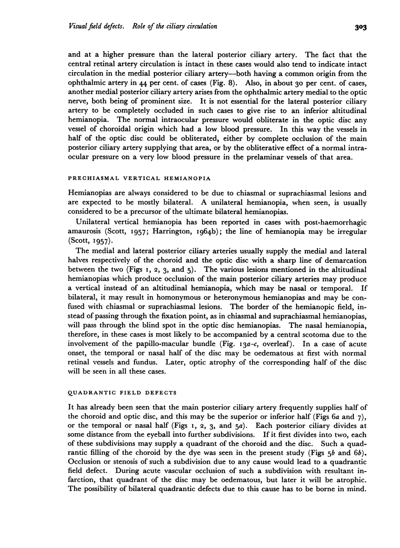
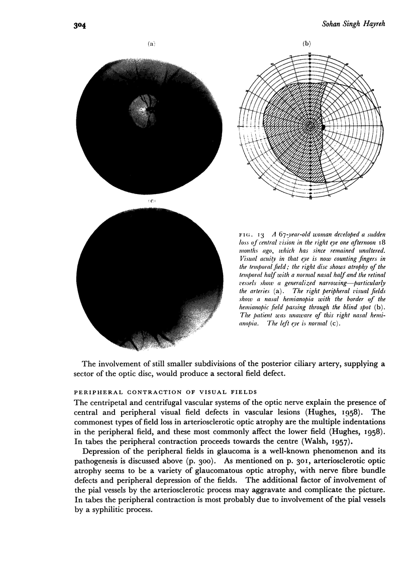
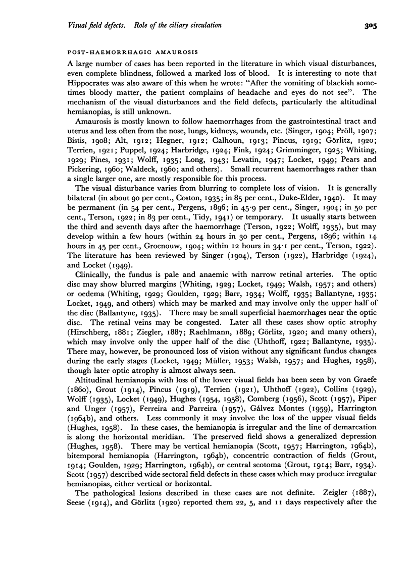
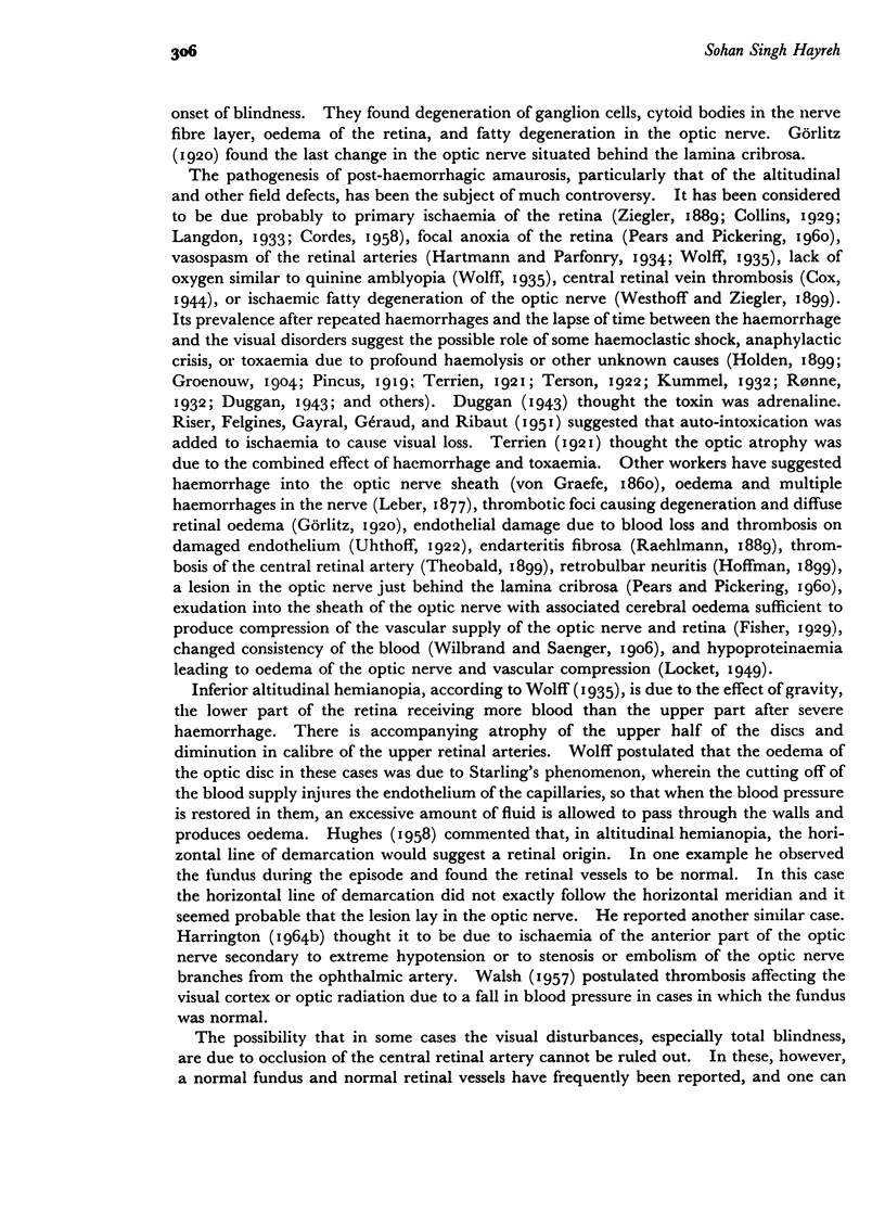
Images in this article
Selected References
These references are in PubMed. This may not be the complete list of references from this article.
- CRISTINI G. Common pathological basis of the nervous ocular symptoms in chronic glaucoma. Br J Ophthalmol. 1951 Jan;35(1):11–20. doi: 10.1136/bjo.35.1.11. [DOI] [PMC free article] [PubMed] [Google Scholar]
- Cullen J. F. Ischaemic optic neuropathy. Trans Ophthalmol Soc U K. 1967;87:759–774. [PubMed] [Google Scholar]
- DE FERREIRA C., PARREIRA F. Hemianópsia bilateral inferior por hemorragia uterina. Gaz Med Port. 1957 May-Jun;10(3):357–369. [PubMed] [Google Scholar]
- Evans P. J. THE UNDERLYING CAUSES OF GLAUCOMA: Including notes on the lines of enquiry which have been pursued, with suggestions as to future research in clinic and laboratory. Br J Ophthalmol. 1939 Dec;23(12):745–783. doi: 10.1136/bjo.23.12.745. [DOI] [PMC free article] [PubMed] [Google Scholar]
- GAFNER F., GOLDMANN H. Experimentelle Untersuchungen über den Zusammenhang von Augendrucksteigerung und Gesichtsfeldschädigung. Ophthalmologica. 1955 Dec;130(6):357–377. doi: 10.1159/000302700. [DOI] [PubMed] [Google Scholar]
- Hamasaki D. I., Fujino T. Effect of intraocular pressure on ocular vessels. Filling with India ink. Arch Ophthalmol. 1967 Sep;78(3):369–379. doi: 10.1001/archopht.1967.00980030371021. [DOI] [PubMed] [Google Scholar]
- Kalvin H. N., Hamasaki D. I., Gass J. D. Experimental glaucoma in monkeys. II. Studies of intraocular vascularity during glaucoma. Arch Ophthalmol. 1966 Jul;76(1):94–103. doi: 10.1001/archopht.1966.03850010096018. [DOI] [PubMed] [Google Scholar]
- LOCKET S. Blindness associated with haemorrhage. Br J Ophthalmol. 1949 Sep;33(9):543–555. doi: 10.1136/bjo.33.9.543. [DOI] [PMC free article] [PubMed] [Google Scholar]
- McGuire W. P. The Effect of Dicumarol on the Visual Fields in Glaucoma. A Preliminary Report. Trans Am Ophthalmol Soc. 1948;46:96–126. [PMC free article] [PubMed] [Google Scholar]
- PARIN P. Opticusatrophie durch Arterio-Sklerose der Carotis interna. Schweiz Arch Neurol Psychiatr. 1951;67(1):139–174. [PubMed] [Google Scholar]
- PEARS M. A., PICKERING G. W. Changes in the fundus oculi after haemorrhage. Q J Med. 1960 Apr;29:153–178. [PubMed] [Google Scholar]
- PETERS W. Uber die Neuritis optici arteriosklerotischer Genese. Klin Monbl Augenheilkd Augenarztl Fortbild. 1958;132(3):363–377. [PubMed] [Google Scholar]
- PIPER H. F., UNGER L. Hemianopsia horizontalis inferior bei akuten Durchblutungsstörungen des Sehnerven. Ophthalmologica. 1957 Sep;134(3):169–180. doi: 10.1159/000303200. [DOI] [PubMed] [Google Scholar]
- REDSLOB E. Le glaucome primaire vu a travers son anatomie pathologique. Ann Ocul (Paris) 1955 Sep;188(9):781–826. [PubMed] [Google Scholar]
- RISER, FELGINES, GAYRAL, GERAUD, RIBAUT L'amaurose post-hémorragique. Rev Otoneuroophtalmol. 1951 Apr;23(3):175–181. [PubMed] [Google Scholar]
- WYBAR K. C. A study of the choroidal circulation of the eye in man. J Anat. 1954 Jan;88(1):94–98. [PMC free article] [PubMed] [Google Scholar]


