Abstract
Following virus infection of the central nervous system, microglia, the ontogenetic and functional equivalents of macrophages in somatic tissues, act as sources of chemokines, thereby recruiting peripheral leukocytes into the brain parenchyma. In the present study, we have systemically examined the growth characteristics of rabies virus (RV) in microglia and the activation of cellular signaling pathways leading to chemokine expression upon RV infection. In RV-inoculated microglia, the synthesis of the viral genome and the production of virus progenies were significantly impaired, while the expression of viral proteins was observed. Transcriptional analyses of the expression profiles of chemokine genes revealed that RV infection, but not exposure to inactivated virions, strongly induces the expression of CXC chemokine ligand 10 (CXCL10) and CC chemokine ligand 5 (CCL5) in microglia. RV infection triggered the activation of signaling pathways mediated by mitogen-activated protein kinases, including p38, extracellular signal-regulated kinases 1 and 2 (ERK1/2), and c-Jun N-terminal kinase, and nuclear factor κB (NF-κB). RV-induced expression of CXCL10 and CCL5 was achieved by the activation of p38 and NF-κB pathways. In contrast, the activation of ERK1/2 was found to down-regulate CCL5 expression in RV-infected microglia, despite the fact that it was involved in partial induction of CXCL10 expression. Furthermore, NF-κB signaling upon RV infection was augmented via a p38-mediated mechanism. Taken together, these results indicate that the strong induction of CXCL10 and CCL5 expression in microglia is precisely regulated by the activation of multiple signaling pathways through the recognition of RV infection.
Microglia, the ontogenetic and functional equivalents of macrophages in somatic tissues (10), exert a central role in immune surveillance and host defense against infectious agents in the central nervous system (CNS) (50). Microglia act as scavengers (phagocytes) and antigen-presenting cells in the CNS, control the proliferation of astrocytes, and produce soluble factors associated with an immunologic response (16, 59). Under normal conditions, microglia exist in a quiescent state lacking many of the effector functions and receptor expression patterns observed in macrophages within other tissues. However, in response to brain infection, microglia readily transform into an activated state, acquiring numerous if not all of the macrophage properties required to launch effective immune responses (2). Upon activation, microglia respond to viral infections through a highly regulated network of cytokines and chemokines, which subsequently facilitate the recruitment of peripheral leukocytes into the CNS and orchestrate a multicellular immune response against the infectious agent (2).
Leukocyte recruitment into the CNS is a multistep process that can be mediated by chemokines. Chemokines are low-molecular-weight and structurally related molecules that are divided into four subfamilies, designated C, CC, CXC, and CX3C chemokine ligands based on the positions of their cysteine residues (65). These molecules control trafficking and recirculation of the leukocyte population among the blood vessels, lymph, lymphoid organs, and tissues, a process important in host immune surveillance and in acute and chronic inflammatory responses (51). A growing body of evidence suggests that CNS-resident cells secrete various kinds of chemokines upon injury or infection and that peripheral leukocytes, such as lymphocytes, monocytes, and natural killer cells, transmigrate toward the chemokine gradient, cross the blood-brain barrier, and gain access to the brain parenchyma (11).
Expression of most chemokines is regulated primarily at the level of transcription through activation of a specific set of transcription factors, such as nuclear factor κB (NF-κB) and interferon (IFN) regulatory factors (32). It has also been shown that signal transduction pathways mediated by the mitogen-activated protein kinase (MAPK) family, including extracellular signal-regulated kinases 1 and 2 (ERK1/2), c-Jun N-terminal kinase (JNK), and p38, contribute to the activation of transcription factors (22, 33). ERK1/2 is activated primarily by stimulation with growth factors, cytokines, and phagocytosis, while p38 and JNK are involved in the MAPK activation induced by environmental stresses, such as bacterial endotoxins, proinflammatory cytokines, osmotic shock, UV irradiation, and virus infections (15, 28).
Rabies virus (RV) is a negative-strand RNA virus belonging to the Rhabdoviridae family, genus Lyssavirus. Most RV strains are highly neurotropic, which usually results in a fatal infection in warm-blooded animals, and viral replication occurs primarily in neurons as a cellular target (27). In vitro, it has also been reported that some RV strains can infect nonneuronal CNS-resident cells, including microglial cells (47, 49, 60). Considering the cell tropism of RV, which is restricted to neurons, it is unlikely that microglia support the productive replication of RV in the infected CNS. However, based on the previous data showing that virions and viral antigens are detected in microglia in the RV-infected brain (60), it is possible that microglial cells can engulf RV virions released from the infected neurons. Recently, we have shown that endocytosis of inactivated RV virions, as well as infectious viruses, triggers the activation of the ERK1/2-mediated signaling cascade, leading to chemokine expression in cells of the macrophage lineage, despite the fact that these cells have extremely low susceptibility to RV infection (35). From these lines of evidence, we postulated that RV infection of microglia might activate an undefined signaling pathway that leads to chemokine production. Still, the molecular mechanism underlying cellular responses of microglia to RV infection remains totally unknown.
In the present study, we have systemically examined the growth characteristics of RV in microglia and the activation of cellular signaling pathways leading to chemokine expression. We demonstrate that the viral genome synthesis and the production of progeny virus are significantly impaired in microglia, while RV can enter these cell types and express viral proteins. We also show that RV infection of microglia strongly induces the gene expression and protein production of two chemokines, CXC chemokine ligand 10 (CXCL10) and CC chemokine ligand 5 (CCL5). Furthermore, our data indicate that the RV-induced production of CXCL10 and CCL5 is positively and negatively regulated by the activation of cellular signaling pathways mediated by p38, ERK1/2, and NF-κB.
MATERIALS AND METHODS
Reagents and antibodies.
Highly purified bovine serum albumin (fatty acid free), 4′,6-diamidino-2-phenylindole (DAPI), 1,4-diazabicyclo-2,2,2-octane (DABCO), and bafilomycin A1 (BA1) were purchased from Sigma (St. Louis, MO). Chemical inhibitors and inactive analogues, U0124, U0126, SB202190, SB202474, BAY 11-7082, and caffeic acid phenethyl ester (CAPE), were obtained from EMD Biosciences, Inc. (San Diego, CA). SP600125 was purchased from Biomol (Plymouth Meeting, PA). Fluorescein isothiocyanate (FITC)-conjugated monoclonal antibodies (MAbs) specific for RV nucleoprotein (N) were obtained from Centocor, Inc. (Malvern, PA). Rabbit antibodies against inhibitory NF-κB α (IκBα), the p65 subunit of NF-κB, phosphorylated ERK1/2 (p-ERK1/2), and phosphorylated JNK (p-JNK) were purchased from Santa Cruz Biotechnology (Hercules, CA). Antibodies specific for ERK1/2, p38, JNK, and α-tubulin, as well as horseradish peroxidase- or FITC-linked secondary antibodies, were purchased from Sigma. Antibodies against phosphorylated forms of p38 (p-p38) and IκBα (p-IκBα) were obtained from New England Biolabs (Beverly, MA). Granulocyte macrophage colony-stimulating factor was obtained from Genzyme (Cambridge, MA).
Cells.
A murine microglial cell line, Ra2, was established by spontaneous immortalization of primary microglia from normal brain tissue (53, 61). Ra2 cells closely resemble primary microglia with respect to morphology, phagocytic functions, expression of microglia-specific molecules, and high migrating activity to the brain (18, 21, 53, 61). Ra2 cells were cultivated in Eagle's minimum essential medium (EMEM) supplemented with 10% heat-inactivated fetal calf serum (FCS) (Invitrogen, Carlsbad, CA), insulin (5 μg/ml), 0.2% glucose, granulocyte macrophage colony-stimulating factor (2 ng/ml), penicillin (100 U/ml), and streptomycin (100 μg/ml). Murine neuroblastoma (NA; C1300 clone) cells were cultured in RPMI1640 medium containing 10% FCS and the above-named antibiotics. All cell cultures were maintained at 37°C in a humidified incubator containing 5% CO2 in air.
Viruses.
The highly neurovirulent RV strain, challenge virus standard-11 (CVS-11), and the attenuated strain high egg passage (HEP)-Flury were propagated in NA cells as previously described (57). Preparation of RV virions was performed as described before (35). Briefly, virions in the culture supernatant of RV-infected NA cells were purified by polyethylene glycol (no. 6000) precipitation, followed by sucrose density ultracentrifugation. Finally, RV virions were further purified and resuspended in EMEM by using ultrafiltration with an Amicon Ultra-15 centrifugal filter device (Millipore, Billerica, MA) according to the manufacturer's instructions. Virus titers were determined by a focal infectivity assay using the FITC-coupled anti-N protein MAbs (35). Alternatively, purified virions were inactivated by UV irradiation for 15 min just prior to the experiments.
Immunofluorescence.
Ra2 cells, which had been plated in 24-well culture plates (3 × 10⁵ cells/well), were incubated with or without 2 focus-forming units (FFU) per cell of CVS-11 virus suspended in EMEM containing 0.5% FCS, 0.2% glucose, and the above-named antibiotics (hereafter called test medium) for 2 h at 37°C, washed, and overlaid with culture medium. Fluorescent staining of cultured cells was performed as described in previous papers (34, 36, 57). Briefly, the cells were fixed with 3% formaldehyde in phosphate-buffered saline (PBS) for 10 min and then permeabilized with 0.2% Triton X-100 in PBS for 5 min. The cells were stained with FITC-conjugated MAbs specific for viral N protein and with DNA dye (DAPI; 0.1 μg/ml). Samples were overlaid with a solution containing 90% glycerol, 2.3% DABCO, and 20 mM Tris-HCl (pH 8.0) and were examined under a fluorescence microscope (Eclipse TE200; Nikon, Tokyo, Japan). The percentages of N-positive cells were calculated with reference to the total cell number (more than 100 cells per sample). For indirect immunofluorescence of NF-κB p65, the fixed cells were labeled with anti-p65 antibodies and stained with FITC-coupled secondary antibodies and with DAPI. The samples were observed under a confocal laser scanning microscope (LSM510; Zeiss, Heidelberg, Germany).
Measurement of virus production.
Cells were incubated with or without virus, washed, and overlaid with culture medium as described above. At different times after inoculation, culture supernatants were separated by centrifugation at 5,000 × g for 5 min and subjected to virus titration on NA cell monolayers.
Real-time PCR analysis of RV genome replication.
In order to assess the replication of the viral RNA genome, cells were mock infected or infected with the CVS-11 strain of RV at a multiplicity of infection (MOI) of 2 (2 FFU/cell) as described above. After incubation for the indicated times, total RNA was isolated from the 1.5 × 10⁶ cells by using an RNA extraction reagent (Isogen; Nippon Gene, Tokyo, Japan), and the first-strand cDNAs were generated by reverse transcription (RT) reaction as described previously (35). In order to obtain cDNAs from RV genome and cellular mRNAs, the RT reaction was performed by using an oligonucleotide primer termed N5-a (57) and oligo(dT) primers, respectively. PCR primers for the amplification of GAPDH (glyceraldehydes-3-phosphate dehydrogenase) cDNA as an internal control of RNA preparation have been published elsewhere (38). A pair of primers termed SYBR RV1 (5′ TCAGGGCTGGTATCGTTTACAGGG 3′) and SYBR RV2 (5′ GGATGAAATAAGAGTGAGGAACAGC 3′) was designed to amplify the viral cDNA sequence, and their specificity was confirmed by using a plasmid which contains viral genomic cDNA from RV-infected cells as a PCR template (data not shown). Real-time PCR was performed on each cDNA preparation using the above-mentioned primers, a LightCycler DX400 (Roche, Penzberg, Germany), and LightCycler DNA Master SYBR Green I (Roche) according to the manufacturer's protocol. The relative amounts of viral cDNAs were normalized with reference to those of GAPDH cDNAs.
Measurement of cell viability.
Cell viability of the RV-infected cells was determined as described in the previous report (34). Briefly, Ra2 cells were mock infected or infected with CVS-11 virus at an MOI of 2 (2 FFU/cell) and stained with trypan blue solution at a final concentration of 0.2% (wt/vol). The percentages of cells excluding trypan blue dye were calculated with reference to the total cell number (more than 50 cells per sample).
RT-PCR analysis of chemokine gene expression.
Semiquantitative RT-PCR analysis to determine the transcription profiles of chemokine genes has been described in the previous report (35). Ra2 cells were plated at a density of 3 × 10⁶ in the 60-mm-diameter dishes and incubated in test media with or without CVS-11 virus (2 FFU/cell), which had been untreated or inactivated with UV irradiation, for 2 h at 37°C. The cells were then washed, overlaid with test media, and further incubated at 37°C. After a 20-h incubation period, total RNA was extracted, and then the first-strand cDNAs were obtained by using the oligo(dT) primer as described above. Oligonucleotides were synthesized on the basis of the published primers for cDNA amplification of chemokines (25, 29, 38), and their specificities and optimum PCR conditions have been described previously. To control for possible contamination of genomic DNAs in total RNA extracts, primer sequences for GAPDH were separated by introns so that the amplified products from genomic DNAs would be longer than the amplified cDNAs (38). The absence of contaminating DNA was also verified by PCR using RNA preparations not treated with reverse transcriptase. PCRs were performed with serially fivefold-diluted cDNA preparations, and the amplified products were separated by using 1% agarose gel electrophoresis and visualized by ethidium bromide staining (35).
Measurement of chemokine production.
The protein levels of CXCL10 and CCL5 in culture supernatants were determined by enzyme-linked immunosorbent assay (ELISA) as described previously (35). Ra2 cells were plated at a density of 3 × 10⁶ in the 60-mm-diameter dishes and incubated in the absence or presence of RV, which had been untreated or inactivated with UV irradiation for 1 h at 37°C. The cells were washed and overlaid with test medium, and then the culture fluids were separated after incubation of the cells for the appropriate times. The protein levels of CXCL10 and CCL5 were determined by using a Quantikine mouse IP-10 immunoassay kit (R&D Systems Inc., Minneapolis, MN) and a mouse RANTES ELISA kit (BioSource International, Inc., Camarillo, CA), respectively. As to the inhibition of vacuolar pH acidification, cells were preincubated in the presence of BA1 for 1 h at 37°C at a concentration that did not cause any cytotoxicity and were infected with CVS-11 virus. The cells were incubated in the test media with or without BA1, and the protein levels of CXCL10 and CCL5 were determined as described above.
Western blot analysis.
The activation of MAPK and NF-κB pathways were measured by immunoblotting using antibodies against phosphorylated forms of ERK1/2, p38, JNK, and IκBα as described elsewhere (35). Ra2 cells were incubated in test media with or without RV virions (2 FFU/cell), which had been untreated or inactivated with UV irradiation, washed, and overlaid with test media as described above. At the appropriate time points, the cells were washed with PBS and lysed directly with lysis buffer containing 50 mM Tris-HCl (pH 7.4), 150 mM NaCl, 1 mM EDTA, 1% Triton X-100, protease inhibitors (Complete Mini; Roche), and phosphatase inhibitor cocktails (Sigma). Extracts were clarified by centrifugation at 12,000 × g for 20 min at 4°C. Each sample, containing 10 μg of proteins, was separated under reducing conditions in 0.4% sodium dodecyl sulfate-12% polyacrylamide gels and was transferred to polyvinylidene difluoride membranes (Millipore). The blots were blocked for 1 h with 2% bovine serum albumin in Tris-buffered saline (20 mM Tris-HCl, pH 7.4, 0.15 M NaCl) containing 0.1% Tween 20 (TBST) and incubated with the primary antibodies in TBST. The proteins were reacted with the horseradish peroxidase-linked secondary antibodies and visualized with enhanced-chemiluminescence Western blotting detection reagent (Amersham Biosciences, Piscataway, NJ) and photographed using an enhanced-chemiluminescence mini camera (Amersham Biosciences). For quantification of IκBα phosphorylation, the digital images of each blot were prepared, and the band densities were measured by using Scion Image (Scion Corp., Frederick, MD) according to the manufacturer's recommendations.
Inhibition of cellular signaling pathway.
Inhibition of MAPK and IκB-NF-κB signaling in microglia was carried out as described elsewhere (19, 62, 64). Briefly, cells were incubated for 1 h at 37°C in test media containing U0126, SB202190, SP600125, BAY 11-7082, or CAPE just prior to the experiment and were subjected to the above-mentioned analyses in the presence of these inhibitors. Under the assay conditions, these inhibitors did not induce any cytotoxic effects as judged by a dye exclusion test using trypan blue (34).
Statistics.
The significance of differences between groups was statistically determined by Student's t test.
RESULTS
Infection of microglia by RV.
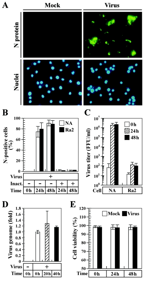
Previous studies demonstrated that some RV strains are able to infect cultured microglia and that the efficiency of viral multiplication in infected cells depends on virus strains (49). Although an attenuated Evelyn-Rotnycki-Abelseth strain, which had been adapted to tissue culture, grew well in primary microglia, the productive replication of pathogenic RV, such as a street isolate of bat origin, was strictly restricted in these cell types (49). We initially examined the overall growth performance of CVS-11, a highly neurovirulent laboratory strain of RV, in microglial Ra2 cells (Fig. 1). Figure 1A shows the expression pattern of the viral N protein, which accumulates in the cytoplasm of RV-infected cells, in microglia. Cells were inoculated with CVS-11 virus at an MOI of 2 (2 FFU/cell), and the viral N protein and cell nuclei were stained with anti-N MAbs and with the DNA dye DAPI, respectively, at 24 h postinfection. As shown in Fig. 1A (right), the majority of the RV-inoculated Ra2 cells exhibited a strong signal of the N protein, while anti-N MAbs did not react with the mock-inoculated cells (Fig. 1A, left). Figure 1B shows the percentages of N-positive cells during the course of viral infection of microglial Ra2 and neuroblastoma NA cells. When Ra2 cells were inoculated with CVS-11, the fluorescent signal of N proteins was observed in about 80 and 90% of the total cell population at 24 and 48 h postinfection, respectively (Fig. 1B). The plating efficiency of CVS-11 in Ra2 cells was at a level similar to that seen in neuronal NA cells, and its infectivity was readily diminished by UV irradiation (Fig. 1B). These data suggest that CVS-11 virus is capable of entering microglial cells and that virus-encoded proteins were expressed in these cell types.
FIG. 1.
Growth characteristics of RV in microglia. (A) Indirect immunofluorescence assay of viral N protein in RV-inoculated cells. Microglia were incubated with CVS-11 virus and then further incubated. After 24 h of incubation, the cells were fixed and stained for N protein (top) and cell nuclei (bottom). The experiments were repeated three times, and representative areas of each culture are shown. Magnification, ×200. (B) Infectivity of CVS-11 virus in microglia. Neuroblastoma (NA) and microglial (Ra2) cells were incubated with or without CVS-11 virus, which had been untreated or inactivated with UV irradiation (Inact.). After incubation for the indicated times, the percentages of N-positive cells were determined with reference to the total cell number as described in the text. The values are averages from three independent experiments, and the error bars indicate standard deviations. (C) Production of progeny virus in RV-inoculated microglia. Cells were infected with CVS-11 virus, and immediately thereafter (0 h) and at the indicated time points, virus progenies in culture supernatant fluids were titrated. The data are averages from three independent experiments, and the error bars indicate standard deviations. (D) Replication of the viral genome in RV-infected microglia. Ra2 cells were mock infected or infected with CVS-11 virus, and the relative amounts of viral cDNA were measured by using real-time PCR analyses immediately thereafter (0 h) or 20 and 40 h after infection. The results are shown as n-fold increases in the amount of viral cDNAwith reference to the cDNA levels in RV-inoculated cells at 0 h postinfection. Mean values and standard errors from the results of three independent experiments are shown. (E) Viability of microglia following RV infection. Cells were mock infected or infected with CVS-11 virus, and the percentages of cells excluding trypan blue dye were calculated with reference to the total cell number at the time points indicated. Values are averages from three separate experiments, and the error bars represent standard deviations.
Defective growth of RV in microglia.
We next assessed the growth characteristics of CVS-11 virus in Ra2 cells. The cells were inoculated with virus, and the culture supernatants were subjected to virus titration (Fig. 1C). In either cell type, low levels of infectious virions, which might be detached from the cells, were detected in culture supernatants at 0 h postinfection. In neuronal NA cells, CVS-11 exhibited a marked increase in virus titers, reaching a near-plateau level at 24 h postinfection (Fig. 1C). In contrast, virus titers in culture supernatant fluids from RV-inoculated cells at 24 and 48 h postinfection were slightly higher than those at 0 h postinfection, and these differences were not statistically significant, suggesting that the production of virus progenies of CVS-11 in microglia is severely impaired. To further assess a defective growth of RV in microglia, Ra2 cells were infected with CVS-11, and the relative amounts of negative-strand RV genome in the infected cells were determined by real-time PCR analyses. As shown in Fig. 1D, the amounts of viral genome in RV-infected microglia at 20 and 40 h postinfection were about 1.3- and 1.2-fold higher than that at 0 h postinfection, respectively, but these differences were not statistically significant, indicating that the viral genome synthesis of CVS-11 is impaired in microglia. In the experiments shown in Fig. 1E, the viability of microglia infected with CVS-11 was examined by using a dye exclusion assay as described above. The dye exclusion capability of RV-infected cells was at the level similar to that of mock-infected cells at all the time points tested. These observations indicate that CVS-11 infection does not induce any cytotoxicity in microglia.
RV infection stimulates chemokine gene expression in microglia.
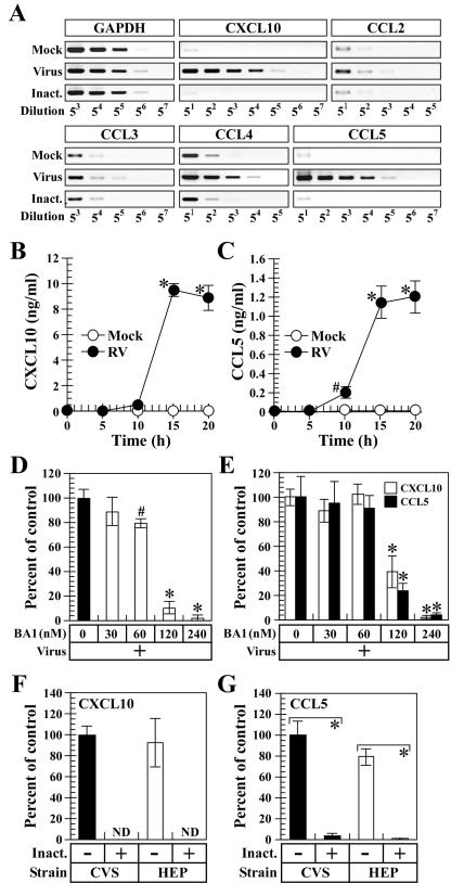
Recently, we have shown that endocytic processing of inactivated RV virion, as well as infectious virus, triggers the activation of a cellular signaling pathway, leading to the selective expression of the chemokine CXCL10 in macrophages (35). To examine whether RV selectively induces CXCL10 expression in microglia, Ra2 cells were inoculated with live CVS-11 virus or exposed to UV-inactivated virion, and the expression profiles of multiple chemokine genes were examined by RT-PCR 20 h after incubation. Figure 2 shows the transcription patterns of two major chemokine subfamilies, CXC and CC of chemokine ligands in microglia. When Ra2 cells were inoculated with infectious CVS-11 virus, the strong signals of amplified products of CXCL10, CCL4, and CCL5 were observed at highly diluted concentrations of PCR templates compared to those of mock-inoculated cells, while CVS-11 infection had little or no effect on the expression of CCL2 and CCL3. In particular, the expression of CXCL10 and CCL5 genes in RV-infected microglia was approximately 3,000-fold higher than that in mock-infected cells. We also examined the transcription patterns of the other chemokines, such as CXCL2, CXCL9, CXCL11, and CX3CL1, in RV-infected microglia, but the expression levels of these chemokine genes were not affected or were less affected by RV infection (data not shown). The chemokine expression patterns in Ra2 cells, which had been exposed to UV-inactivated RV virions, were similar to those seen in mock-treated cells. Thus, these results indicate that RV infection, but not exposure to inactivated virions, greatly induces the expression of CXCL10 and CCL5 in microglia. To assess whether the enhanced expression of CXCL10 and CCL5 mRNAs in RV-infected cells correlates with protein production, we measured the levels of CXCL10 (Fig. 2B) and CCL5 (Fig. 2C) production in microglia upon RV infection. When Ra2 cells were infected with CVS-11 virus, the onsets of chemokine production were detected 10 h after infection, and the amounts of chemokines increased steeply between 10 and 15 h after infection, reaching near-plateau levels at 20 h postinfection. In the experiments shown in Fig. 2D and E, we examined whether virion uncoating is required for the RV-induced expression of CXCL10 and CCL5 in microglia. BA1 is a selective and potent inhibitor of vacuolar H+-ATPase, prevents endosomal acidification (13), and is applicable to the suppression of an acid-induced fusion of lyssaviral envelope with cell membrane (24). As shown in Fig. 2D, when Ra2 cells were treated with BA1 ranging from 60 to 240 nM and then infected with CVS-11, the virus infectivity was significantly impaired in a dose-dependent manner. Figure 2E shows the RV-induced expression of CXCL10 and CCL5 in BA1-treated Ra2 cells. Pretreatment with BA1 at concentrations of 120 and 240 nM significantly diminished the expression of either chemokine in RV-infected Ra2 cells, suggesting that the virion uncoating is necessary for RV-induced chemokine expression in microglia. In order to examine whether viral gene expression is required for the RV-induced chemokine expression in microglia, we compared the chemokine production in Ra2 cells, which had been exposed to UV-inactivated virions, to that in RV-infected cells. As shown in Fig. 2F and G, the CVS-11-induced expression of CXCL10 and CCL5 was nearly completely abrogated by UV inactivation of virions. To examine whether the RV-induced chemokine expression depends on virus strains, we measured the expression of CXCL10 and CCL5 in microglia infected with HEP-Flury, a tissue culture-adapted and highly attenuated RV strain. In Ra2 cells, the induction of CXCL10 (Fig. 2F) and CCL5 (Fig. 2G) production was also observed following incubation with infectious HEP-Flury virus but not with inactivated virions. Taken together, these data demonstrate that the RV-induced expression of CXCL10 and CCL5 is triggered at the stage after viral gene expression and is not dependent on the virus strains.
FIG. 2.
Chemokine responses in microglia against RV infection. (A) Transcription patterns of chemokine genes in microglia following RV infection. Cells were incubated in the absence (Mock) or presence of infectious (Virus) or UV-inactivated (Inact.) CVS-11 virions and were subjected to RT-PCR analyses for the chemokine gene expression by using serially fivefold-diluted cDNA preparations as described in the text. The data are from one of three separate experiments with similar results. (B and C) Time course of RV-induced chemokine production by microglia. Cells were mock infected or infected with CVS-11 virus. After additional incubation for the indicated times, the protein contents of CXCL10 (B) and CCL5 (C) in culture supernatants were determined by using ELISA. (D and E) Effects of lysosomotropic agents on virus infectivity and chemokine expression in microglia. Ra2 cells, which had been treated with the indicated doses of BA1, were infected with CVS-11 virus and were further incubated in the presence or absence of BA1. After a 24-h incubation period, virus infectivity (D) and chemokine expression (E) were determined as described in the text. The results are shown as percentages of virus infectivity and chemokine expression with reference to the values for the drug-untreated controls. (F and G) RV-induced expression of chemokines isdependent on viral gene expression. Ra2 cells were incubated with neurovirulent CVS-11 (CVS) and attenuated HEP-Flury (HEP), which had been untreated (−) or inactivated with UV irradiation (+). At 20 h postinfection, protein contents of CXCL10 (F) and CCL5 (G) were determined. The percentages of chemokine expression were calculated with reference to the protein levels in culture fluids of the CVS-11-infected cells. For each panel, mean values and standard deviations from three separate experiments are shown, and statistically significant differences are indicated by asterisks (P < 0.01) and pound signs (P < 0.05).
RV infection activates MAPK signaling in microglia.
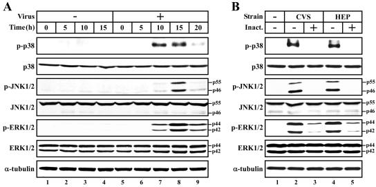
The enhanced production of CXCL10 and CCL5 in RV-infected microglia implies the possibility that RV infection may stimulate the cellular signaling pathway underlying chemokine expression. Considering the important roles of MAPK-mediated signaling pathways in host defense, we examined the activation of three major MAPK subfamilies, p38, JNK, and ERK1/2, in microglia upon RV infection (Fig. 3). In order to assess the activation of MAPK signaling pathways in microglia during the course of RV infection, cells were mock infected or infected with CVS-11, and the degrees of MAPK phosphorylation were examined by Western blotting. To avoid an additional effect of growth factors on MAPK activation, the cells were incubated in test medium containing extremely low concentration of FCS that did not induce MAPK phosphorylation. As shown in Fig. 3A, when microglia was infected with CVS-11 virus, the strong signals of p-p38 were detected between 10 and 15 h after infection (Fig. 3A, lanes 7 and 8). In RV-infected cells, the amounts of p-JNK1/2 and p-ERK1/2 were also increased between 10 and 20 h (Fig. 3A, lanes 7 to 9), and the maximal induction of either MAPK was seen at 15 h postinfection (Fig. 3A, lane 8). The increased levels of MAPK phosphorylation were not due to the enhanced production of these molecules or the difference in protein extracts loaded, as the protein levels of total amounts of MAPKs, as well as α-tubulin, in each sample were comparable. As shown in Fig. 3B, the RV-induced phosphorylation of each MAPK was also observed for microglia infected with attenuated HEP-Flury virus (Fig. 3B, lane 4). When virions were inactivated, RV-induced MAPK activation was nearly completely diminished (Fig. 3B, lanes 3 and 5). Thus, these data indicate that RV infection, but not exposure to inactivated virions, induces the activation of cellular signaling pathways mediated by multiple MAPK subfamilies, p38, ERK1/2, and JNK.
FIG. 3.
RV infection activates MAPK signaling pathways in microglia. (A) Time course of MAPK activation in microglia following RV infection. Cells were mock infected (−) or infected with CVS-11 (+). At the indicated time points, cells were harvested, and equal amounts of protein extracts from each sample (10 μg/lane) were loaded on the gel. The protein levels of phosphorylated and total MAPKs were analyzed by Western blotting as described in the text. The amounts of α-tubulin were also assessed to monitor the equal loadings of protein extracts. (B) Viral gene expression is required for RV-induced MAPK activation. Cells were incubated with CVS-11 and HEP-Flury virions, which had been untreated (−) or inactivated with UV irradiation (+) (Inact.), washed, and further incubated for 15 h. The phosphorylation levels of MAPKs were examined as described for panel A. The data are from one of three separate experiments with similar results.
RV-induced MAPK activation regulates chemokine expression in microglia.
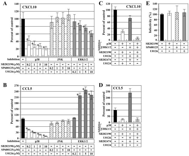
Based on the above-mentioned results, we hypothesized that the activation of MAPK signaling pathways underlies the up-regulation of CXCL10 and CCL5 expression in RV-infected microglia. To examine this possibility, we assessed the effects of MAPK inhibitors on the induction of chemokine expression in RV-infected microglia. Ra2 cells were pretreated with increasing doses of MAPK inhibitors and then infected with CVS-11 virus. Of the inhibitors tested, p38 inhibitor SB202190 significantly reduced the RV-induced expression of CXCL10 in a dose-dependent manner, and the maximal concentration of this compound (10 μM) diminished the expression level of CXCL10 by about 80% (Fig. 4A). Treatment with the ERK1/2 inhibitor U0126 resulted in a partial but statistically significant diminution of CXCL10 expression upon RV infection, while the JNK inhibitor SP600125 had little or no effect on the CXCL10 expression levels (Fig. 4A). These observations suggest that RV-induced CXCL10 expression is partly achieved through the activation of p38 and ERK1/2 pathways. Figure 4B shows CCL5 production in RV-infected microglia in the presence of MAPK inhibitors. The inhibition of p38 led to a considerable diminution of RV-induced expression of CCL5, and the amounts of CCL5 were not affected, or were less affected, by the treatment with JNK inhibitor (Fig. 4B). Interestingly, treatment with ERK1/2 inhibitor at concentrations ranging from 1 to 10 μM led to an up-regulation of CCL5 expression in RV-infected cells (Fig. 4B). These data suggest that the p38-mediated signaling pathway partly contributes to the RV-induced expression of CCL5 but that ERK1/2 activation participates in the down-regulation of CCL5 production in microglia. To examine the regulation of the RV-induced expression of CXCL10 and CCL5 through the p38 and ERK1/2 pathways in more detail, we measured chemokine production in RV-infected microglia under conditions in which both the p38 and ERK1/2 pathways were disrupted. In these experiments, SB202474 and U0124 (inactive analogues of SB202190 and U0126, respectively) were used to control the nonspecific effect of simultaneous treatments with SB202190 and U0126. As shown in Fig. 4C, simultaneous treatments with SB202190 and U0126 additively reduced the RV-induced CXCL10 expression compared to that with SB202190 and U0124 (P < 0.05) or with SB202474 and U0126 (P < 0.01) (Fig. 4C), suggesting that the p38 and ERK1/2 pathways individually facilitate CXCL10 expression in microglia in response to RV infection. Figure 4D shows RV-induced CCL5 expression in microglia, which had been treated with p38 and ERK1/2 inhibitors in combination. Under conditions in which both the p38 and ERK1/2 pathways were inhibited, CCL5 expression was at a level similar to that seen in microglia treated with p38 inhibitor alone, suggesting that ERK1/2 activation participates in the down-regulation of an excessive production of CCL5 in response to RV infection. In addition, CVS-11 virus was able to infect microglia normally even when the cells were treated with maximal doses of MAPK inhibitors tested (10 μM). Taken together, our findings indicate that the RV-induced expression of CXCL10 and CCL5 in microglia is positively and negatively regulated by the activation of the p38 and ERK1/2 pathways.
FIG. 4.
MAPK signaling pathways regulate RV-induced chemokine expression in microglia. (A and B) Effects of MAPK inhibitors on chemokine expression in RV-infected cells. Ra2 cells were untreated (−) or treated with the indicated doses of SB202190, SP600125, and U0126 just prior to infection with CVS-11 virus. At 15 h postinfection, the protein contents of CXCL10 (A) and CCL5 (B) were determined. The results are shown as percentages of chemokine expression with reference to the values for the drug-untreated control. (C and D) Effect of simultaneous inhibitions of p38 and ERK1/2 pathways on RV-induced chemokine expression. The cells were untreated (−) or treated with SB202190 and U0126 (2.5 μM each) individually or in combination prior to infection with CVS-11 virus. SB202474 and U0124 were also used as controls to verify the specificities of SB202190 and U0126. After a 15-h incubation period, the protein levels of CXCL10 (C) and CCL5 (D) were determined. The percentages of chemokine expression were calculated with reference to the values for the drug-untreated control. (E) Effects of MAPK inhibitors on virus infectivity. Cells were pretreated with or without the indicated compounds (10 μM each) and infected with CVS-11 virus. At 24 h postinfection, virus infectivity was determined as described in the text. The results are shown as percentages of infectivity with reference to the values for the drug-untreated control. For each panel, the data are averages from three independent experiments, and the error bars represent standard deviations. Statistically significant differences are indicated by asterisks (P < 0.01) and pound signs (P < 0.05).
RV infection activates the IκB-NF-κB pathway in microglia.
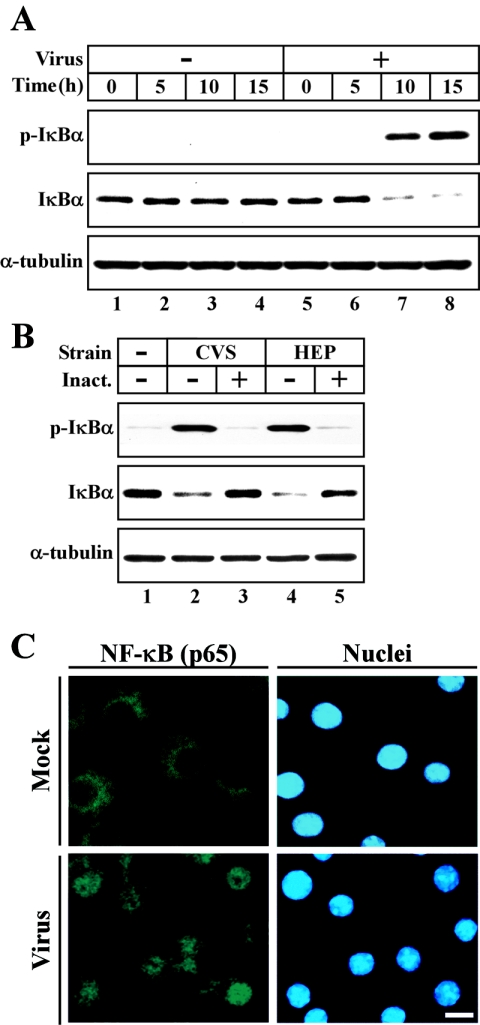
Analyses of the RV-induced expression of CXCL10 and CCL5 in the presence of MAPK inhibitors have revealed that the p38 and ERK1/2 pathways regulate the production of these chemokines. In particular, we found that p38 activation is responsible for the RV-induced expression of these chemokines. However, RV-induced chemokine expression was not completely blocked by MAPK inhibitors even at maximal concentrations that did not cause cytotoxicity. These lines of evidence imply that another signaling pathway may also contribute to the chemokine responses of microglia to RV infection. NF-κB plays critical roles in transcriptional regulation of numerous genes involved in host defense mechanisms (3). This transcription factor is normally found in the cytoplasm in a latent form associated with IκB, of which various isoforms exist (23). Following the appropriate stimulus, IκB is phosphorylated by IκB kinase, ubiquitinated, and degraded by proteasomes. Degradation of IκB exposes the nuclear localization signal of NF-κB, and then NF-κB migrates to the nucleus and activates transcription (23). Since it remains unclear whether RV infection triggers the activation of the NF-κB pathway in mammalian cells, we examined the onset of NF-κB activation in RV-infected microglia (Fig. 5). Ra2 cells were uninfected or infected with CVS-11, and the phosphorylation and degradation of IκBα proteins were examined by Western blotting. In RV-infected cells, the signals of p-IκBα were detected between 10 and 15 h after infection (Fig. 5A, lanes 7 and 8), and only low, but detectable, levels of total IκBα were observed in RV-infected microglia at these time points (Fig. 5A, lanes 7 and 8). The alterations in the amounts of p-IκBα and total IκBα were not due to the difference in protein extracts loaded, as the protein levels of α-tubulin in each sample were comparable. As shown in Fig. 5B, the RV-induced phosphorylation and degradation of IκBα proteins were also observed for microglia, which had been infected with attenuated HEP-Flury virus (Fig. 5B, lane 4), and were markedly disrupted by the UV inactivation of RV virions (Fig. 5B, lanes 3 and 5). To confirm the NF-κB activation, microglia were mock infected or infected with CVS-11 virus, and the nuclear translocation of the NF-κB p65 subunit was examined by confocal laser scanning microscopy (Fig. 5C). In mock-infected cells, the p65 molecules appeared to be dispersed in the cytoplasmic region (Fig. 5C, top). In contrast, when cells were infected with CVS-11, fluorescent signals of p65 were found primarily in the nuclei (Fig. 5C, bottom). These data demonstrate that RV infection of microglia results in the activation of the NF-κB signaling pathway.
FIG. 5.
RV infection activates the IκB-NF-κB signaling cascade in microglia. (A) Time course of IκBα phosphorylation and degradation in microglia following RV infection. Cells were mock infected (−) or infected with CVS-11 virus (+). After additional incubation for the indicated times, cells were harvested and subjected to Western blot analyses. The data are from one of three individual experiments with similar results. (B) Viral gene expression is required for the RV-induced phosphorylation of IκBα. Cells were incubated with or without CVS-11 and HEP-Flury virions, which had been untreated (−) or inactivated with UV irradiation (+) (Inact.). At 15 h postincubation, Western blot analyses were carried out as described for panel A. The data are from one of two individual experiments with similar results. (C) Nuclear translocation of NF-κB p65 in microglia infected with RV. Ra2 cells were mock infected (top) or infected with CVS-11 virus (bottom). At 15 h postinfection, the cells were fixed, and p65 proteins (left) and cell nuclei (right) were stained as described in the text. All images are confocal sections taken through the center of the cells. The experiments were repeated three times, and representative areas of each culture are shown. White bar, 10 μm.
RV infection stimulates chemokine expression via the IκB-NF-κB signaling pathway.
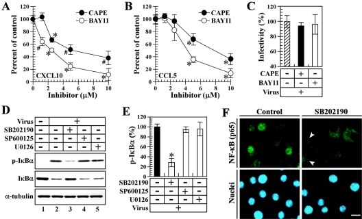
We assessed whether the activation of the NF-κB signaling cascade is responsible for CXCL10 and CCL5 production in RV-infected microglia. Ra2 cells were preincubated with the increasing doses of CAPE, a chemical compound that has been shown to inhibit the nuclear translocation of NF-κB (37), or BAY 11-7082, an inhibitor of IκB phosphorylation (45), and then RV-induced chemokine expression in microglia was examined as described above. In the presence of either inhibitor, the RV-induced expression of CXCL10 (Fig. 6A) and CCL5 (Fig. 6B) was significantly abrogated in a dose-dependent fashion without affecting virus infectivity (Fig. 6C). Thus, these results indicate that RV infection stimulates CXCL10 and CCL5 production in microglia through activation of the IκB-NF-κB signaling cascade. We further investigated whether the activation of MAPK pathways is associated with NF-κB signaling in RV-infected microglia. Ra2 cells, which had been pretreated with MAPK inhibitors, were infected with CVS-11 virus, and the amounts of p-IκBα and total IκBα were examined by using Western blot analysis. Of the chemical compounds tested, the p38 inhibitor SB202190 partially but not completely diminished IκBα phosphorylation and degradation in RV-infected microglia (Fig. 6D, lane 3). As shown in Fig. 6E, the RV-induced phosphorylation of IκBα in SB202190-treated cells was reduced by about 72% compared to that in the untreated control. In the experiments shown in Fig. 6F, we examined the effect of p38 inhibitor on subcellular localization patterns of NF-κB p65 in RV-infected microglia. When Ra2 cells were pretreated with SB202190 and infected with CVS-11 virus, the RV-induced nuclear translocation of p65 proteins was inhibited (Fig. 6F, right) but not completely blocked (Fig. 6F, upper right panel). Taken together, these results suggest that the p38-mediated pathway is required for the efficient activation of NF-κB signaling in microglia in response to RV infection.
FIG. 6.
RV infection stimulates chemokine expression via the NF-κB signaling pathway. (A and B) Effects of IκB and NF-κB inhibitors on the RV-induced expression of chemokines. Cells were untreated or treated with the indicated doses of CAPE and BAY 11-7082 and were infected with CVS-11 virus. At 15 h postinfection, the expression levels of CXCL10 (A) and CCL5 (B) were determined as described in the text. The results are shown as percentages of chemokine expression with reference to the values for the drug-untreated control. (C) Effects of IκB and NF-κB inhibitors on virus infectivity. Ra2 cells were pretreated with or without the indicated inhibitors (10 μM each) and infected with CVS-11 virus. At 24 h postinfection, the cells were fixed, and the percentages of virus plating were determined with reference to the values for the drug-untreated control. (D) IκB-NF-κB signaling is augmented by p38 activation in RV-infected microglia. Ra2 cells were pretreated with or without MAPK inhibitors (10 μM each) and infected with CVS-11 virus. After a 15-h incubation period, the cells were subjected to Western blot analyses. The data are from one of three separate experiments with similar results. (E) Quantitative analyses of IκBα phosphorylation in microglia following RV infection. The digital images of each blot shown in panel D were prepared, and the density of each band was quantified by image analysis. The percentages of band densities of p-IκBα were calculated with reference to the values for the drug-untreated control. For panels A, B, C, and E, mean values and standard deviations from the results of three individual experiments are shown, and significant differences are indicated by asterisks (P < 0.01) and pound signs (P < 0.05). (F) Effect of the p38 inhibitor on subcellular localization patterns of NF-κB p65 in RV-infected microglia. Cells were preincubated in the absence (left) or presence (right) of SB202190 and then infected with CVS-11 virus. At 15 h postinfection, the cells were stained for NF-κB p65 and cell nuclei. All images were taken through the centers of the cells. Similar results were obtained from two other experiments. Arrowheads indicate the p65 localization mentioned in the text. White bar, 10 μm.
DISCUSSION
Microglia are resident immune effector cells within the CNS and are hence likely to encounter infectious agents at very early stages of infection, as well as at later stages, when peripheral leukocytes, such as lymphocytes and monocytes, are recruited into the brain parenchyma (26). Recruitment of leukocytes into the CNS is usually preceded by chemokine production from microglia and other CNS-resident cells, which is the first line of defense against neurotropic viruses (55).
It is well known that RV replication within the CNS occurs primarily in neurons and not in other CNS-resident cells, such as microglia and astrocytes. However, in vitro and in vivo studies provide evidence for the onset of viral gene expression in glial cells (17, 47, 49, 60), implying that although RV virions were taken up by glial cells, in which virus gene expression occurs, the production of virus progenies is impaired at the later stage of the viral replicative cycle. It is also reported that RV infection affects the characteristics of glial cells. Prosniak and colleagues have demonstrated that the mRNA expression levels of neuroleukin and the two isoforms of fibroblast growth factor-homologous factor 4 are significantly induced in RV-infected astrocytes in vitro and in vivo (47).
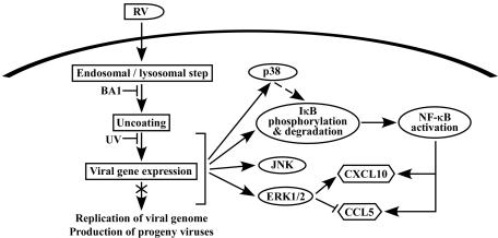
In the present study, as an initial step toward understanding the cellular response of microglia to RV infection, we have systemically examined the growth characteristics of RV and the activation of cellular signaling pathways leading to chemokine expression in these cell types. On the basis of our data, we provide a model for the cellular signaling events underlying microglial activation in response to RV infection (Fig. 7). We suggest here that RV virions are taken up by microglia into endosomal-lysosomal compartments and that virion uncoating occurs in the acidic environment of these vesicles. Although the virus-encoded proteins can be expressed in cytoplasms, the synthesis of the viral genome and the production of virus progenies were significantly impaired. As to the cellular response of microglia to RV infection, we also observed that the expression of two chemokines, CXCL10 and CCL5, is notably induced in RV-infected microglia. Furthermore, the data obtained here demonstrate that RV infection stimulates multiple signaling pathways mediated by NF-κB, p38, JNK, and ERK1/2 in microglia and that viral gene expression is required for the activation of these signal-transducing molecules. NF-κB-dependent signal transduction is a key process leading to the strong induction of CXCL10 and CCL5 expression in RV-infected microglia, and this signaling is indirectly augmented via the activation of the p38-mediated pathway. Our data also indicate that ERK1/2, but not JNK, partly contributes to the induction of CXCL10 expression and that it acts as a down-regulator of the excessive production of CCL5 in response to RV infection.
FIG. 7.
Schematic model for the chemokine responses of microglia to RV infection. The model is based on the data in the present study and previous publications (see the text for the appropriate references). RV infection of microglia is initiated by the uptake of virions into endosomal-lysosomal compartments. Although microglia allows RV uncoating and expression of virus-encoded proteins, the replication of the viral genome and the production of virus progenies are impaired. RV infection of microglia leads to the activation of cellular signaling pathways mediated by MAPKs and IκB-NF-κB, and this process is triggered at the stage after viral gene expression. NF-κB signaling, which is additively augmented by p38 activation, is crucial for the expression of CXCL10 and CCL5 in the RV-infected microglia. The ERK1/2 pathway also participates in the induction of CXCL10 expression, while it acts as a down-regulator of the excessive production of CCL5 upon RV infection.
A recent study focusing on the overall expression profiles of host genes in the RV-infected brain has revealed that the predominant effect of RV infection is the down-regulation of gene expression (48). Consistent with these results in vivo, in the earlier study using culture of neuronal cells, we were unable to demonstrate marked changes in expression patterns of multiple host genes following RV infection (35). However, some reports indicate the RV-induced gene expression of a subset of cellular proteins, such as soluble factors and signal-transducing molecules, in the CNS (14, 48, 52). Galelli and colleagues demonstrated that the gene expression levels of chemokines, including CXCL10 and CCL5, are induced in mononuclear cell populations of the CNS infected with RV (14). Considering the potent chemotactic effects of CXCL10 and CCL5 on leukocytes, such as T cells and monocytes (39, 54), it is likely that the production of these chemokines is associated with the infiltration of mononuclear cells into the RV-infected CNS, as was reported previously (5, 14). Still, little information is available concerning the type of CNS-resident cells, which are able to produce these chemokines in response to RV infection. Our findings in the current study demonstrate the possibility that CNS-resident cells can produce CXCL10 and CCL5 via the recognition of RV infection. As for the activation of signal-transducing molecules evoked by RV infection, Dietzschold and colleagues previously suggested that the DNA-binding activities of AP-1, which is a downstream target of JNK pathway, and NF-κB are enhanced in the brains of RV-infected animals (9). The data obtained here provide direct evidence for the RV-induced activation of intracellular signaling pathways in CNS-resident cells.
Recent extensive studies have indicated that microglia intrinsically produces CXCL10 and CCL5 upon infection with a variety of neurotropic viruses, including cytomegalovirus (6), human immunodeficiency virus (58), herpes simplex virus (30), Newcastle disease virus (12, 63), Theiler's murine encephalomyelitis virus (42, 43), and Japanese encephalitis virus (7). However, the precise role of cell signaling molecules, especially that of MAPK subfamilies, in the virus-induced expression of these chemokines in microglia remains poorly understood. As for MAPK signaling being responsible for CXCL10 expression, a recent report suggests that the induction of CXCL10 production in microglia upon CMV infection is partly mediated by the p38 pathway (6). The results obtained in the present study indicate that ERK1/2, along with p38, is required for CXCL10 expression in RV-infected microglia. We also demonstrate that p38 and ERK1/2 participate in a different process leading to CXCL10 expression in microglia following RV infection, as the simultaneous inhibition of p38 and ERK1/2 additively decreased the CXCL10 levels. A previous study using culture of epithelial cells from peripheral tissue suggested that adenovirus entry into the cells induces CXCL10 expression via both the p38 and ERK1/2 pathways (62). Our findings are unique in that the activation of these MAPK pathways leading to CXCL10 expression is triggered at the step after virus entry, because UV-inactivated RV virions failed to induce MAPK phosphorylation.
As to CCL5 expression via MAPK signaling, it has been previously shown that the expression of this chemokine is induced via the p38 and ERK1/2 pathways in alveolar epithelial cells infected with respiratory syncytial virus (44). It has also been reported that p38, as well as JNK, is required for CCL5 production in bronchial epithelial cells following infection with influenza virus (28). On the other hand, a recent study focusing on herpes simplex virus-induced chemokine expression indicates that CCL5 responses in macrophages and fibroblasts against virus infection are induced by the p38-independent mechanism (31). From these studies, the contribution of MAPK signaling to CCL5 expression is considered to be dependent on the types of cells and viruses. With respect to the CCL5 response to neurotropic virus infection, Chen et al. (7) have demonstrated that the Japanese encephalitis virus-induced CCL5 production in mixed microglia-astrocyte cultures is partially induced via ERK1/2 activation. However, it remains unclear whether other MAPK pathways are responsible for virus-induced CCL5 production by microglia. The data shown in the present study constitute a body of evidence for virus-induced CCL5 expression through activation of the p38 signaling pathway in microglia. Furthermore, as a striking feature of chemokine expression in RV-infected microglia, we found that the activation of the ERK1/2 pathway negatively regulates virus-induced CCL5 expression. In view of our evidence that p38, as well as NF-κB, activates CCL5 expression in virus-infected microglia, it appears paradoxical that ERK1/2 inhibits the expression of this chemokine. However, the analysis of the kinetics of MAPK phosphorylation in RV-infected microglia demonstrated that p38 phosphorylation precedes the maximal activation of ERK1/2, rendering it likely that the ERK1/2 pathway acts as a negative regulator of CCL5 expression. Previously, it has been suggested that the CCL5 response of microglia to human immunodeficiency virus infection is attenuated by the activation of p38 but not of ERK1/2 (58). Considering the strong chemotactic effect of CCL5 on a broad range of inflammatory cells (1, 54) and the lack of evidence for the MAPK-mediated down-regulation of CCL5 production in other cell types upon virus infection, it can be postulated that the MAPK signal pathways intrinsically adjust the level of CCL5 expression in microglia via recognition of virus infection, thereby controlling excessive leukocyte trafficking into the brain parenchyma.
An important body of knowledge has been accumulated about the chemokine expression mediated by the NF-κB pathway (32), while the NF-κB-dependent expression of CXCL10 and CCL5 in virus-infected microglia has been addressed in only a few studies (6, 7). In the present study, we show that NF-κB signaling pathway acts as a cardinal mediator of CXCL10 and CCL5 expression in virus-infected microglia, and our observation is consistent with earlier results from other investigators. As a remarkable feature of NF-κB-meditated chemokine expression, we found that NF-κB signaling can be augmented by p38 activation in RV-infected microglia. We also observed that the onset of this signal transduction was not completely blocked even in the absence of p38 activation, suggesting that the RV-induced expression of CXCL10 and CCL5 is achieved by two distinct mechanisms, the direct activation of NF-κB evoked by the recognition of virus infection and the p38-dependent transactivation of NF-κB signaling, both of which are required to fully activate chemokine expression in microglia. It is probable that the p38-independent activation of NF-κB in RV-infected microglia is mediated by the other signal-transducing molecules, such as phosphatidylinositol 3-kinase and double-stranded-RNA-activated protein kinase (33). Recent studies have demonstrated that the p38-dependent pathway activates NF-κB in response to stimuli, such as proinflammatory cytokines and environmental stress (8, 20, 56). However, much remains to be understood with regard to the convergence of these pathways during virus infection. Since IκB is not a substrate for p38 (56), it is likely that p38 may activate one or more of the signaling molecules upstream of IκB in the NF-κB cascade. Alternatively, as the p38 pathway participates in the induction of proinflammatory cytokines, such as interleukin 1β (33), which can activate the NF-κB pathway (4), it is possible that the RV-induced activation of p38 may facilitate proinflammatory cytokine expression, which subsequently induces NF-κB activation in an autocrine action. The strong induction of CXCL10 and CCL5 expression in RV-infected microglia may also be explained by a positive transcriptional synergy between NF-κB and the other transcription factors. It is well known that both CXCL10 and CCL5 are IFN-inducible chemokines (32), and in preliminary experiments, we observed that RV infection of microglia led to the induction of IFN-α and -β (data not shown). Accumulating evidence has indicated a transcriptional synergy between NF-κB and IFN-responsive transcription factors, such as signal transducer and activator of transcription (40, 41, 46). Thus, it is likely that IFNs secreted from RV-infected microglia stimulates IFN signaling in an autocrine manner, which subsequently facilitates chemokine gene expression in cooperation with NF-κB.
Overall, our study presented here provides an insight into the precise roles of intracellular signaling pathways mediated by MAPK and NF-κB in the regulation of chemokine expression in microglia through recognition of neurotropic virus infection.
Acknowledgments
This work was supported by a grant for research on health sciences focusing on drug innovation from the Japan Health Sciences Foundation; by a grant from the Ichiro Kanehara Foundation; by a grant-in-aid for scientific research from the Ministry of Education, Science, Sports and Culture of Japan; and by a grant for research on emerging and reemerging infectious diseases from the Ministry of Health, Labor and Welfare of Japan.
REFERENCES
- 1.Alam, R., S. Stafford, P. Forsythe, R. Harrison, D. Faubion, M. A. Lett-Brown, and J. A. Grant. 1993. RANTES is a chemotactic and activating factor for human eosinophils. J. Immunol. 150:3442-3448. [PubMed] [Google Scholar]
- 2.Aloisi, F. 2001. Immune function of microglia. Glia 36:165-179. [DOI] [PubMed] [Google Scholar]
- 3.Baeuerle, P. A., and T. Henkel. 1994. Function and activation of NF-kappa B in the immune system. Annu. Rev. Immunol. 12:141-179. [DOI] [PubMed] [Google Scholar]
- 4.Bowie, A., and L. A. O'Neill. 2000. The interleukin-1 receptor/Toll-like receptor superfamily: signal generators for pro-inflammatory interleukins and microbial products. J. Leukoc. Biol. 67:508-514. [DOI] [PubMed] [Google Scholar]
- 5.Camelo, S., M. Lafage, and M. Lafon. 2000. Absence of the p55 Kd TNF-alpha receptor promotes survival in rabies virus acute encephalitis. J. Neurovirol. 6:507-518. [DOI] [PubMed] [Google Scholar]
- 6.Cheeran, M. C., S. Hu, W. S. Sheng, P. K. Peterson, and J. R. Lokensgard. 2003. CXCL10 production from cytomegalovirus-stimulated microglia is regulated by both human and viral interleukin-10. J. Virol. 77:4502-4515. [DOI] [PMC free article] [PubMed] [Google Scholar]
- 7.Chen, C. J., J. H. Chen, S. Y. Chen, S. L. Liao, and S. L. Raung. 2004. Upregulation of RANTES gene expression in neuroglia by Japanese encephalitis virus infection. J. Virol. 78:12107-12119. [DOI] [PMC free article] [PubMed] [Google Scholar]
- 8.Craig, R., A. Larkin, A. M. Mingo, D. J. Thuerauf, C. Andrews, P. M. McDonough, and C. C. Glembotski. 2000. p38 MAPK and NF-kappa B collaborate to induce interleukin-6 gene expression and release. Evidence for a cytoprotective autocrine signaling pathway in a cardiac myocyte model system. J. Biol. Chem. 275:23814-23824. [DOI] [PubMed] [Google Scholar]
- 9.Dietzschold, B., K. Morimoto, and D. C. Hooper. 2001. Mechanisms of virus-induced neuronal damage and the clearance of viruses from the CNS. Curr. Top. Microbiol. 253:145-155. [DOI] [PubMed] [Google Scholar]
- 10.Dixon, D. W., L. A. Mattiace, K. Kure, K. Hutchins, X. Lyman, and C. F. Brosnan. 1991. Microglia in human disease, with an emphasis on acquired immune deficiency syndrome. Lab. Investig. 64:135-156. [PubMed] [Google Scholar]
- 11.Eugenin, E. A., and J. W. Berman. 2003. Chemokine-dependent mechanisms of leukocyte trafficking across a model of the blood-brain barrier. Methods 29:351-361. [DOI] [PubMed] [Google Scholar]
- 12.Fisher, S. N., P. Vanguri, H. S. Shin, and M. L. Shin. 1995. Regulatory mechanisms of MuRantes and CRG-2 chemokine gene induction in central nervous system glial cells by virus. Brain Behav. Immun. 9:331-344. [DOI] [PubMed] [Google Scholar]
- 13.Gagliardi, S., M. Rees, and C. Farina. 1999. Chemistry and structure activity relationships of bafilomycin A1, a potent and selective inhibitor of the vacuolar H+-ATPase. Curr. Med. Chem. 6:1197-1212. [PubMed] [Google Scholar]
- 14.Galelli, A., L. Baloul, and M. Lafon. 2000. Abortive rabies virus central nervous infection is controlled by T lymphocyte local recruitment and induction of apoptosis. J. Neurovirol. 6:359-372. [DOI] [PubMed] [Google Scholar]
- 15.Garrington, T. P., and G. L. Johnson. 1999. Organization and regulation of mitogen-activated protein kinase signaling pathways. Curr. Opin. Cell Biol. 11:211-218. [DOI] [PubMed] [Google Scholar]
- 16.Gonzalez-Scarano, F., and G. Baltuch. 1999. Microglia as mediators of inflammatory and degenerative diseases Annu. Rev. Neurosci. 22:219-240. [DOI] [PubMed] [Google Scholar]
- 17.Guigoni, C., and P. Coulon. 2002. Rabies virus is not cytolytic for rat spinal motoneurons in vitro. J. Neurovirol. 8:306-317. [DOI] [PubMed] [Google Scholar]
- 18.Inoue, H., M. Sawada, A. Ryo, H. Tanahashi, T. Wakatsuki, A. Hada, N. Kondoh, K. Nakagaki, K. Takahashi, A. Suzumura, M. Yamamoto, and T. Tabira. 1999. Serial analysis of gene expression in a microglial cell line. Glia 28:265-271. [DOI] [PubMed] [Google Scholar]
- 19.Jaramillo, M., D. C. Gowda, D. Radzioch, and M. Olivier. 2003. Hemozoin increases IFN-gamma-inducible macrophage nitric oxide generation through extracellular signal-regulated kinase- and NF-kappa B-dependent pathways. J. Immunol. 171:4243-4253. [DOI] [PubMed] [Google Scholar]
- 20.Jaspers, I., J. M. Samet, S. Erzurum, and W. Reed. 2000. Vanadium-induced kappaB-dependent transcription depends upon peroxide-induced activation of the p38 mitogen-activated protein kinase. Am. J. Respir. Cell Mol. Biol. 23:95-102. [DOI] [PubMed] [Google Scholar]
- 21.Kanazawa, T., M. Sawada, K. Kato, K. Yamamoto, H. Mori, and R. Tanaka. 2000. Differentiated regulation of allo-antigen presentation by different types of murine microglial cell lines. J. Neurosci. Res. 62:383-388. [DOI] [PubMed] [Google Scholar]
- 22.Karin, M. 1998. Mitogen-activated protein kinase cascades as regulators of stress responses. Ann. N. Y. Acad. Sci. 851:139-146. [DOI] [PubMed] [Google Scholar]
- 23.Karin, M., and Y. Ben Neriah. 2000. Phosphorylation meets ubiquitination: the control of NF-κB activity. Annu. Rev. Immunol. 18:621-663. [DOI] [PubMed] [Google Scholar]
- 24.Kassis, R., F. Larrous, J. Estaquier, and H. Bourhy. 2004. Lyssavirus matrix protein induces apoptosis by a TRAIL-dependent mechanisms involving caspase-8 activation. J. Virol. 78:6543-6555. [DOI] [PMC free article] [PubMed] [Google Scholar]
- 25.Kawakami, K., K. Shibuya, M. H. Qureshi, T. Zhang, Y. Koguchi, M. Tohyama, Q. Xie, S. Naoe, and A. Saito. 1999. Chemokine responses and accumulation of inflammatory cells in the lungs of mice infected with highly virulent Cryptococcus neoformans: effects of interleukin-12. FEMS Immunol. Med. Microbiol. 25:391-402. [DOI] [PubMed] [Google Scholar]
- 26.Kreutzberg, G. W. 1996. Microglia: a sensor for pathological events in the CNS. Trends Neurosci. 19:312-318. [DOI] [PubMed] [Google Scholar]
- 27.Kristensson, K., D. K. Dastur, D. K. Manghani, H. Tsiang, and M. Bentivoglio. 1996. Rabies: interactions between neurons and viruses. A review of the history of Negri inclusion bodies. Neuropathol. Appl. Neurobiol. 22:179-187. [PubMed] [Google Scholar]
- 28.Kujime, K., S. Hashimoto, Y. Gon, K. Shimizu, and T. Horie. 2000. p38 mitogen-activated protein kinase and c-jun-NH2-terminal kinase regulate RANTES production by influenza virus-infected human bronchial epithelial cells. J. Immunol. 164:3222-3228. [DOI] [PubMed] [Google Scholar]
- 29.Lacroix-Lamande, S., R. Mancassola, M. Naciri, and F. Laurent. 2002. Role of gamma interferon in chemokine expression in the ileum of mice and in a murine intestinal epithelial cell line after Cryptosporidium parvum infection. Infect. Immun. 70:2090-2099. [DOI] [PMC free article] [PubMed] [Google Scholar]
- 30.Lokensgard, J. R., S. Hu, W. Sheng, M. vanOijen, D. Cox, M. C. Cheeran, and P. K. Peterson. 2001. Robust expression of TNF-alpha, IL-1beta, RANTES, and IP-10 by human microglial cells during nonproductive infection with herpes simplex virus. J. Neurovirol. 7:208-219. [DOI] [PubMed] [Google Scholar]
- 31.Melchjorsen, J., and S. R. Paludan. 2003. Induction of RANTES/CCL5 by herpes simplex virus is regulated by nuclear factor kappa B and interferon regulatory factor 3. J. Gen. Virol. 84:2491-2495. [DOI] [PubMed] [Google Scholar]
- 32.Melchjorsen, J., L. N. Sorensen, and S. R. Paludan. 2003. Expression and function of chemokines during viral infections: from molecular mechanisms to in vivo function. J. Leukoc. Biol. 74:331-343. [DOI] [PMC free article] [PubMed] [Google Scholar]
- 33.Mogensen, T. H., and S. R. Paludan. 2001. Molecular pathways in virus-induced cytokine production. Microbiol. Mol. Biol. Rev. 65:131-150. [DOI] [PMC free article] [PubMed] [Google Scholar]
- 34.Nakamichi, K., D. Kuroki, Y. Matsumoto, and H. Otsuka. 2001. Bovine herpesvirus 1 glycoprotein G is required for prevention of apoptosis and efficient viral growth in rabbit kidney cells. Virology 279:488-498. [DOI] [PubMed] [Google Scholar]
- 35.Nakamichi, K., S. Inoue, T. Takasaki, K. Morimoto, and I. Kurane. 2004. Rabies virus stimulates nitric oxide production and CXC chemokine ligand 10 expression in macrophages through activation of extracellular signal-regulated kinases 1 and 2. J. Virol. 78:9376-9388. [DOI] [PMC free article] [PubMed] [Google Scholar]
- 36.Nakamichi, K., Y. Matsumoto, and H. Otsuka. 2002. Bovine herpesvirus 1 glycoprotein G is necessary for maintaining cell-to-cell junctional adherence among infected cells. Virology 294:22-30. [DOI] [PubMed] [Google Scholar]
- 37.Natarajan, K., S. Singh, T. R. Burke, Jr., D. Grunberger, and B. B. Aggarwal. 1996. Caffeic acid phenethyl ester is a potent and specific inhibitor of activation of nuclear transcription factor NF-kappa B. Proc. Natl. Acad. Sci. USA 93:9090-9095. [DOI] [PMC free article] [PubMed] [Google Scholar]
- 38.Neumann, B., N. Zantl, A. Veihelmann, K. Emmanuilidis, K. Pfeffer, C. D. Heidecke, and B. Holzmann. 1999. Mechanisms of acute inflammatory lung injury induced by abdominal sepsis. Int. Immunol. 11:217-227. [DOI] [PubMed] [Google Scholar]
- 39.Neville, L. F., G. Mathiak, and O. Bagasra. 1997. The immunobiology of interferon-gamma inducible protein 10 kD (IP-10): a novel, pleiotropic member of the C-X-C chemokine superfamily. Cytokine Growth Factor Rev. 8:207-219. [DOI] [PubMed] [Google Scholar]
- 40.Nguyen, V. T., and E. N. Benveniste. 2002. Critical role of tumor necrosis factor-alpha and NF-kappa B in interferon-gamma-induced CD40 expression in microglia/macrophages. J. Biol. Chem. 277:13796-13803. [DOI] [PubMed] [Google Scholar]
- 41.Ohmori, Y., R. D. Schreiber, and T. A. Hamilton. 1997. Synergy between interferon-gamma and tumor necrosis factor-alpha in transcriptional activation is mediated by cooperation between signal transducer and activator of transcription 1 and nuclear factor kappaB. J. Biol. Chem. 272:14899-14907. [DOI] [PubMed] [Google Scholar]
- 42.Olson, J. K., and S. D. Miller. 2004. Microglia initiate central nervous system innate and adaptive immune responses through multiple TLRs. J. Immunol. 173:3916-3924. [DOI] [PubMed] [Google Scholar]
- 43.Palma, J. P., and B. S. Kim. 2001. Induction of selected chemokines in glial cells infected with Theiler's virus. J. Neuroimmunol. 117:166-170. [DOI] [PubMed] [Google Scholar]
- 44.Pazdrak, K., B. Olszewska-Pazdrak, T. Liu, R. Takizawa, A. R. Brasier, R. P. Garofalo, and A. Casola. 2002. MAPK activation is involved in posttranscriptional regulation of RSV-induced RANTES gene expression. Am. J. Physiol. Lung Cell Mol. Physiol. 283:L364-L372. [DOI] [PubMed] [Google Scholar]
- 45.Pierce, J., W., R. Schoenleber, G. Jesmok, J. Best, S. A. Moore, T. Collins, M. E. Gerritsen. 1997. Novel inhibitors of cytokine-induced IκBα phosphorylation and endothelial cell adhesion molecule expression show anti-inflammatory effects in vivo. J. Biol. Chem. 272:21096-21103. [DOI] [PubMed] [Google Scholar]
- 46.Pine, R. 1997. Convergence of TNFalpha and IFNgamma signalling pathways through synergistic induction of IRF-1/ISGF-2 is mediated by a composite GAS/kappaB promoter element. Nucleic Acids Res. 25:4346-4354. [DOI] [PMC free article] [PubMed] [Google Scholar]
- 47.Prosniak, M., A. Zborek, G. S. Scott, A. Roy, T. W. Phares, H. Koprowski, and D. C. Hooper. 2003. Differential expression of growth factors at the cellular level in virus-infected brain. Proc. Natl. Acad. Sci. USA 100:6765-6770. [DOI] [PMC free article] [PubMed] [Google Scholar]
- 48.Prosniak, M., D. C. Hooper, B. Dietzschold, and H. Koprowski. 2001. Effect of rabies virus infection on gene expression in mouse brain. Proc. Natl. Acad. Sci. USA 98:2758-2763. [DOI] [PMC free article] [PubMed] [Google Scholar]
- 49.Ray, N. B., C. Power, W. P. Lynch, L. C. Ewalt, and D. L. Lodmell. 1997. Rabies viruses infect primary cultures of murine, feline, and human microglia and astrocytes. Arch. Virol. 142:1011-1019. [DOI] [PMC free article] [PubMed] [Google Scholar]
- 50.Rock, R. B., G. Gekker, S. Hu, W. S. Sheng, M. Cheeran, J. R. Lokensgard, and P. K. Peterson. 2004. Role of microglia in central nervous system infections. Clin. Microbiol. Rev. 17:942-964. [DOI] [PMC free article] [PubMed] [Google Scholar]
- 51.Rossi, D., and A. Zlotnik. 2000. The biology of chemokines and their receptors. Annu. Rev. Immunol. 18:217-242. [DOI] [PubMed] [Google Scholar]
- 52.Saha, S., and P. N. Rangarajan. 2003. Common host genes are activated in mouse brain by Japanese encephalitis and rabies viruses. J. Gen. Virol. 84:1729-1735. [DOI] [PubMed] [Google Scholar]
- 53.Sawada, M., F. Imai, H. Suzuki, M. Hayakawa, T. Kanno, and T. Nagatsu. 1998. Brain-specific gene expression by immortalized microglial cell-mediated gene transfer in the mammalian brain. FEBS Lett. 433:37-40. [DOI] [PubMed] [Google Scholar]
- 54.Schall, T. J., J. B. Jongstra, J. Dyer, J. Jorgensen, C. Clayberger, M. M. Davis, and A. M. Krensky. 1988. A human T cell-specific molecule is a member of a new gene family. J. Immunol. 141:1018-1025. [PubMed] [Google Scholar]
- 55.Schneider-Schaulies, J., U. Liebert, R. Dorries, and V. Meullen. 1997. Establishment and control of viral infections of the nervous system, p. 576-610. In R. W. Keane and W. F. Hickey (ed.), Immunology of the nervous system. Oxford University Press, New York, N.Y.
- 56.Schulze-Osthoff, K., D. Ferrari, K. Riehemann, and S. Wesselborg. 1997. Regulation of NF-kappa B activation by MAP kinase cascades. Immunobiology 198:35-49. [DOI] [PubMed] [Google Scholar]
- 57.Shoji, Y., S. Inoue, K. Nakamichi, I. Kurane, T. Sakai, and K. Morimoto. 2004. Generation and characterization of P gene-deficient rabies virus. Virology 318:295-305. [DOI] [PubMed] [Google Scholar]
- 58.Si, Q., M. O. Kim, M. L. Zhao, N. R. Landau, H. Goldstein, and S. Lee. 2002. Vpr- and Nef-dependent induction of RANTES/CCL5 in microglial cells. Virology 301:342-353. [DOI] [PubMed] [Google Scholar]
- 59.Streit, W. J., M. B. Graeber, and G. W. Kreutzberg. 1988. Functional plasticity of microglia: a review. Glia 1:301-307. [DOI] [PubMed] [Google Scholar]
- 60.Sugamata, M., M. Miyazawa, S. Mori, G. J. Spangrude, L. C. Ewalt, and D. L. Lodmell. 1992. Paralysis of street rabies-infected mice is dependent on T lymphocytes. J. Virol. 66:1252-1260. [DOI] [PMC free article] [PubMed] [Google Scholar]
- 61.Suzumura, A., M. Sawada, and T. Takayanagi. 1998. Production of interleukin-12 and expression of its receptors by murine microglia. Brain Res. 787:139-142. [DOI] [PubMed] [Google Scholar]
- 62.Tibbles, L. A., J. C. Spurrell, G. P. Bowen, Q. Liu, M. Lam, A. K. Zaiss, S. M. Robbins, M. D. Hollenberg, T. J. Wickham, and D. A. Muruve. 2002. Activation of p38 and ERK signaling during adenovirus vector cell entry lead to expression of the C-X-C chemokine IP-10. J. Virol. 76:1559-1568. [DOI] [PMC free article] [PubMed] [Google Scholar]
- 63.Vanguri, P., and J. M. Farber. 1994. IFN and virus-inducible expression of an immediate early gene, crg-2/IP-10, and a delayed gene, I-A alpha in astrocytes and microglia. J. Immunol. 152:1411-1418. [PubMed] [Google Scholar]
- 64.Wu, D., M. Marko, K. Claycombe, K. E. Paulson, and S. N. Meydani. 2003. Ceramide-induced and age-associated increase in macrophage COX-2 expression is mediated through up-regulation of NF-kappa B activity. J. Biol. Chem. 278:10983-10992. [DOI] [PubMed] [Google Scholar]
- 65.Zlotnik, A., and O. Yoshie. 2000. Chemokines: a new classification system and their role in immunity. Immunity 12:121-127. [DOI] [PubMed] [Google Scholar]


