

Abstract
In a prospective echocardiographic study of over 2000 pregnancies, 24 fetuses were found to have dilatation of the right ventricle and pulmonary artery when these structures were compared with those of the left heart. In 18 of these cases the diagnosis of coarctation or interruption of the aorta was correctly inferred from these findings. On direct echocardiographic examination of the aortic arch, arch hypoplasia or interruption of the aortic arch was recognisable prenatally in 10 of these 18 cases, most readily between 20-30 weeks' gestation. In 11 of 12 cases of aortic arch anomaly studied by Doppler echocardiography, a reduction in blood flow in the ascending aorta was demonstrated. The intracardiac appearances were suggestive of coarctation but the Doppler findings were within normal limits in two further cases which later proved to be normal. A diaphragmatic hernia was found in four fetuses and the intracardiac echocardiography and Doppler findings were the same as those found in fetuses with aortic coarctation. Five cases of coarctation were overlooked on the prenatal echocardiogram but these were found at follow up of the infants. Thus coarctation of the aorta can be diagnosed prenatally, although the condition may be missed or incorrectly predicted by the criteria examined in the present study. Reduced aortic blood flow is a demonstrable feature of aortic arch abnormalities but it is found in other conditions that are not associated with coarctation.
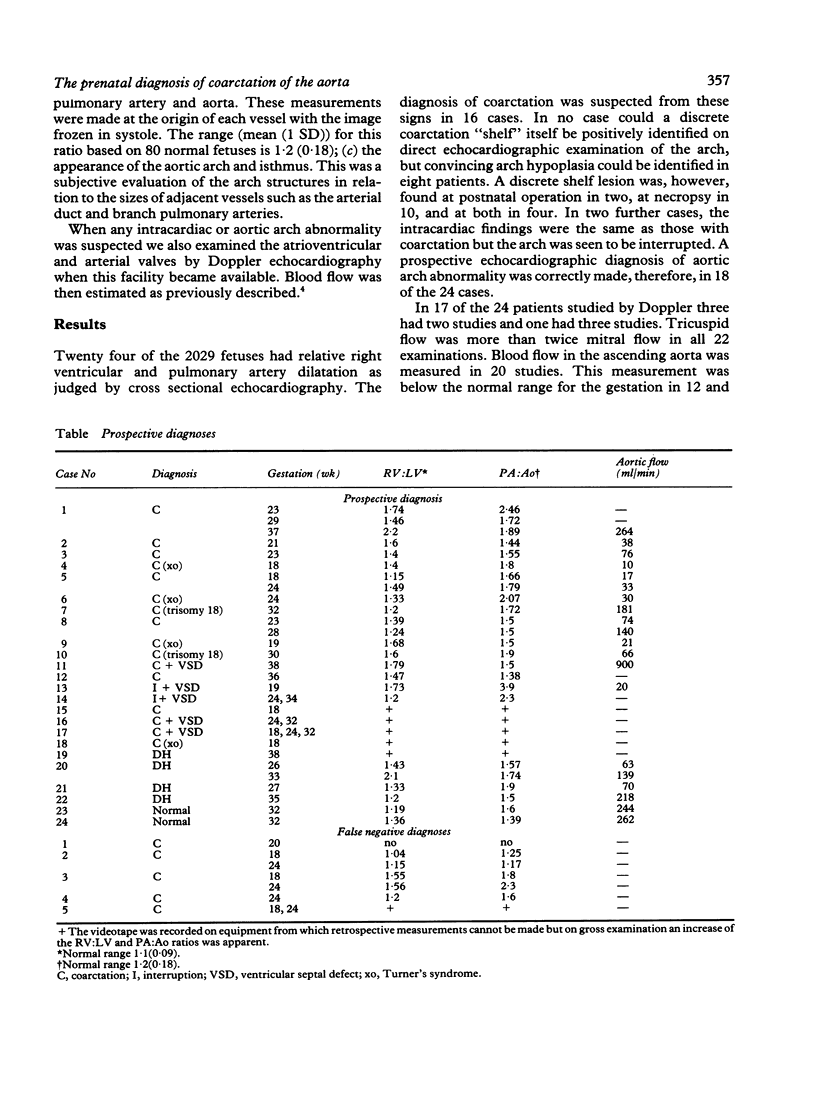
Full text
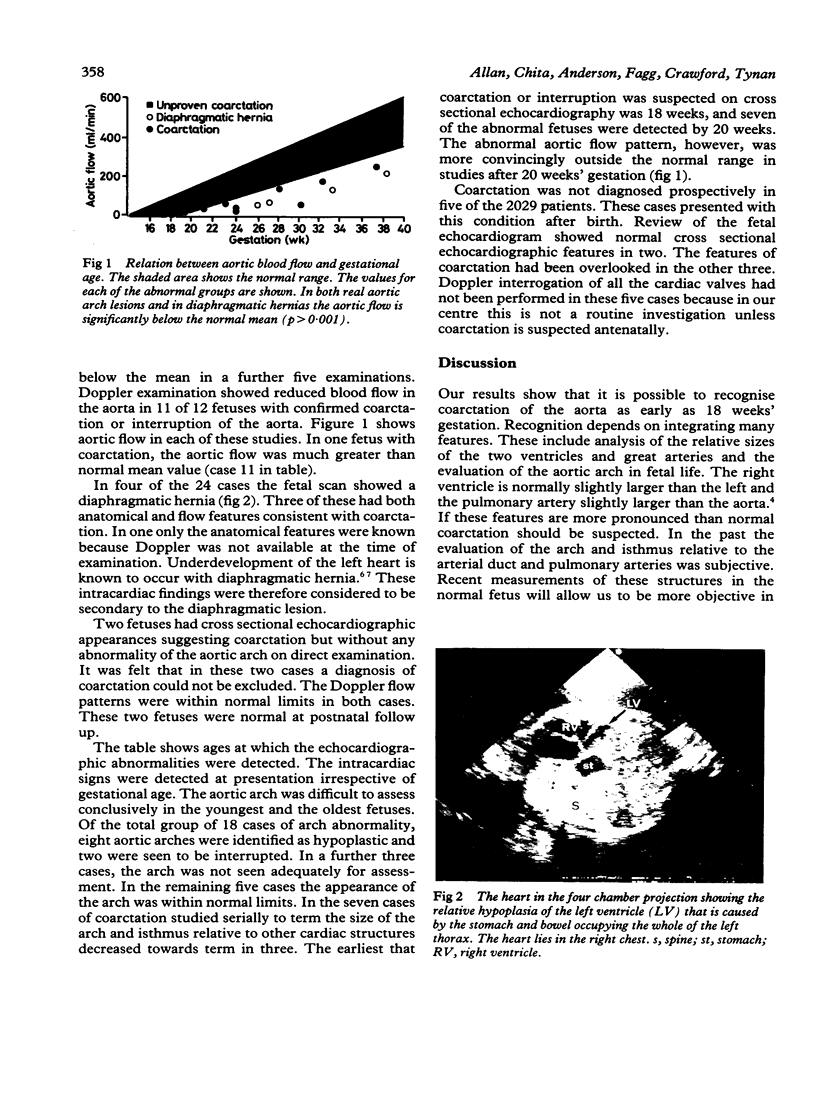
PDF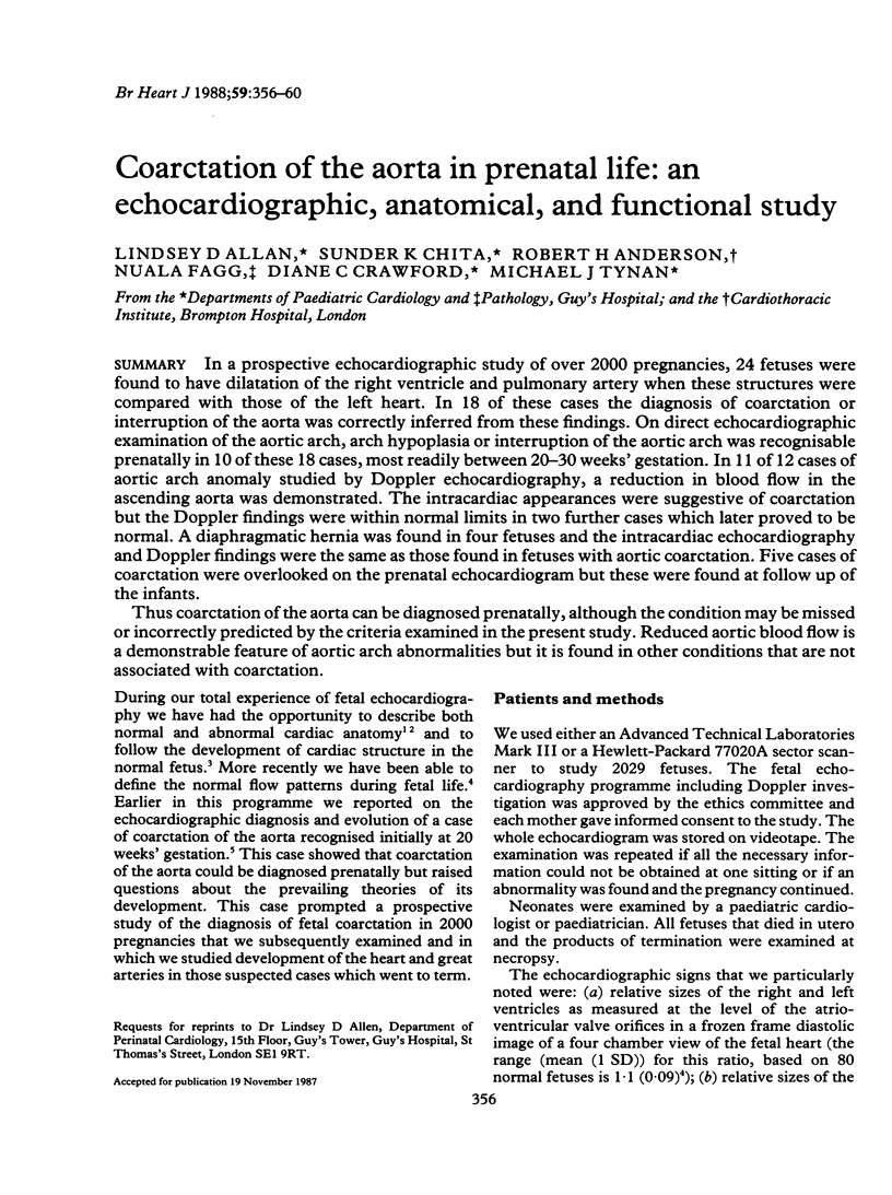
Images in this article
Selected References
These references are in PubMed. This may not be the complete list of references from this article.
- Allan L. D., Chita S. K., Al-Ghazali W., Crawford D. C., Tynan M. Doppler echocardiographic evaluation of the normal human fetal heart. Br Heart J. 1987 Jun;57(6):528–533. doi: 10.1136/hrt.57.6.528. [DOI] [PMC free article] [PubMed] [Google Scholar]
- Allan L. D., Crawford D. C., Anderson R. H., Tynan M. J. Echocardiographic and anatomical correlations in fetal congenital heart disease. Br Heart J. 1984 Nov;52(5):542–548. doi: 10.1136/hrt.52.5.542. [DOI] [PMC free article] [PubMed] [Google Scholar]
- Allan L. D., Crawford D. C., Tynan M. Evolution of coarctation of the aorta in intrauterine life. Br Heart J. 1984 Oct;52(4):471–473. doi: 10.1136/hrt.52.4.471. [DOI] [PMC free article] [PubMed] [Google Scholar]
- Allan L. D., Joseph M. C., Boyd E. G., Campbell S., Tynan M. M-mode echocardiography in the developing human fetus. Br Heart J. 1982 Jun;47(6):573–583. doi: 10.1136/hrt.47.6.573. [DOI] [PMC free article] [PubMed] [Google Scholar]
- Allan L. D., Tynan M. J., Campbell S., Wilkinson J. L., Anderson R. H. Echocardiographic and anatomical correlates in the fetus. Br Heart J. 1980 Oct;44(4):444–451. doi: 10.1136/hrt.44.4.444. [DOI] [PMC free article] [PubMed] [Google Scholar]
- Crawford D. C., Drake D. P., Kwaitkowski D., Chapman M. G., Allan L. D. Prenatal diagnosis of reversible cardiac hypoplasia associated with congenital diaphragmatic hernia: implications for postnatal management. J Clin Ultrasound. 1986 Nov-Dec;14(9):718–721. doi: 10.1002/jcu.1870140910. [DOI] [PubMed] [Google Scholar]
- Siebert J. R., Haas J. E., Beckwith J. B. Left ventricular hypoplasia in congenital diaphragmatic hernia. J Pediatr Surg. 1984 Oct;19(5):567–571. doi: 10.1016/s0022-3468(84)80105-0. [DOI] [PubMed] [Google Scholar]