

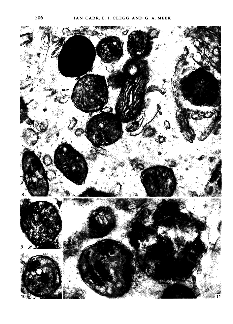
Full text
PDF
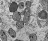
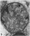
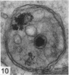
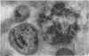
Images in this article
Selected References
These references are in PubMed. This may not be the complete list of references from this article.
- BAWA S. R. FINE STRUCTURE OF THE SERTOLI CELL OF THE HUMAN TESTIS. J Ultrastruct Res. 1963 Dec;52:459–474. doi: 10.1016/s0022-5320(63)80078-7. [DOI] [PubMed] [Google Scholar]
- BESSIS M., BRETON-GORIUS J. Accumulation de granules ferrugineux dans les mitochondries des érythroblastes. C R Hebd Seances Acad Sci. 1957 Jun 3;244(23):2846–2847. [PubMed] [Google Scholar]
- CAULFIELD J. B., SCHRAG P. E. ELECTRON MICROSCOPIC STUDY OF RENAL CALCIFICATION. Am J Pathol. 1964 Mar;44:365–381. [PMC free article] [PubMed] [Google Scholar]
- CLEGG E. J., MACMILLAN E. W. THE UPTAKE OF VITAL DYES AND PARTICULATE MATTER BY THE SERTOLI CELLS OF THE RAT TESTIS. J Anat. 1965 Apr;99:219–229. [PMC free article] [PubMed] [Google Scholar]
- D AGOSTINO A. N. AN ELECTRON MICROSCOPIC STUDY OF CARDIAC NECROSIS PRODUCED BY 9 ALPHA-FLUOROCORTISOL AND SODIUM PHOSPHATE. Am J Pathol. 1964 Oct;45:633–644. [PMC free article] [PubMed] [Google Scholar]
- D AGOSTINO A. N. AN ELECTRON MICROSCOPIC STUDY OF SKELETAL AND CARDIAC MUSCLE OF THE RAT POISONED BY PLASMOCID. Lab Invest. 1963 Nov;12:1060–1071. [PubMed] [Google Scholar]
- FARRANT J. L. An electron microscopic study of ferritin. Biochim Biophys Acta. 1954 Apr;13(4):569–576. doi: 10.1016/0006-3002(54)90376-5. [DOI] [PubMed] [Google Scholar]
- GARDNER P. J., HOLYOKE E. A. FINE STRUCTURE OF THE SEMINIFEROUS TUBULE OF THE SWISS MOUSE. I. THE LIMITING MEMBRANE, SERTOLI CELL, SPERMATOGONIA, AND SPERMATOCYTES. Anat Rec. 1964 Dec;150:391–404. doi: 10.1002/ar.1091500407. [DOI] [PubMed] [Google Scholar]
- KARRER H. E. Electron microscopic study of the phagocytosis process in lung. J Biophys Biochem Cytol. 1960 Apr;7:357–366. doi: 10.1083/jcb.7.2.357. [DOI] [PMC free article] [PubMed] [Google Scholar]
- LACY D. Certain aspects of testis structure and function. Br Med Bull. 1962 Sep;18:205–208. doi: 10.1093/oxfordjournals.bmb.a069979. [DOI] [PubMed] [Google Scholar]
- LEBLOND C. P., CLERMONT Y. Definition of the stages of the cycle of the seminiferous epithelium in the rat. Ann N Y Acad Sci. 1952 Nov 20;55(4):548–573. doi: 10.1111/j.1749-6632.1952.tb26576.x. [DOI] [PubMed] [Google Scholar]
- MUIR A. R., GOLBERG L. The tissue response to iron-dextran; an electron-microscope study. J Pathol Bacteriol. 1961 Oct;82:471–482. [PubMed] [Google Scholar]
- NIEMI M., KORMANO M. CYCLICAL CHANGES IN AND SIGNIFICANCE OF LIPIDS AND ACID PHOSPHATASE ACTIVITY IN THE SEMINIFEROUS TUBULES OF THE RAT TESTIS. Anat Rec. 1965 Feb;151:159–170. doi: 10.1002/ar.1091510207. [DOI] [PubMed] [Google Scholar]
- OETTLE A. G., HARRISON R. G. The histological changes produced in the rat testis by temporary and permanent occlusion of the testicular artery. J Pathol Bacteriol. 1952 Apr;64(2):273–297. doi: 10.1002/path.1700640204. [DOI] [PubMed] [Google Scholar]
- PEACHEY L. D. ELECTRON MICROSCOPIC OBSERVATIONS ON THE ACCUMULATION OF DIVALENT CATIONS IN INTRAMITOCHONDRIAL GRANULES. J Cell Biol. 1964 Jan;20:95–111. doi: 10.1083/jcb.20.1.95. [DOI] [PMC free article] [PubMed] [Google Scholar]
- RICHTER G. W. The cellular transformation of injected colloidal iron complexes into ferritin and hemosiderin in experimental animals; a study with the aid of electron microscopy. J Exp Med. 1959 Feb 1;109(2):197–216. doi: 10.1084/jem.109.2.197. [DOI] [PMC free article] [PubMed] [Google Scholar]
- TRUMP B. F., GOLDBLATT P. J., STOWELL R. E. STUDIES ON NECROSIS OF MOUSE LIVER IN VITRO. ULTRASTRUCTURAL ALTERATIONS IN THE MITOCHONDRIA OF HEPATIC PARENCHYMAL CELLS. Lab Invest. 1965 Apr;14:343–371. [PubMed] [Google Scholar]
- VILAR O., PEREZ DEL CERRO M. I., MANCINI R. E. The Sertoli cell as a "bridge cell" between the basal membrane and the germinal cells. Histochemical and electron microscope observations. Exp Cell Res. 1962 Jun;27:158–161. doi: 10.1016/0014-4827(62)90056-3. [DOI] [PubMed] [Google Scholar]