Full text
PDF
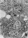
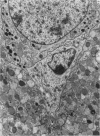
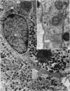
Images in this article
Selected References
These references are in PubMed. This may not be the complete list of references from this article.
- Abe K., Nicholson W. E., Liddle G. W., Island D. P., Orth D. N. Radioimmunoassay of beta-MSH in human plasma and tissues. J Clin Invest. 1967 Oct;46(10):1609–1616. doi: 10.1172/JCI105653. [DOI] [PMC free article] [PubMed] [Google Scholar]
- Bargmann W., Lindner E., Andres K. H. Uber Synapsen an endokrinen Epithelzellen und die Definition sekretorischer Neurone. Untersuchungen am Zwischenlappen der Katzenhypophyse. Z Zellforsch Mikrosk Anat. 1967;77(2):282–298. [PubMed] [Google Scholar]
- Howe A., Maxwell D. S. Electron microscopy of the pars intermedia of the pituitary gland in the rat. Gen Comp Endocrinol. 1968 Aug;11(1):169–185. doi: 10.1016/0016-6480(68)90118-4. [DOI] [PubMed] [Google Scholar]
- KARKUN J. N., KAR A. B., SEN D. P. SOME EXTRA-PIGMENTARY EFFECTS OF MELANOPHORESTIMULATING HORMONE. Ann Biochem Exp Med. 1963 Jul;23:253–260. [PubMed] [Google Scholar]
- KNOWLES F. NEUROENDOCRINE CORRELATIONS AT THE LEVEL OF ULTRASTRUCTURE. Arch Anat Microsc Morphol Exp. 1965 Jan-Mar;54:343–357. [PubMed] [Google Scholar]
- KRIVOY W. A., LANE M., KROEGER D. C. The actions of certain polypeptides on synaptic transmission. Ann N Y Acad Sci. 1963 Feb 4;104:312–329. doi: 10.1111/j.1749-6632.1963.tb17676.x. [DOI] [PubMed] [Google Scholar]
- Kastin A. J., Kullander S., Borglin N. E., Dahlberg B., Dyster-Aas K., Krakau C. E., Ingvar D. H., Miller M. C., 3rd, Bowers C. Y., Schally A. V. Extrapigmentary effects of melanocyte-stimulating hormone in amenorrhoeic women. Lancet. 1968 May 11;1(7550):1007–1010. doi: 10.1016/s0140-6736(68)91113-6. [DOI] [PubMed] [Google Scholar]
- Kastin A. J., Schally A. V., Viosca S., Miller M. C., 3rd MSH activity in plasma and pituitaries of rats after various treatments. Endocrinology. 1969 Jan;84(1):20–27. doi: 10.1210/endo-84-1-20. [DOI] [PubMed] [Google Scholar]
- Knowles F., Vollrath L. A functional relationship between neurosecretory fibres and pituicytes in the eel. Nature. 1965 Dec 25;208(5017):1343–1343. doi: 10.1038/2081343a0. [DOI] [PubMed] [Google Scholar]
- Kobayashi Y. Functional morphology of the pars intermedia of the rat hypophysis as revealed with the electron microscope. II. Correlation of the pars intermedia with the hypophyseo-adrenal axis. Z Zellforsch Mikrosk Anat. 1965 Oct 12;68(2):155–171. doi: 10.1007/BF00342425. [DOI] [PubMed] [Google Scholar]
- PALAY S. L., McGEE-RUSSELL S. M., GORDON S., Jr, GRILLO M. A. Fixation of neural tissues for electron microscopy by perfusion with solutions of osmium tetroxide. J Cell Biol. 1962 Feb;12:385–410. doi: 10.1083/jcb.12.2.385. [DOI] [PMC free article] [PubMed] [Google Scholar]
- Palade G. E. Structure and function at the cellular level. JAMA. 1966 Nov 21;198(8):815–825. [PubMed] [Google Scholar]
- SABATINI D. D., BENSCH K., BARRNETT R. J. Cytochemistry and electron microscopy. The preservation of cellular ultrastructure and enzymatic activity by aldehyde fixation. J Cell Biol. 1963 Apr;17:19–58. doi: 10.1083/jcb.17.1.19. [DOI] [PMC free article] [PubMed] [Google Scholar]
- Taleisnik S., Tomatis M. E. Melanocyte-stimulating hormone-releasing and inhibiting factors in two hypothalamic extracts. Endocrinology. 1967 Oct;81(4):819–825. doi: 10.1210/endo-81-4-819. [DOI] [PubMed] [Google Scholar]
- Tomatis M. E., Taleisnik S. Pituitary melanocyte-stimulating hormone and its hypothalamic factors during the estrous cycle. Acta Physiol Lat Am. 1968;18(1):96–101. [PubMed] [Google Scholar]
- Vincent D. S., Kumar T. C. Electron microscopic studies on the pars intermedia of the ferret. Z Zellforsch Mikrosk Anat. 1969;99(2):185–197. doi: 10.1007/BF00342220. [DOI] [PubMed] [Google Scholar]


