Abstract
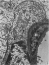
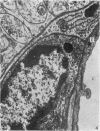
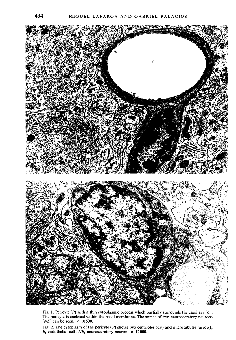
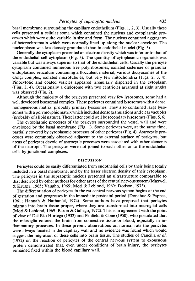
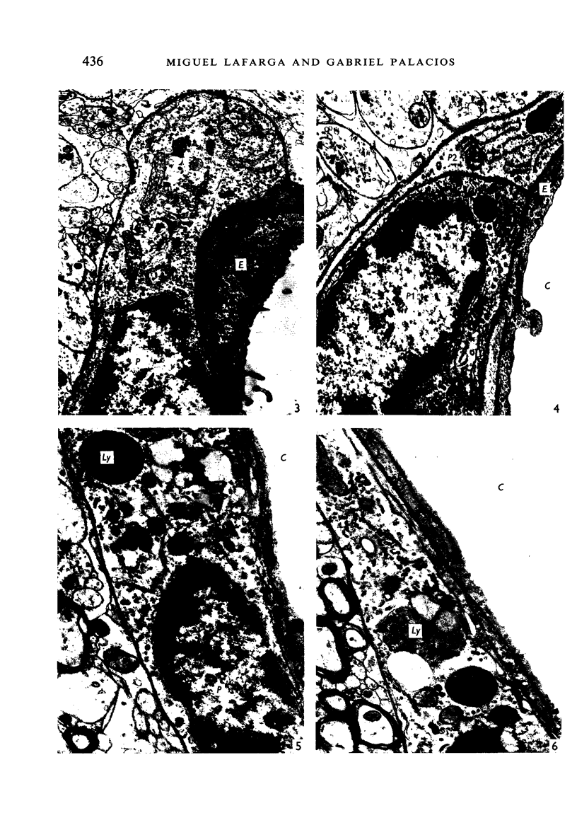
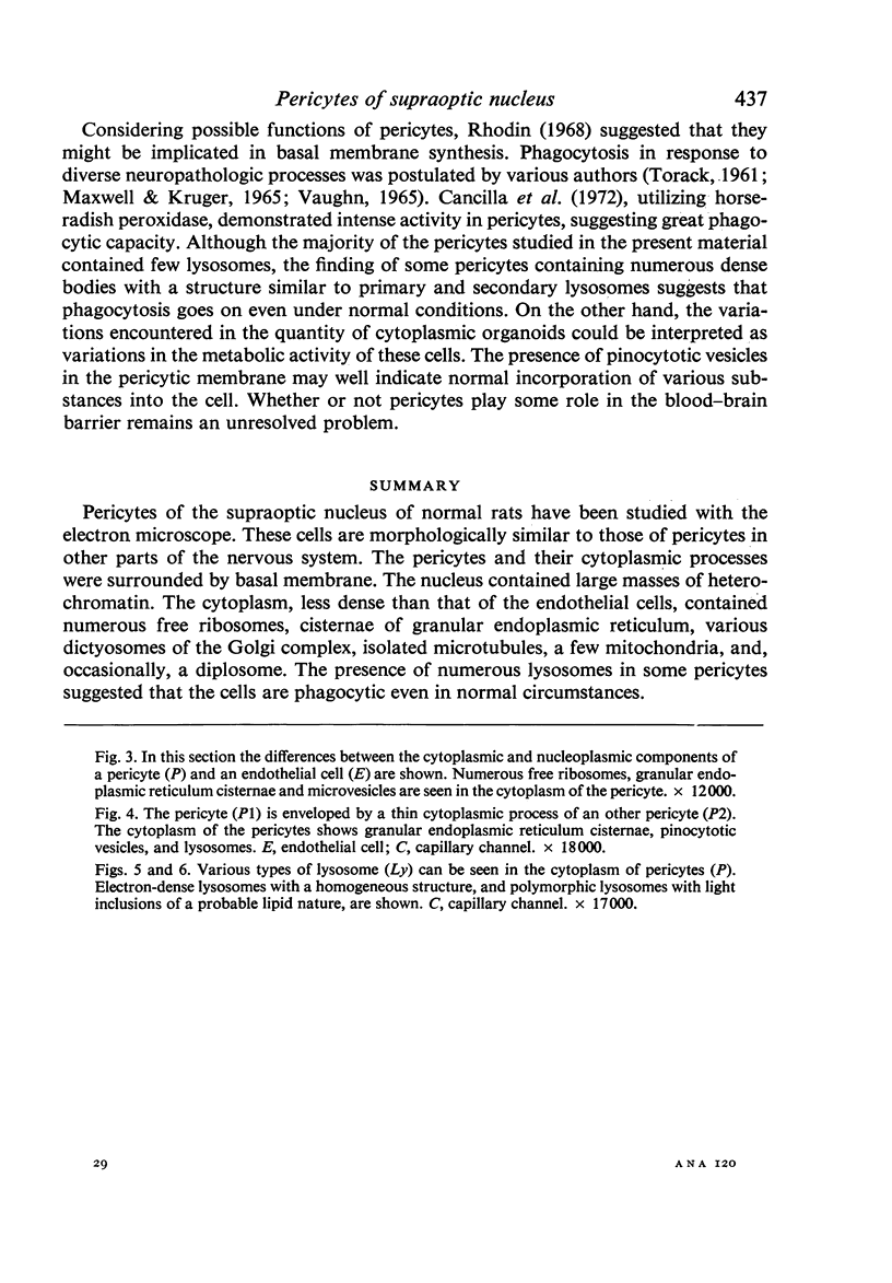
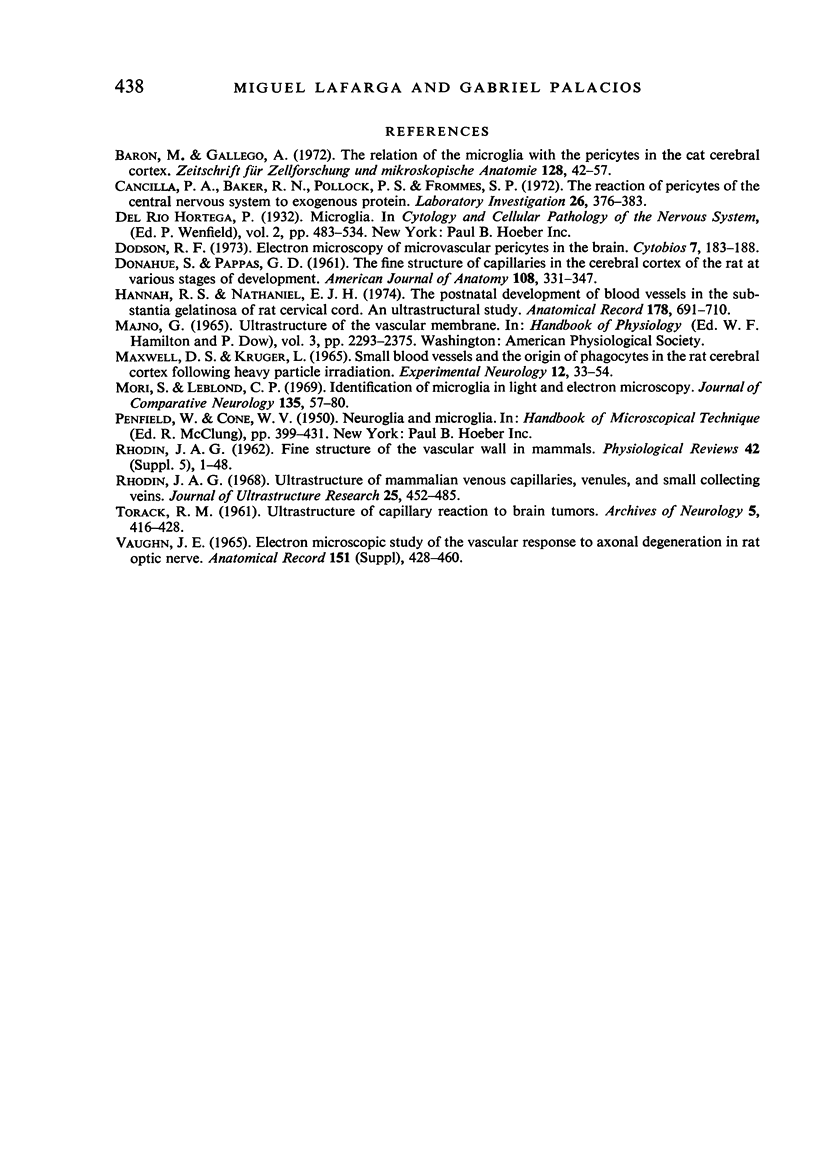
Pericytes of the supraoptic nucleus of normal rats have been studied with the electron microscope. These cells are morphologically similar to those of pericytes in other parts of the nervous system. The pericytes and their cytoplasmic processes were surrounded by basal membrane. The nucleus contained large masses of heterochromatin. The cytoplasm, less dense than that of the endothelial cells, contained numerous free ribosomes, cisternae of granular endoplasmic reticulum, various dictyosomes of the Golgi complex, isolated microtubules, a few mitochondria, and, occasionally, a diplosome. The presence of numerous lysosomes in some pericytes suggested that the cells are phagocytic even in normal circumstances.
Full text
PDF
Images in this article
Selected References
These references are in PubMed. This may not be the complete list of references from this article.
- BISHOP D. W. Sperm motility. Physiol Rev. 1962 Jan;42:1–59. doi: 10.1152/physrev.1962.42.1.1. [DOI] [PubMed] [Google Scholar]
- Barón M., Gallego A. The relation of the microglia with the pericytes in the cat cerebral cortex. Z Zellforsch Mikrosk Anat. 1972;128(1):42–57. doi: 10.1007/BF00306887. [DOI] [PubMed] [Google Scholar]
- Cancilla P. A., Baker R. N., Pollock P. S., Frommes S. P. The reaction of pericytes of the central nervous system to exogenous protein. Lab Invest. 1972 Apr;26(4):376–383. [PubMed] [Google Scholar]
- DONAHUE S., PAPPAS G. D. The fine structure of capillaries in the cerebral cortex of the rat at various stages of development. Am J Anat. 1961 May;108:331–347. doi: 10.1002/aja.1001080307. [DOI] [PubMed] [Google Scholar]
- Hannah R. S., Nathaniel E. J. The postnatal development of blood vessels in the substantia gelatinosa of rat cervical cord--an ultrastructural study. Anat Rec. 1974 Apr;178(4):691–709. doi: 10.1002/ar.1091780404. [DOI] [PubMed] [Google Scholar]
- MAXWELL D. S., KRUGER L. SMALL BLOOD VESSELS AND THE ORIGIN OF PHAGOCYTES IN THE RAT CEREBRAL CORTEX FOLLOWING HEAVY PARTICLE IRRADIATION. Exp Neurol. 1965 May;12:33–54. doi: 10.1016/0014-4886(65)90097-x. [DOI] [PubMed] [Google Scholar]
- Mori S., Leblond C. P. Identification of microglia in light and electron microscopy. J Comp Neurol. 1969 Jan;135(1):57–80. doi: 10.1002/cne.901350104. [DOI] [PubMed] [Google Scholar]
- Rhodin J. A. Ultrastructure of mammalian venous capillaries, venules, and small collecting veins. J Ultrastruct Res. 1968 Dec;25(5):452–500. doi: 10.1016/s0022-5320(68)80098-x. [DOI] [PubMed] [Google Scholar]
- TORACK R. M. Ultrastructure of capillary reaction to brain tumors. Arch Neurol. 1961 Oct;5:416–428. doi: 10.1001/archneur.1961.00450160066004. [DOI] [PubMed] [Google Scholar]


