Abstract
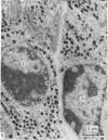
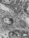
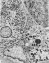
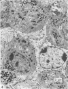
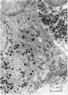
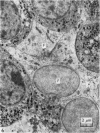
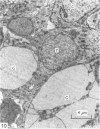
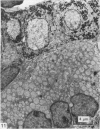
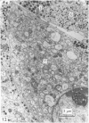
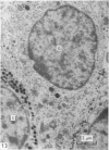
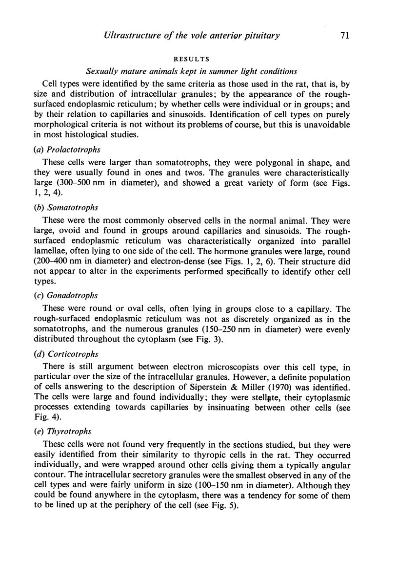
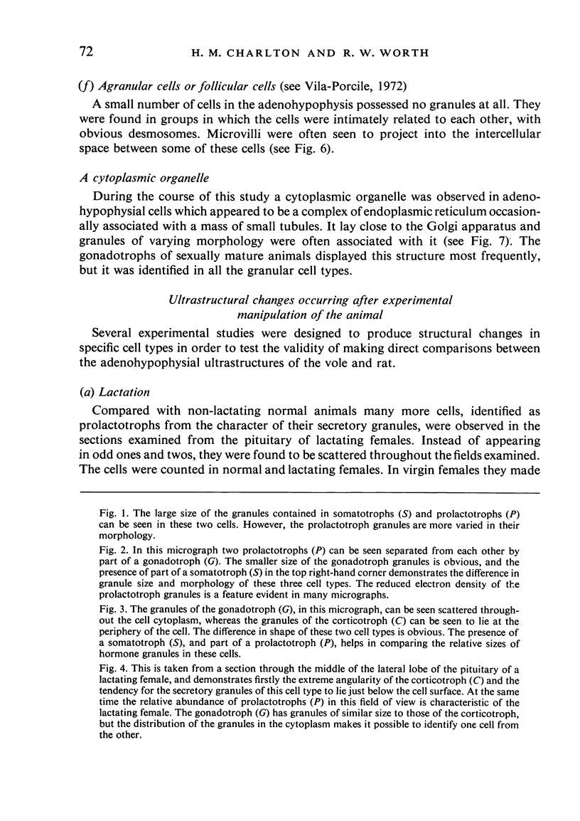
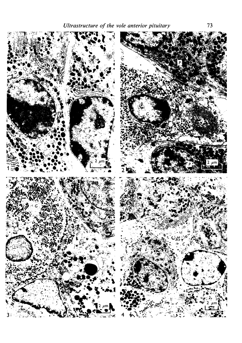
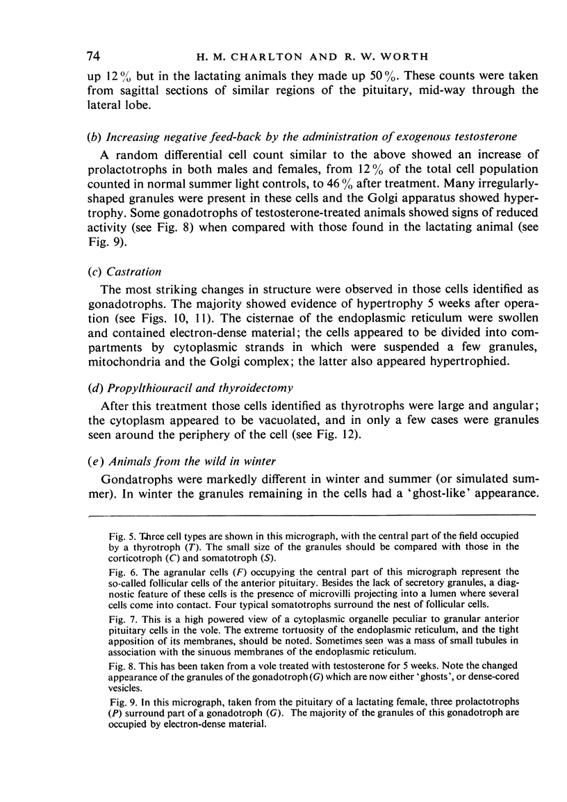
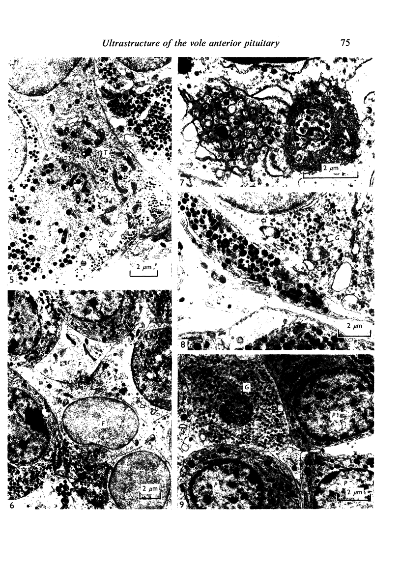
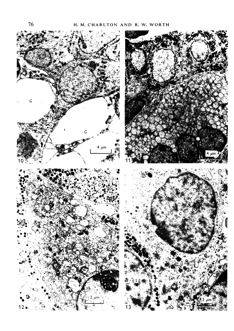
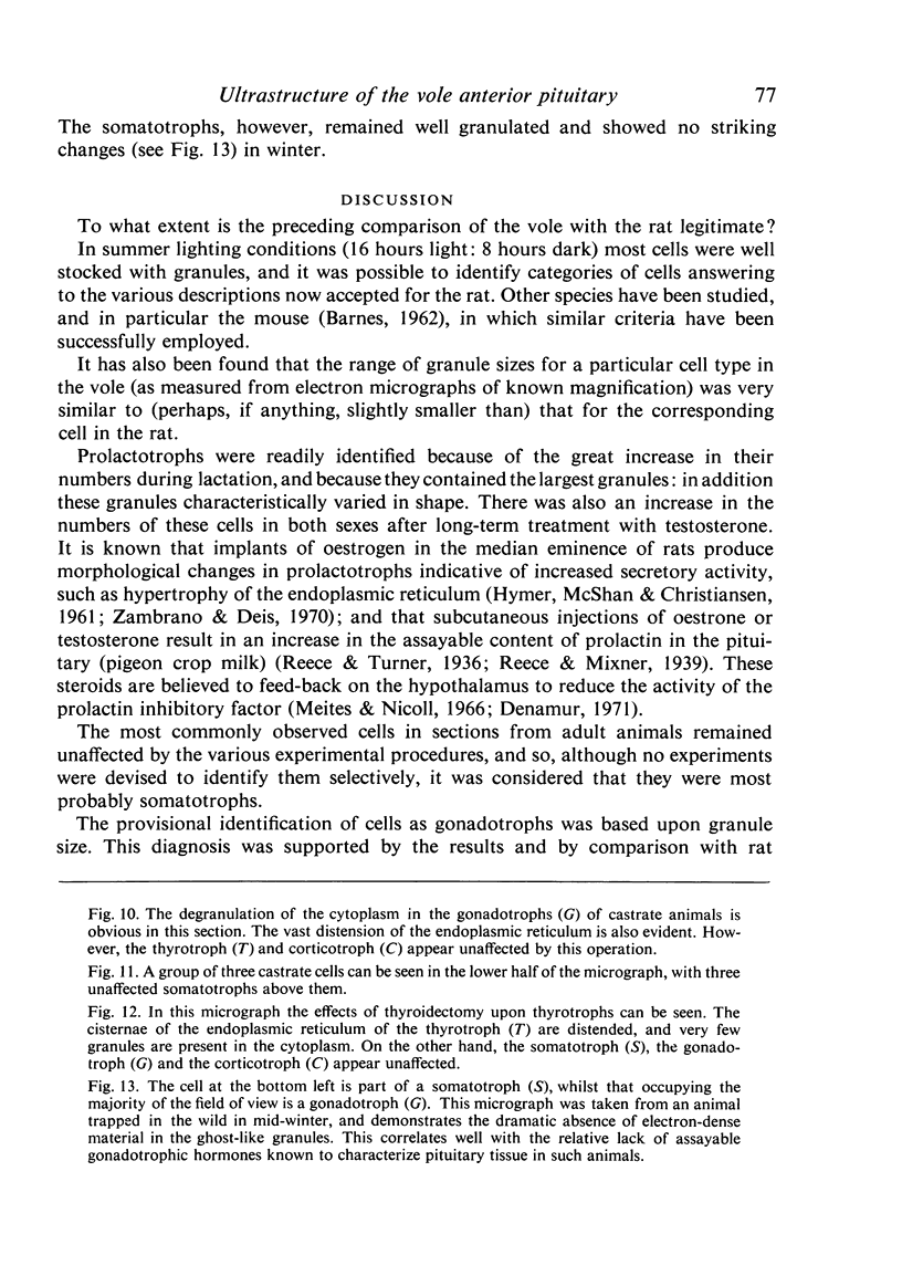
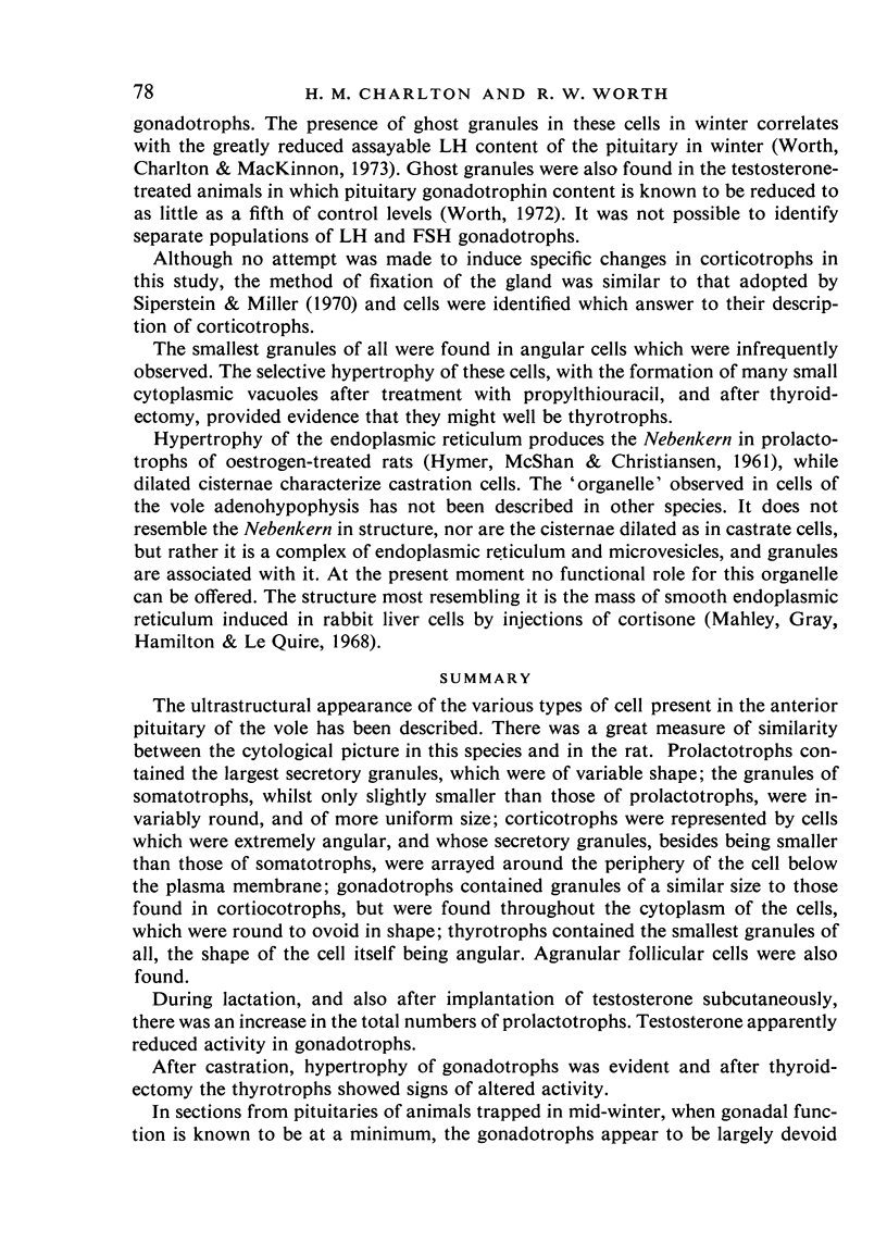
The ultrastructural appearance of the various types of cell present in the anterior pituitary of the vole has been described. There was a great measure of similarity between the cytological picture in this species and in the rat. Prolactotrophs contained the largest secretory granules, which were of variable shape; the granules of somatotrophs, whilst only slightly smaller than those of prolactotrophs, were invariably round, and of more uniform size; corticotrophs were represented by cells which were extremely angular, and whose secretory granules, besides being smaller than those of somatotrophs, were arrayed around the periphery of the cell below the plasma membrane; gonadotrophs contained granules of a similar size to those found in cortiocotrophs, but were found throughout the cytoplasm of the cells, whic were round to ovoid in shape; thyrotrophs contained the smallest granules of all, the shape of the cell itself bein angular...
Full text
PDF
Images in this article
Selected References
These references are in PubMed. This may not be the complete list of references from this article.
- BARNES B. G. Electron microscope studies on the secretory cytology of the mouse anterior pituitary. Endocrinology. 1962 Oct;71:618–628. doi: 10.1210/endo-71-4-618. [DOI] [PubMed] [Google Scholar]
- CLARKE J. R., FORSYTH I. A. SEASONAL CHANGES IN THE ADENOHYPOPHYSIS OF THE VOLE (MICROTUS AGRESTIS). Gen Comp Endocrinol. 1964 Jun;4:243–252. doi: 10.1016/0016-6480(64)90018-8. [DOI] [PubMed] [Google Scholar]
- Denamur R. Reviews of the progress of dairy science. Section A. Physiology. Hormonal control of lactogenesis. J Dairy Res. 1971 Jun;38(2):237–264. doi: 10.1017/s0022029900019348. [DOI] [PubMed] [Google Scholar]
- HYMER W. C., McSHAN W. H., CHRISTIANSEN R. G. Electron microscopic studies of anterior pituitary glands from lactating and estrogen-treated rats. Endocrinology. 1961 Jul;69:81–90. doi: 10.1210/endo-69-1-81. [DOI] [PubMed] [Google Scholar]
- Meites J., Nicoll C. S. Adenohypophysis:prolactin. Annu Rev Physiol. 1966;28:57–88. doi: 10.1146/annurev.ph.28.030166.000421. [DOI] [PubMed] [Google Scholar]
- Moriarty G. C. Adenohypophysis: ultrastructural cytochemistry. A review. J Histochem Cytochem. 1973 Oct;21(10):855–894. doi: 10.1177/21.10.855. [DOI] [PubMed] [Google Scholar]
- Siperstein E. R., Miller K. J. Further cytophysiologic evidence for the dentity of the cells that produce adrenocorticotrophic hormone. Endocrinology. 1970 Mar;86(3):451–486. doi: 10.1210/endo-86-3-451. [DOI] [PubMed] [Google Scholar]
- VENABLE J. H., COGGESHALL R. A SIMPLIFIED LEAD CITRATE STAIN FOR USE IN ELECTRON MICROSCOPY. J Cell Biol. 1965 May;25:407–408. doi: 10.1083/jcb.25.2.407. [DOI] [PMC free article] [PubMed] [Google Scholar]
- Vila-Porcile E. Le réseau des cellules folliculo-stellaires et les follicules de l'adénohypophyse du rat (pars distalis. Z Zellforsch Mikrosk Anat. 1972;129(3):328–369. [PubMed] [Google Scholar]
- Worth R. W., Charlton H. M., Mackinnon P. C. Field and laboratory studies on the control of luteinizing hormone secretion and gonadal activity in the vole, Microtus agrestis. J Reprod Fertil Suppl. 1973 Dec;19:89–99. [PubMed] [Google Scholar]
- Zambrano D., Deis R. P. The adenohypophysis of female rats after hypothalamic oestradiol implants: an electron microscopic study. J Endocrinol. 1970 May;47(1):101–110. doi: 10.1677/joe.0.0470101. [DOI] [PubMed] [Google Scholar]


