Abstract
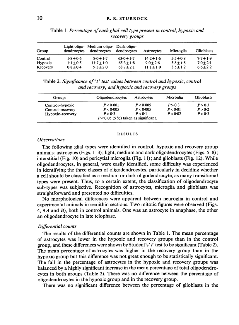
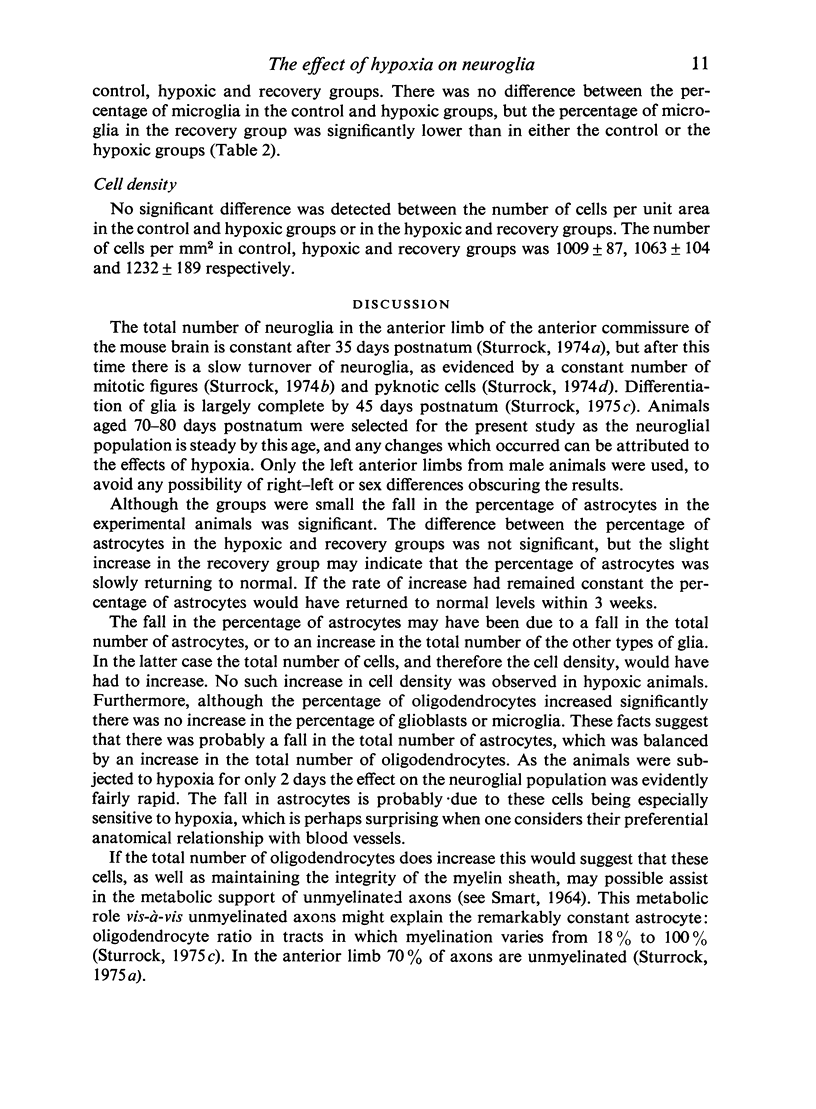
Two experimental and one control group of 70-80 day old mice were used in this study. The two experimental groups were subjected to hypoxia for 2 days in a decompression chamber at 390 mmHg. The animals in one experimental group were killed on removal from the chamber (hypoxic group) while those in the other (recovery group) were allowed to recover at sea-level atmospheric pressure for one week before being killed. Semithin, toluidine blue stained sections from the anterior limb of the anterior commissure were examined to find whether any quantitative changes occurred in the neuroglia with hypoxic stress. The following changes were observed: (1) The percentage of astrocytes in the hypoxic and recovery groups was significantly (P less than 0-005) lower than in the control group. (2) The percentage of oligodendrocytes in the hypoxic and recovery groups was significantly (P less than 0-001) higher than in the control group. (3) The percentage of microglia in the recovery groups was significantly (P less than 0-02) lower than in either of the other two groups. (4) The percentage of astrocytes in the recovery group was slightly (2-1%) higher than in the hypoxic group, and although not statistically significant, this result suggested that a slow return to normal might be occurring. (5) Little change was observed in cell density. The possible significance of these changes is discussed. I should like to express my indebtedness to Dr E.J. Clegg of the Department of Anatomy, Sheffield University, for the use of the decompression chamber, for his advice and help in the preparation of the control and experimental animals, and also for his hospitality throughout the duration of the experiments. Thanks are also due to Mrs Sheila Ramsay for her careful preparation of the perfusing fluids, and to Mrs Dawn Alexander for typing the manuscript.
Full text
PDF
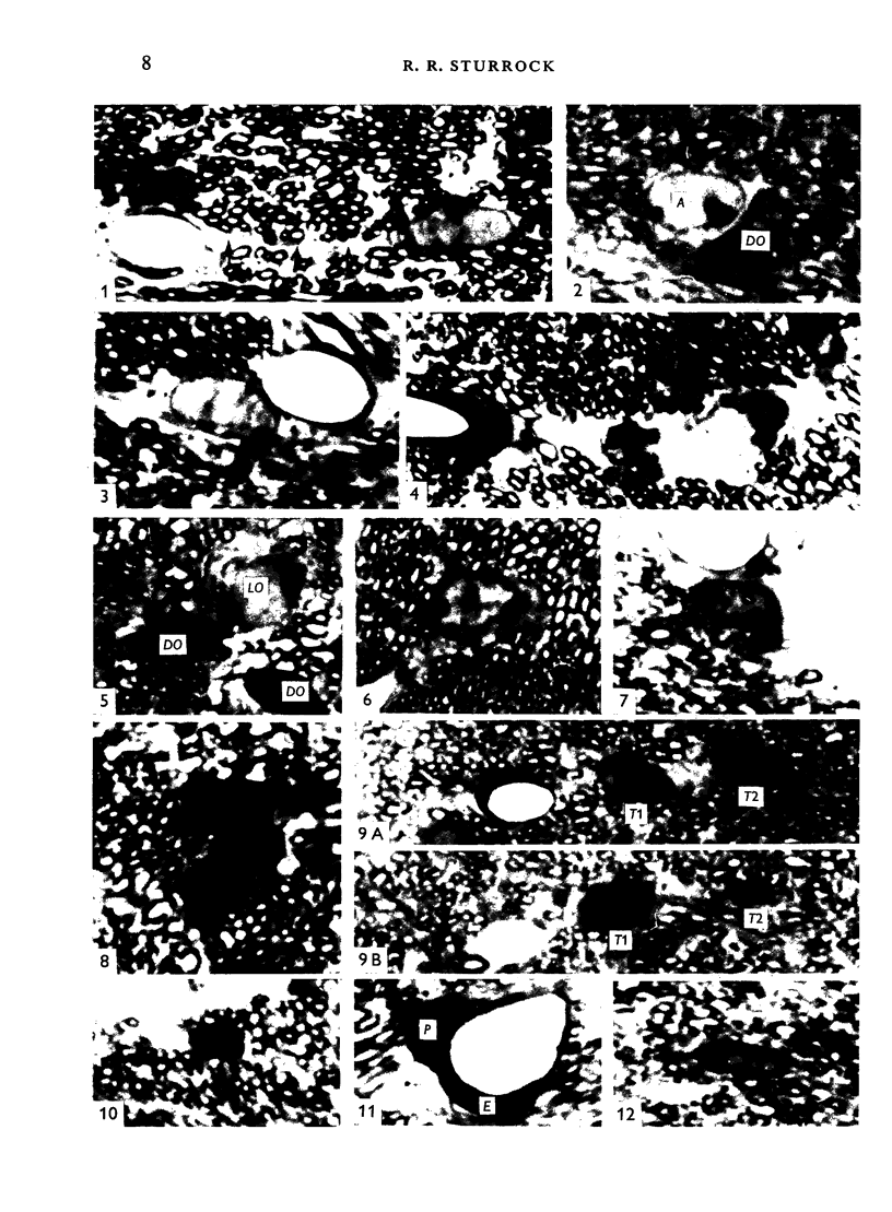
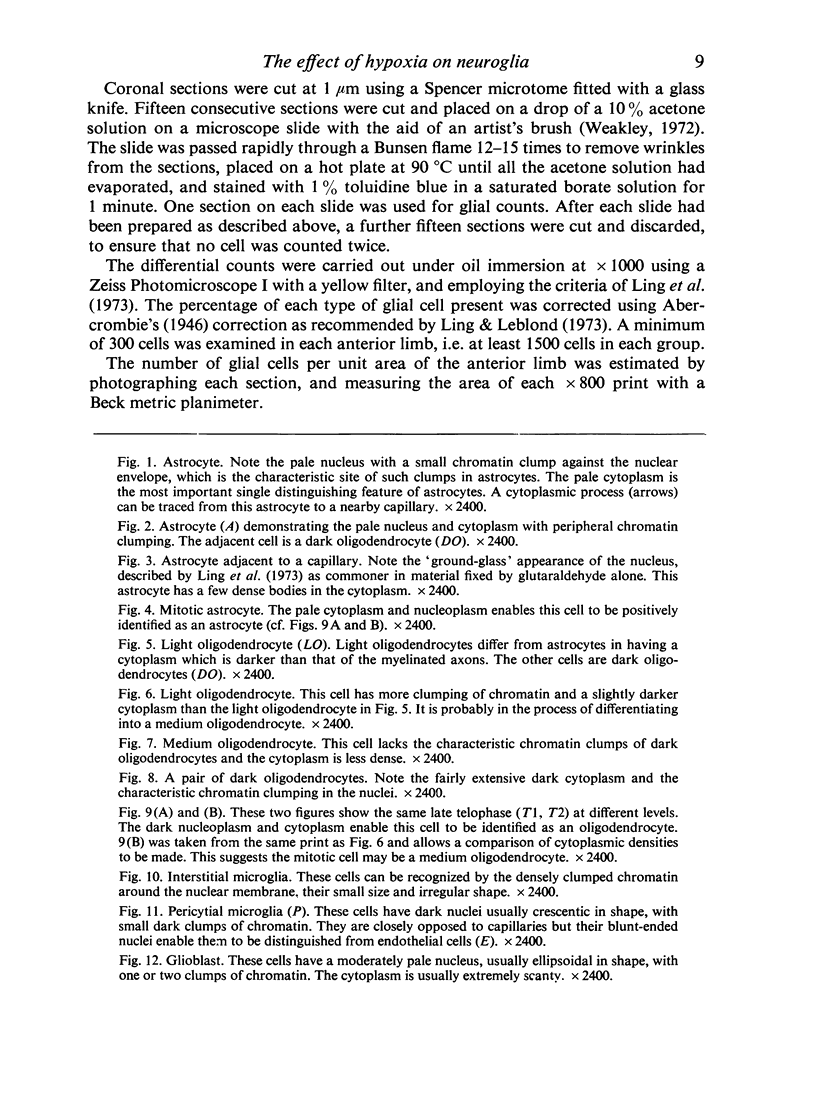
Images in this article
Selected References
These references are in PubMed. This may not be the complete list of references from this article.
- Griffin R., Illis L. S., Mitchell J. Identification of neuroglia by light and electronmicroscopy. Acta Neuropathol. 1972;22(1):7–12. doi: 10.1007/BF00687546. [DOI] [PubMed] [Google Scholar]
- Hunter C., Clegg E. J. Changes in body weight of the growing and adult mouse in response to hypoxic stress. J Anat. 1973 Feb;114(Pt 2):185–199. [PMC free article] [PubMed] [Google Scholar]
- Ling E. A., Leblond C. P. Investigation of glial cells in semithin sections. II. Variation with age in the numbers of the various glial cell types in rat cortex and corpus callosum. J Comp Neurol. 1973 May 1;149(1):73–81. doi: 10.1002/cne.901490105. [DOI] [PubMed] [Google Scholar]
- Ling E. A., Paterson J. A., Privat A., Mori S., Leblond C. P. Investigation of glial cells in semithin sections. I. Identification of glial cells in the brain of young rats. J Comp Neurol. 1973 May 1;149(1):43–71. doi: 10.1002/cne.901490104. [DOI] [PubMed] [Google Scholar]
- Mori S., Leblond C. P. Electron microscopic features and proliferation of astrocytes in the corpus callosum of the rat. J Comp Neurol. 1969 Oct;137(2):197–225. doi: 10.1002/cne.901370206. [DOI] [PubMed] [Google Scholar]
- Mori S., Leblond C. P. Electron microscopic identification of three classes of oligodendrocytes and a preliminary study of their proliferative activity in the corpus callosum of young rats. J Comp Neurol. 1970 May;139(1):1–28. doi: 10.1002/cne.901390102. [DOI] [PubMed] [Google Scholar]
- Mori S., Leblond C. P. Identification of microglia in light and electron microscopy. J Comp Neurol. 1969 Jan;135(1):57–80. doi: 10.1002/cne.901350104. [DOI] [PubMed] [Google Scholar]
- Sturrock R. R. A light and electron microscopic study of proliferation and maturation of fibrous astrocytes in the optic nerve of the human embryo. J Anat. 1975 Apr;119(Pt 2):223–234. [PMC free article] [PubMed] [Google Scholar]
- Sturrock R. R. A light microscope study of glial necrosis with age in the anterior limb of the anterior commissure of the pre and postnatal mouse. J Anat. 1974 Jul;117(Pt 3):469–474. [PMC free article] [PubMed] [Google Scholar]
- Sturrock R. R. A quantitative electron microscopic study of myelination in the anterior limb of the anterior commissure of the mouse brain. J Anat. 1975 Feb;119(Pt 1):67–75. [PMC free article] [PubMed] [Google Scholar]
- Sturrock R. R. Histogenesis of the anterior limb of the anterior commissure of the mouse brain. 3. An electron microscopic study of gliogenesis. J Anat. 1974 Feb;117(Pt 1):37–53. [PMC free article] [PubMed] [Google Scholar]
- Sturrock R. R. Histogenesis of the anterior limb of the anterior commissure of the mouse brain. I. A quantitative study of changes in the glial population with age. J Anat. 1974 Feb;117(Pt 1):17–25. [PMC free article] [PubMed] [Google Scholar]
- Sturrock R. R. Histogenesis of the anterior limb of the anterior commissure of the mouse brain. II. A quantitative study of pre- and postnatal mitosis. J Anat. 1974 Feb;117(Pt 1):27–35. [PMC free article] [PubMed] [Google Scholar]
- Vaughn J. E., Peters A. A third neuroglial cell type. An electron microscopic study. J Comp Neurol. 1968 Jun;133(2):269–288. doi: 10.1002/cne.901330207. [DOI] [PubMed] [Google Scholar]


