

Abstract
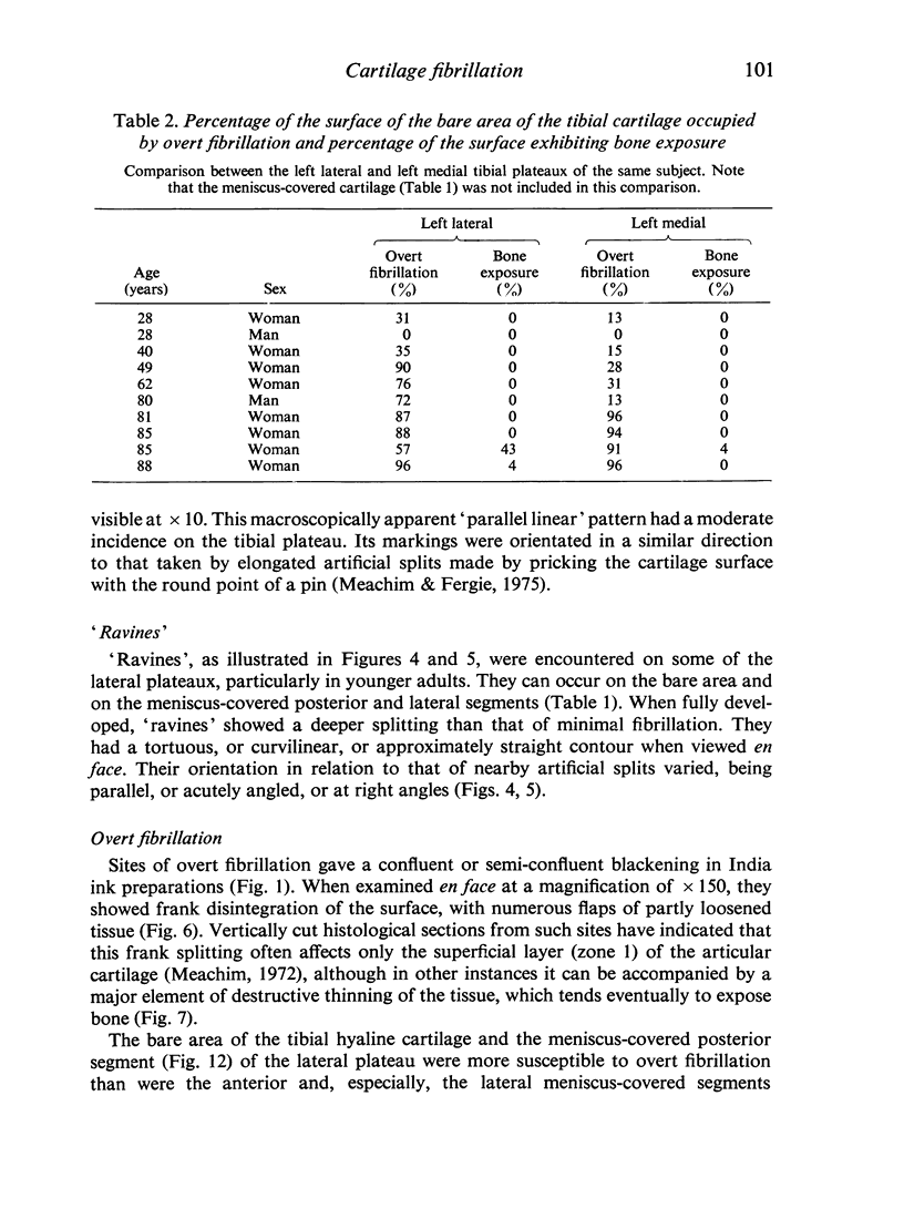
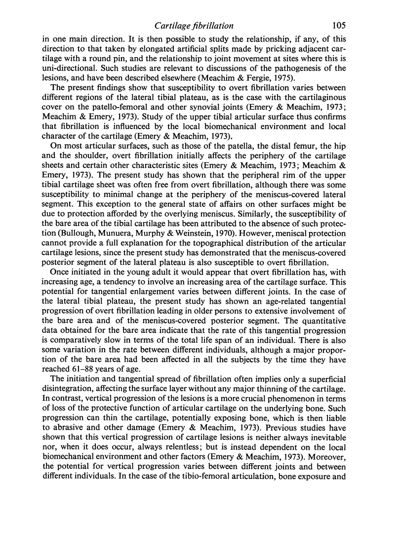
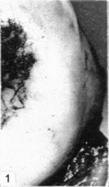
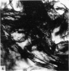
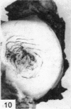
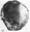
A study has been made of the state at necropsy of the hyaline articular cartilage of the left tibial plateaux, with particular reference to the lateral plateau, in 47 adult white Europeans (24 men; 23 women) aged 21-88 years. The surface morphology and topographical distribution of the lesions is described for the bare area of the lateral plateau and its meniscus-covered segments, and quantitative point-counting data are presented for the amount, according to age, of overt fibrillation on the bare area. A variety of cartilage lesions was encountered: macroscopically apparent ' parallel linear' minimal fibrillation; other patterns of minimal fibrillation; 'ravines'; overt fibrillation; localized incomplete defects of the cartilage; and full-thickness cartilage loss with bone exposure. Sites of superficial fraying and splitting of the hyaline articular cartilage are a normal finding on adult human tibial plateaux. Especially in younger adults, such sites are often accompanied by large areas of cartilage surface which are still intact. On the lateral plateau, the bare area and the meniscus-covered posterior segment are more susceptible to overt fibrillation than are the meniscus-covered lateral and anterior segments. In contrast to the findings in other synovial joints, the peripheral rim of the upper tibial cartilage sheet is not particularly susceptible to overt fibrillation. Tangential extension of the changes on the lateral plateau leads to widespread involvement of the bare area and the meniscus-covered posterior segment in older subjects. However, vertical progression of the changes, sufficient to give full-thickness cartilage loss with tibio-femoral bone exposure, was seen in only a minority of persons aged over 80 years.
Full text
PDF

Images in this article


Selected References
These references are in PubMed. This may not be the complete list of references from this article.
- Bullough P. G., Munuera L., Murphy J., Weinstein A. M. The strength of the menisci of the knee as it relates to their fine structure. J Bone Joint Surg Br. 1970 Aug;52(3):564–567. [PubMed] [Google Scholar]
- Emery I. H., Meachim G. Surface morphology and topography of patello-femoral cartilage fibrillation in Liverpool necropsies. J Anat. 1973 Oct;116(Pt 1):103–120. [PMC free article] [PubMed] [Google Scholar]
- Longmore R. B., Gardner D. L. Development with age of human articular cartilage surface structure. A survey by interference microscopy of the lateral femoral condyle. Ann Rheum Dis. 1975 Feb;34(1):26–37. doi: 10.1136/ard.34.1.26. [DOI] [PMC free article] [PubMed] [Google Scholar]
- Meachim G., Emery I. H. Cartilage fibrillation in shoulder and hip joints in Liverpool necropsies. J Anat. 1973 Nov;116(Pt 2):161–179. [PMC free article] [PubMed] [Google Scholar]
- Meachim G., Emery I. H. Quantitative aspects of patello-femoral cartilage fibrillation in Liverpool necropsies. Ann Rheum Dis. 1974 Jan;33(1):39–47. doi: 10.1136/ard.33.1.39. [DOI] [PMC free article] [PubMed] [Google Scholar]
- Meachim G., Fergie I. A. Morphological patterns of articular cartilage fibrillation. J Pathol. 1975 Apr;115(4):231–240. doi: 10.1002/path.1711150408. [DOI] [PubMed] [Google Scholar]
- Meachim G. Light microscopy of Indian ink preparations of fibrillated cartilage. Ann Rheum Dis. 1972 Nov;31(6):457–464. doi: 10.1136/ard.31.6.457. [DOI] [PMC free article] [PubMed] [Google Scholar]