Abstract
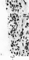
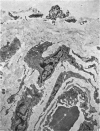
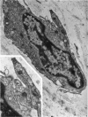
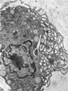
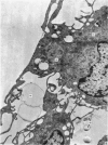
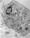
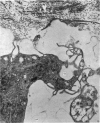
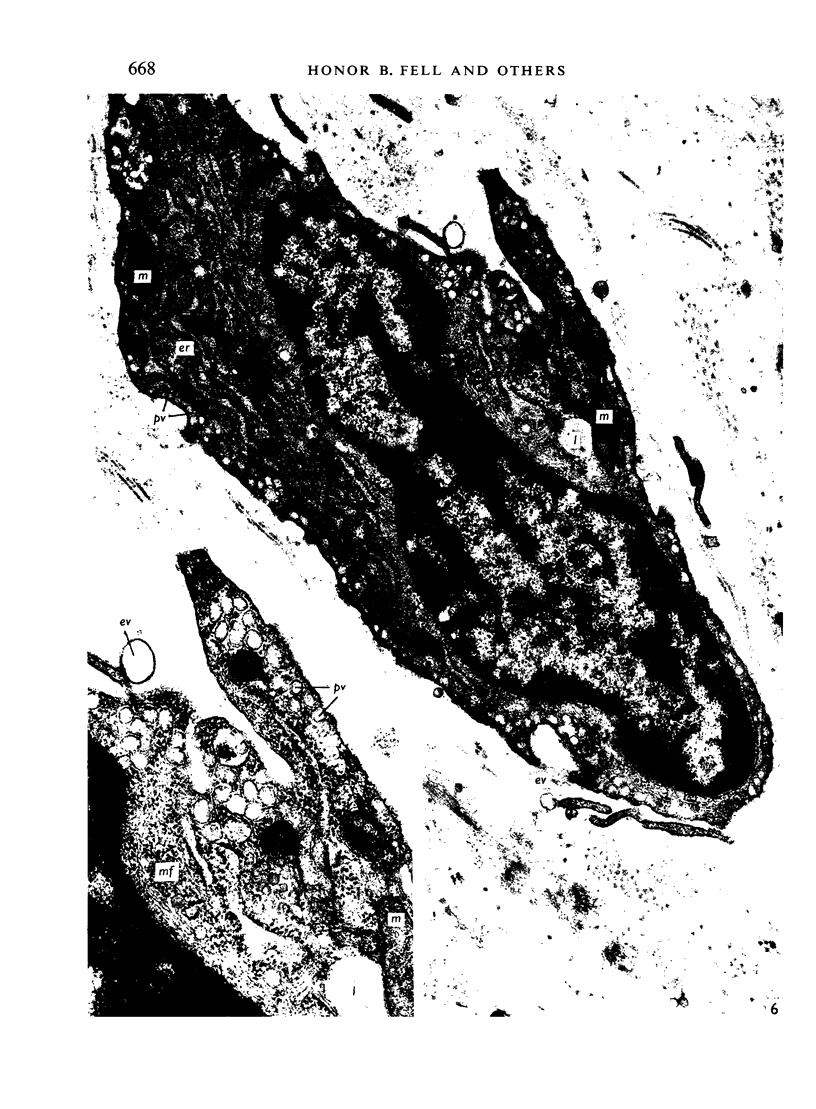
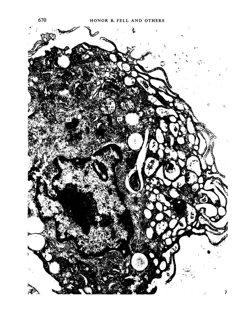
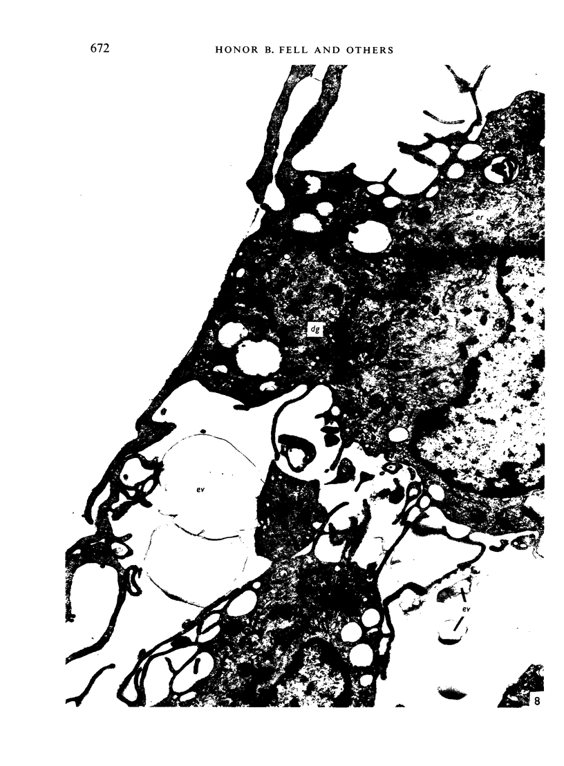
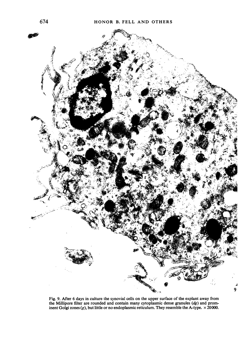
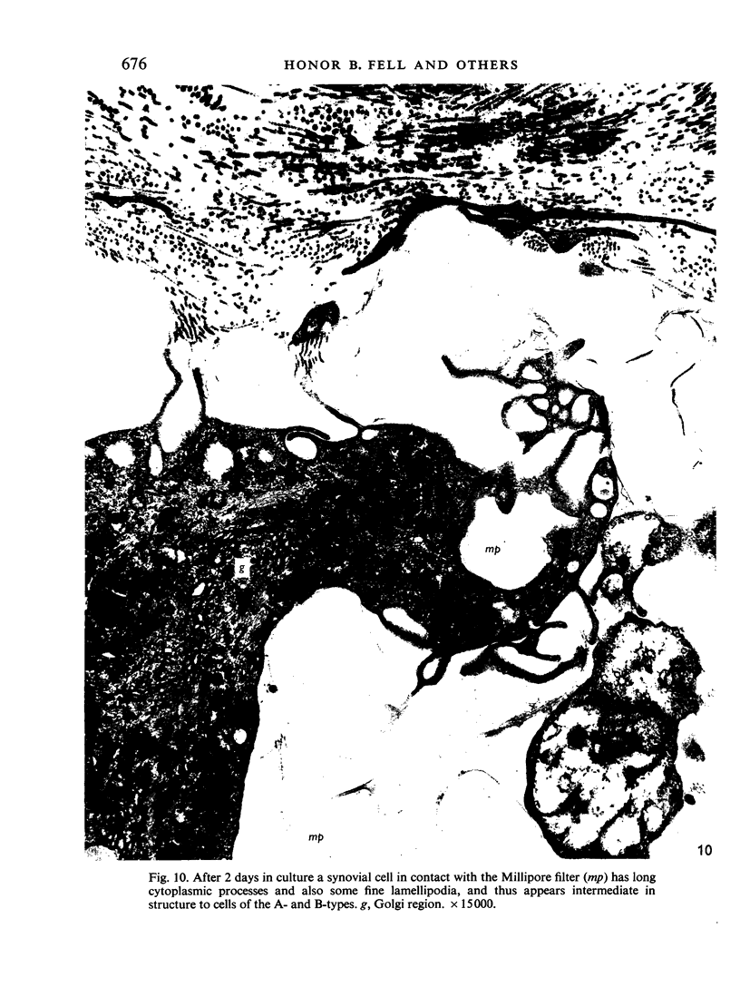
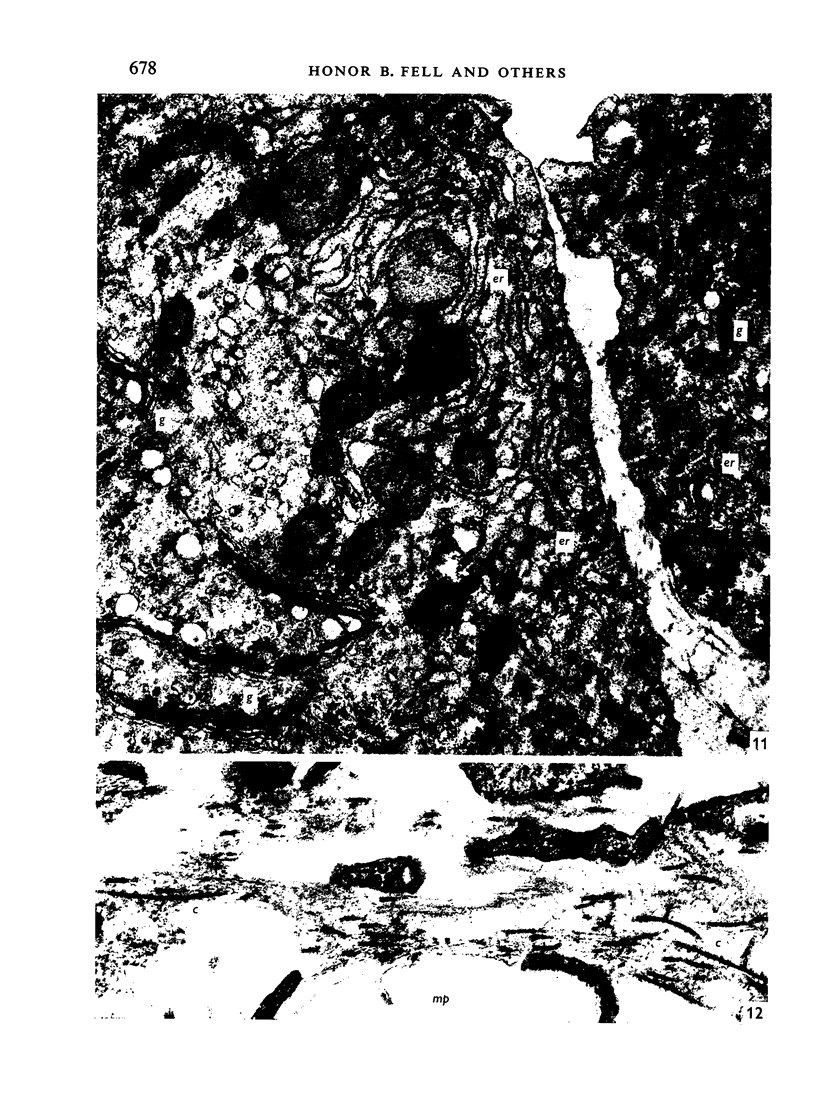
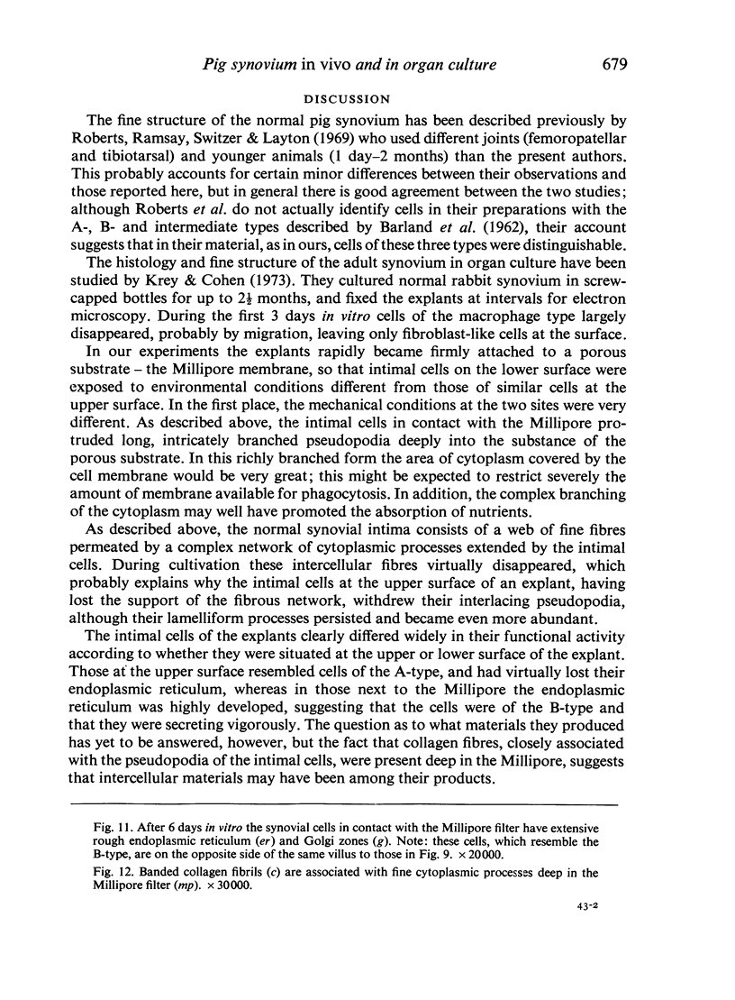
1. The normal synovium of the metacarpophalangeal joints of young pigs was examined by light and electron microscopy with special reference to the superficial layer (intima). 2. Cells of the macrophage-like or A-type (Barland et al. 1962) constituted only a small proportion of the intimal synoviocytes; the majority were of the intermediate and B-types. 3. Synovial villi were explanted on Millipore filters and maintained as organ cultures. The intimal cells in contact with the Millipore formed long branched processes which penetrated deeply into the substrate; these cells, which had a very well-developed endoplasmic reticulum, resembled those of the B-type. The synoviocytes at the upper (free) surface of the villus withdrew their long processes, acquired lamelliform pseudopodia, and their endoplasmic reticulum regressed; they were similar in appearance to the A-type. 4. In the organ cultures the highly branched cells (B-type) next to the Millipore were less phagocytic than the rounded cells (A-type) at the free surface of the villus.
Full text
PDF
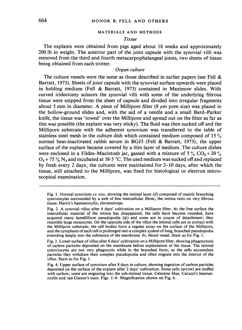
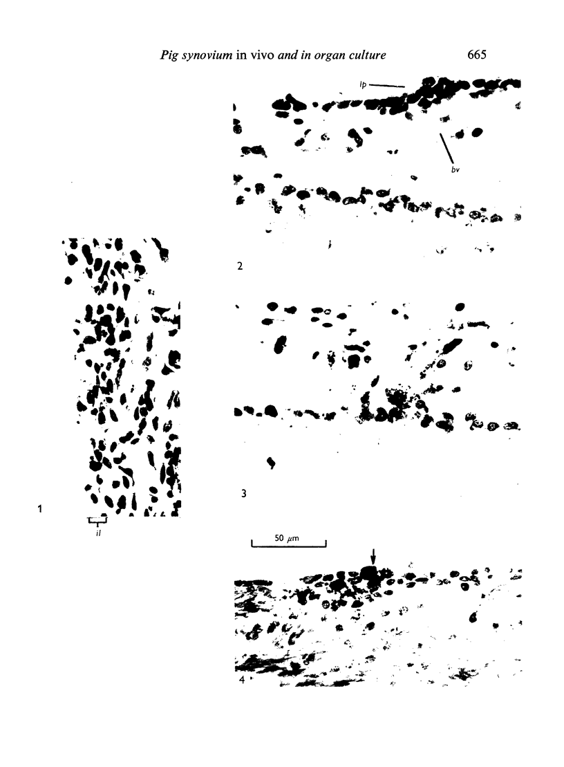
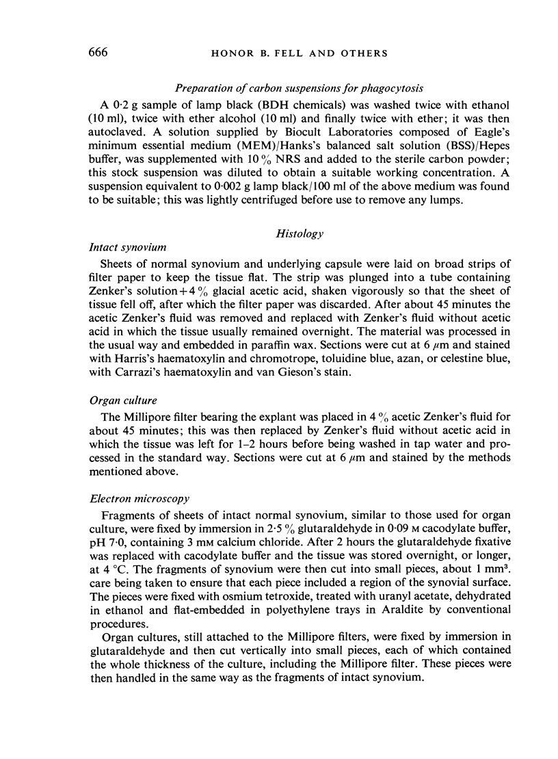
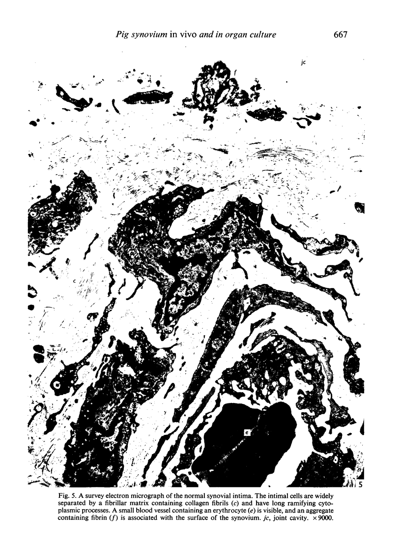
Images in this article
Selected References
These references are in PubMed. This may not be the complete list of references from this article.
- BARLAND P., NOVIKOFF A. B., HAMERMAN D. Electron microscopy of the human synovial membrane. J Cell Biol. 1962 Aug;14:207–220. doi: 10.1083/jcb.14.2.207. [DOI] [PMC free article] [PubMed] [Google Scholar]
- Fell H. B., Barratt M. E. The role of soft connective tissue in the breakdown of pig articular cartilage cultivated in the presence of complement-sufficient antiserum to pig erythrocytes. I. Histological changes. Int Arch Allergy Appl Immunol. 1973;44(3):441–468. doi: 10.1159/000230951. [DOI] [PubMed] [Google Scholar]
- Krey P. R., Cohen A. S. Fine structural analysis of rabbit synovial cells. I. The normal synovium and changes in organ culture. Arthritis Rheum. 1973 May-Jun;16(3):324–340. doi: 10.1002/art.1780160306. [DOI] [PubMed] [Google Scholar]
- Luckenbill L. M., Cohen A. S. Phagocytic function of the avian synovial membrane: a light and electron microscopic study. Arthritis Rheum. 1967 Dec;10(6):517–537. doi: 10.1002/art.1780100605. [DOI] [PubMed] [Google Scholar]
- Roberts E. D., Ramsey F. K., Switzer W. P., Layton J. M. Electron microscopy of procine synovial cell layer. J Comp Pathol. 1969 Jan;79(1):41–45. doi: 10.1016/0021-9975(69)90025-5. [DOI] [PubMed] [Google Scholar]


